
A Novel CsYABBY3‐CsAS1 Feedback Loop Coordinates Trichome Differentiation and Cannabinoid Biosynthesis in Cannabis sativa L.
Abstract
Glandular trichome development typically coincides with specialized‐metabolite production, yet the regulatory mechanisms that synchronizes these processes remain elusive. We uncover a trichome‐enriched CsYABBY3‐CsAS1 transcriptional module that links glandular trichome differentiation with cannabinoid biosynthesis in cannabis. CsYABBY3, a FILAMENTOUS FLOWER (FIL)/YAB3‐family transcription factor, promotes glandular trichome formation and elevates total cannabinoid levels. It directly binds a conserved TAATTAA motif in the promoters of CsPT4 and CsCBDAS, activating their transcription. The R2R3‐MYB CsAS1 can also stimulate cannabinoid pathway gene expression independently, but its activity is enhanced in partnership with CsYABBY3. The two factors physically associate and reciprocally upregulate each other, forming a positive feedback loop that amplifies target‐gene output. A single residue (M199) in CsYABBY3 is required for CsAS1 interaction and cooperative promoter activation. Comparative evidence suggests broadly conserved FIL/YAB3‐AS1 coupling across species, alongside lineage‐specific differences in the molecular basis of complex formation. These findings define an engineerable regulatory circuit that connects trichome fate with specialized metabolism and provide a practical route to improve cannabinoid yield.
Article type: Research Article
Keywords: cannabinoid biosynthesis, feedback loop, FIL/YAB3‐AS1 transcriptional module, trichome differentiation
Affiliations: State Key Laboratory for Quality Ensurance and Sustainable Use of Dao‐di Herbs Institute of Chinese Materia Medica China Academy of Chinese Medical Sciences Beijing China; Key Laboratory of National Forestry and Grassland Administration for Chinese Herbal Medicine College of Life Science Northeast Forestry University Harbin China; Institute of Chinese Materia Medica and Artemisinin Research Center China Academy of Chinese Medical Sciences Beijing China; Lushan Botanical Garden Chinese Academy of Sciences Jiujiang Jiangxi China; College of Science National University of Defense Technology Changsha Hunan China; South China Botanical Garden Chinese Academy of Sciences Guangzhou Guangdong China; Institute of Herbgenomics Chengdu University of Traditional Chinese Medicine Chengdu China
License: © 2026 The Author(s). Advanced Science published by Wiley‐VCH GmbH CC BY 4.0 This is an open access article under the terms of the http://creativecommons.org/licenses/by/4.0/ License, which permits use, distribution and reproduction in any medium, provided the original work is properly cited.
Article links: DOI: 10.1002/advs.75055 | PubMed: 41926629 | PMC: PMC13285160
Relevance: Relevant: mentioned in keywords or abstract
Full text: PDF (60 KB)
Introduction
Cannabis sativa L. is an herbaceous plant in the Cannabinaceae family, known for its medicinal and commercial value due to the production of cannabinoids, a class of terpenophenolic compounds [ref. 1]. These cannabinoids, which are primarily synthesized as acidic precursors such as Δ9‐tetrahydrocannabinolic acid (THCA) and cannabidiol acid (CBDA), are produced in the glandular trichomes of pistillate bracts and sugar leaves [ref. 2]. While Δ9‐tetrahydrocannabinol is known for psychoactive effects, cannabidiol has demonstrated therapeutic properties, such as in the treatment of refractory seizures [ref. 3]. The increasing global demand for cannabinoids has spurred significant interest in optimizing their production in C. sativa, making it critical to understand the regulatory mechanisms governing cannabinoid biosynthesis [ref. 4, ref. 5].
Glandular trichomes are specialized epidermal structures essential for producing secondary metabolites, including cannabinoids. Cannabis trichomes, distributed on flowers, bracts, sugar leaves, and leaves, store phytocannabinoids, and their density correlates with cannabinoid content, making them an ideal system for studying trichome development and plant secondary metabolism [ref. 6, ref. 7]. Cannabinoid biosynthesis originates from the enzymatic conversion of precursor compounds, olivetolic acid (OA) and geranyl diphosphate (GPP), into cannabigerolic acid (CBGA), which is subsequently converted into cannabidiolic acid (CBDA) and tetrahydrocannabinolic acid (THCA) by cannabinoid synthases CsCBDAS and CsTHCAS, respectively [ref. 8, ref. 9, ref. 10]. Emerging research has identified several transcription factors (TFs) that influence cannabinoid biosynthesis and trichome development. For instance, CsMIXTA and CsMYC4 have been shown to regulate trichome initiation and activate genes involved in cannabinoid biosynthesis [ref. 11]. While individual factors controlling trichome density have been identified, the integrated regulatory modules that synchronize the morphological differentiation of glandular trichomes with the specialized metabolic pathways for cannabinoid biosynthesis are not yet understood.
Among the TFs involved in plant development, the YABBY gene family is notable for its role in lateral organ formation and trichome initiation [ref. 12, ref. 13, ref. 14]. YABBY genes, unique to seed plants, encode proteins defined by a C2C2 zinc‐finger domain and a YABBY‐specific domain [ref. 14, ref. 15, ref. 16, ref. 17]. Phylogenetically, the family is classified into five clades: FILAMENTOUS FLOWER (FIL)/YAB3, YAB2, YAB5, CRABS CLAW (CRC), and INNER NO OUTER (INO) [ref. 18, ref. 19]. The FIL/YAB3 subgroup is preferentially expressed in leaves and leaf‐derived organs and contributes to leaf polarity and trichome development [ref. 12, ref. 18, ref. 20, ref. 21]. In Arabidopsis thaliana, YABBY proteins form homodimers or heterodimers with other TFs, such as AtKANADI, to modulate the development of lateral organs and trichome formation [ref. 22]. Emerging evidence further implicates YABBYs in glandular trichome development and the regulation of secondary metabolite accumulation; for example, YABBY genes have been linked to these processes in Mentha spicata and Artemisia annua [ref. 23, ref. 24, ref. 25].
In this study, we unveil a novel synergistic transcriptional module that coordinates trichome differentiation with cannabinoid biosynthesis. Departing from previously characterized single‐factor regulators, we demonstrated that the FIL/YAB3 member CsYABBY3 and the R2R3‐MYB protein CsAS1 establish a reciprocal positive feedback loop. Mechanistically, we identify a critical methionine residue (M199) in CsYABBY3 as indispensable for the physical interaction and functional synergy of this complex. These findings define a previously unknown CsAS1‐CsYABBY3‐CsPT4/CsCBDAS regulatory axis that couples trichome differentiation with specialized metabolism, providing a strategic route to orchestrate cannabinoid production in C. sativa.
Results
Identification and Characterization of the Cannabinoid Biosynthesis‐Related Transcription Factor CsYABBY3
In C. sativa, glandular trichomes are primarily localized on the pistillate bract surfaces and associated bracteal tissues (Figure 1A), functioning as specialized metabolic factories for cannabinoid biosynthesis and storage [ref. 26]. To establish the spatial coordination between trichome distribution and cannabinoid deposition, we performed high‐resolution matrix‐assisted laser desorption/ionization mass spectrometry imaging (MALDI‐MSI), revealing an obvious accumulation of cannabidiol (CBD), cannabigerol (CBG), CBDA, and CBGA predominantly within glandular trichomes (Figure 1B,C; Figures S1A).
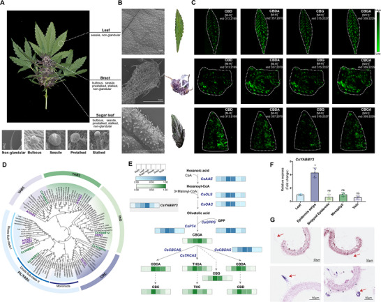
To identify the regulators of cannabinoid biosynthesis, we conducted a weighted gene co‐expression network analysis (WGCNA) using transcriptomes from six organs of C. sativa (cv. DiKu): flower, bracts, sugar leaf, leaf, stem, seed, and root. This analysis recovered a module enriched with cannabinoid biosynthetic genes, including CsPT4, CsCBDAS, and CsGPPS, within which 72 TFs co‐clustered. To ensure the identified regulators are functionally conserved across different genetic backgrounds, we integrated our organ‐level transcriptome data (Group A) with a publicly released trichome‐specific dataset (Group B), covering a total of eight distinct cultivars. By applying a threshold of FPKM > 10, we identified 12 candidate TFs that consistently exhibited both flower/sugar leaves‐specific and trichome enriched expression across all sampled cultivars (Figure S1B,C). Notably, a member of the YABBY family, designated as CsYABBY3, was annotated.
A genome‐wide survey identified five YABBY genes in C. sativa, and phylogenetic analysis indicated that the co‐expressed gene belongs to the FIL/YAB3 clade (Figure 1D; Table S1). The expression pattern of CsYABBY3 correlated with cannabinoid accumulation, particularly total CBD and CBG (Figure 1E; Figure S1D). Given the expression pattern, qRT‐PCR validation showed that only CsYABBY3 was significantly expressed in the trichome‐rich epidermal strips of leaves (Figure 1F; Figure S1E). Domain analysis revealed that CsYABBY3 encodes a 235‐amino acid protein with two conserved domains: an N‐terminal C2C2 zinc finger domain and a C‐terminal YAB domain (Figure S2). Furthermore, we performed RNA in situ hybridization to determine the spatial expression of CsYABBY3 and found that it was predominantly localized to the trichomes in C. sativa (Figure 1G). These findings suggest a potential regulatory role for CsYABBY3, a member of the FIL/YAB3 subgroup, in the biosynthesis of cannabinoids within cannabis trichomes.
CsYABBY3 Coordinated Trichome Differentiation and Cannabinoid Biosynthesis
To elucidate the functional role of CsYABBY3 in CBDA biosynthesis, we transiently overexpressed this TF in C. sativa leaves (Figure 2A) and generated stable transgenic hairy root lines (Figure 2B), utilizing RUBY reporter to validate overexpression. LC‐MS quantification revealed significantly higher total CBD and CBG accumulation in CsYABBY3‐OE leaves and hairy roots than in controls (Figure 2A,B). Consistently, CsYABBY3‐OE in both the transient assay (Figure 2C) and stable hairy root system (Figure 2D) upregulated key cannabinoid biosynthetic genes, including CsGPPS, CsPT4, and CsCBDAS.
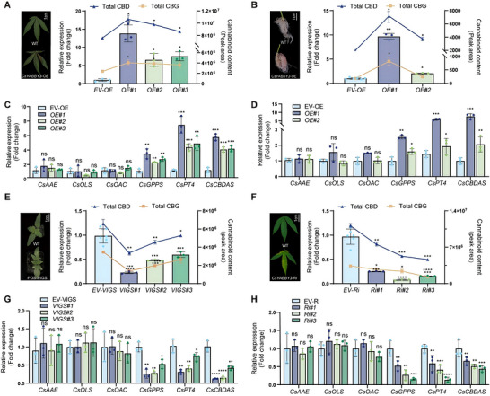
We further employed three loss‐of‐function approaches, including virus‐induced gene silencing (VIGS), RNA interference (RNAi), and antisense oligonucleotide (AsODN). Relative to the TRV2 control group, TRV‐CsYABBY3 lines exhibited significantly reduced CsYABBY3 transcript abundance and, correspondingly, lower total CBD and CBG (Figure 2E). Consistent with the VIGS results, CsYABBY3 suppression by RNAi and AsODN similarly decreased cannabinoid accumulation (Figure 2F; Figure S3B). qRT‐PCR further revealed marked downregulation of CsGPPS, CsPT4, and CsCBDAS in TRV2‐CsYABBY3, CsYABBY3‐RNAi, and CsYABBY3‐AsODN lines compared with their respective controls (Figure 2G,H; Figure S3C). Collectively, these results demonstrate that CsYABBY3 functions as a transcriptional activator that orchestrates pathway gene expression to modulate cannabinoid biosynthesis in C. sativa.
To test whether CsYABBY3 regulates trichome development, we generated CsYABBY3‐OE tomato plants. Scanning electron microscopy (SEM) revealed that the densities of both glandular and non‐glandular trichomes were significantly increased in CsYABBY3‐OE plants relative to wild type (WT) (Figure S4A–E). In two independent CsYABBY3‐OE lines, trichome density increased by 2.5‐fold and 6.5‐fold compared with WT. Given that cannabinoids and monoterpenes share the 2‐C‐methyl‐D‐erythritol 4‐phosphate (MEP) pathway, we profiled terpenoid metabolites in leaves and fruits of CsYABBY3‐OE tomatoes plants. Compared with WT, CsYABBY3‐OE lines accumulated significantly more total terpenoids in both organs and showed increased GPP in fruits (Figure S4F,G). Together, these findings support a role for CsYABBY3 in regulating trichome development and terpenoid biosynthesis, potentially through modulation of the MEP pathway.
CsYABBY3 Activated the Expression of CsPT4 and CsCBDAS in C. sativa by Directly Binding to Their Promoters
Subcellular localization showed that CsYABBY3 was localized in the nucleus (Figure S5). To clarify the regulatory role of CsYABBY3, we performed Y1H assays, which demonstrated that CsYABBY3 directly bound to the full‐length promoters of CsPT4 (CsPT4pro) (Figure 3A). The full‐length promoters of CsCBDAS (CsCBDASpro) exhibited strong self‐activation (Figure S6A). MST analysis further confirmed the direct binding of CsYABBY3 to both CsPT4 and CsCBDAS promoters, with binding affinity (Kd) of 29.66 and 218.46 nm, respectively (Figure 3B). To assess the functional impact of CsYABBY3 binding to these promoters, we conducted Dual‐LUC assays. Co‐expression of 35S:CsYABBY3 and CsPT4pro:LUC or CsCBDASpro:LUC reporter constructs resulted in significantly higher reporter gene expression in N. benthamiana (Figure 3C), suggesting that CsYABBY3 activates the transcription of CsPT4 and CsCBDAS. Collectively, these findings establish CsYABBY3 as a nuclear‐localized transcriptional activator that directly targets CsPT4 and CsCBDAS promoters to drive CBDA biosynthesis.
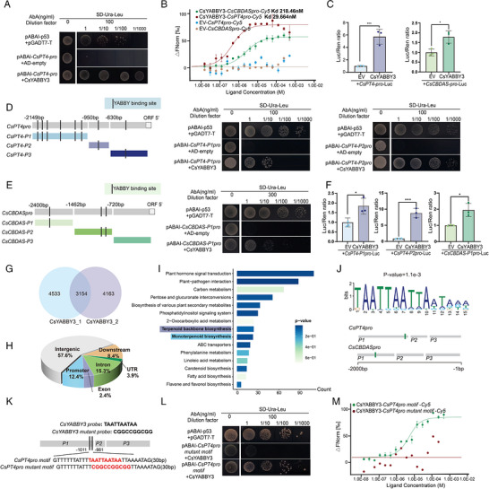
To investigate the cis‐regulatory elements recognized by CsYABBY3 in the ∼2 kb promoter regions upstream of CsPT4 and CsCBDAS, we performed computational predictions of TF binding sites using the JASPAR database. The analysis identified seven potential CsYABBY3‐binding motifs in the CsPT4pro and three in the CsCBDASpro (Figure 3D,E). Based on the motif distribution, we generated three sequential promoter truncation constructs for both CsPT4pro and CsCBDASpro (designated as P1‐P3pro), none of which exhibited transcriptional self‐activation (Figure S6B–D). Y1H and Dual‐LUC assays demonstrated specific binding interactions between CsYABBY3 and the CsPT4‐P1pro, CsPT4‐P2pro, and CsCBDAS‐P1pro fragments (Figure 3D‐F, Supporting Information).
To delineate the precise binding loci of CsYABBY3, we performed DAP‐seq, achieving a 99% mapping rate of clean reads to the reference genome and identifying 3154 CsYABBY3‐binding peaks across all 10 chromosomes (Figure 3G; Figure S7A). Approximately 12.4% of the binding peaks were located within 2 kb upstream of the transcription start site, corresponding to the promoter region (Figure 3H; Figure S7B). KEGG pathway and Gene Ontology (GO) enrichment analyses of binding peak‐associated target genes identified enriched terms related to Terpenoid backbone biosynthesis and Monoterpenoid biosynthesis (Figure 3I), as well as fatty acid metabolic process (Figure S7C). The highest‐scoring cis‐acting element from DAP‐seq data, TAATTAA, was found in both CsPT4pro and CsCBDASpro, indicating that these promoters are direct targets of CsYABBY3 (Figure 3J,K). The sequence‐specific interaction between CsYABBY3 and the core motif (−1011∼−981 bp) in CsPT4‐P1pro was further validated by independent Y1H assays (Figure 3L) and MST analysis (Figure 3M). Importantly, mutation of the TAATTAA motif to CGGCCGGCGG completely abolished the binding in both assays, providing definitive evidence for the motif‐specific recognition of CsYABBY3.
CsAS1 Binds to the M199 Residue of the CsYABBY3 Protein
To identify potential interacting protein partners of CsYABBY3 implicated in the regulation of cannabinoid biosynthesis, we performed yeast two‐hybrid (Y2H) library screening (Table S2). Among the candidates, a trichome‐enriched R2R3‐MYB transcription factor was identified and designated CsAS1 (ASYMMETRIC LEAF1). This interaction was functionally validated through Y2H (Figure 4A) and firefly luciferase complementation imaging (LCI) (Figure 4B). Co‐immunoprecipitation (Co‐IP) experiments further corroborated the interaction between CsAS1‐HA and CsYABBY3‐GFP fusion proteins (Figure 4C). Isothermal Titration Calorimetry (ITC) analysis revealed a Kd of 4.74 ± 0.56 µm with a 1:1 binding stoichiometry (N = 1.06), indicating moderate binding affinity (Figure 4D). Subcellular localization showed nuclear enrichment of CsAS1, consistent with a role as a transcriptional regulator (Figure S8A).
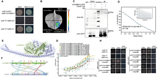
To elucidate the molecular basis of the CsYABBY3‐CsAS1 interaction, we divided the CsYABBY3 coding sequence into three distinct fragments based on the conserved YABBY protein domains: an N‐terminal zinc finger domain (P1), a middle region lacking conserved motifs (P2), and a C‐terminal YABBY domain (P3) (Figure S8A,B). Transcriptional auto‐activation assays confirmed that CsAS1 had no self‐activation activity. Y2H assays revealed a direct interaction between CsAS1 and the C‐terminal YABBY domain (CsYABBY3‐P3). Molecular docking predicted a high‐affinity heterodimeric complex (Figure 4E) with eight putative hydrogen‐bonding sites (#1‐#8) spanning the protein‐protein interface, with bond lengths ranging from 1.5 to 4.2 Å (Figure 4F; Figure S8C).
MST analysis quantified the binding affinities of site‐directed CsYABBY3 interface mutants (#1‐#8; Figure S8) to CsAS1, yielding Kd of 3.22–31.62 µm, compared with 8.05 µm for the wild‐type CsYABBY‐CsAS1 complex (Figure 4G). Among these, mutant #7 exhibited significantly reduced affinity. Y2H assays confirmed that only the CsYABBY3 mutant #7 failed to grow on QDO medium supplemented with X‐α‐Gal (Figure 4H), highlighting residue M199 (site #7) as indispensable for the CsYABBY3‐CsAS1 interaction in vivo.
CsAS1 Is a Positive Regulator of CBDA Biosynthesis
Organs‐specific expression profiling demonstrated strong co‐expression of CsAS1 with CsYABBY3, CsPT4, and CsCBDAS across cannabinoid‐rich organs of different cultivars (Figure S1B). Subcellular localization showed CsAS1 was located in nucleus (Figure S9A). RNA in situ hybridization localized CsAS1 transcripts exclusively to glandular trichomes, mirroring the expression pattern of CsYABBY3 (Figure S9B).
To investigate CsAS1 function, we applied the same framework used for CsYABBY3. CsAS1‐OE lines in transient cannabis leaves (Figure S10A) and hairy root transformation systems (Figure 5A) revealed a significant increase in total CBD and CBG accumulation relative to controls. Correspondingly, transcriptional suppression of CsAS1 by VIGS and AsODN reduced CsAS1 expression and markedly decreased total CBD and CBG (Figure 5B; Figure S10B). Parallel qRT‐PCR analyses demonstrated substantial downregulation of CsPT4 and CsCBDAS in silenced lines (Figure 5A,B, right panels), supporting CsAS1 as a transcriptional activator of these key cannabinoid biosynthetic genes in C. sativa.
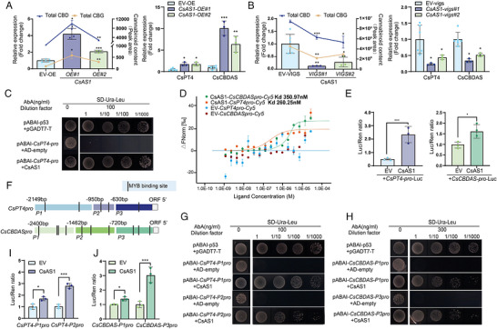
Y1H assays demonstrated direct binding of CsAS1 to CsPT4pro (Figure 5C). MST further quantified this interaction, revealing high‐affinity binding of CsAS1 to CsPT4pro (Kd = 260.25 nm) and CsCBDAS (Kd = 350.97 nm) (Figure 5D). Dual‐LUC assays showed that co‐expression of 35S:CsAS1 with CsPT4pro:LUC or CsCBDASpro:LUC significantly enhanced reporter activity (Figure 5E), confirming CsAS1 as a transcriptional activator. We predicted at least one putative CsAS1‐binding motif in each truncated fragment of CsPT4pro and CsCBDASpro (Figure 5F). Promoter truncation assays indicated that CsAS1 directly bound to CsPT4‐P1pro, CsPT4‐P2pro, CsCBDAS‐P1pro, and CsCBDAS‐P3pro fragments (Figure 5G‐J). Together, these results demonstrate that CsAS1 directly regulates cannabinoid biosynthetic genes at the transcriptional level in C. sativa.
CsYABBY3 and CsAS1 Established a Positive Feedback Loop and Synergistically Regulate CBDA Biosynthesis in C. sativa
To define the regulatory relationship between CsYABBY3 and CsAS1, we mapped the top motifs enriched by DAP‐seq onto their respective promoters (Figure S11A,B). Y1H assays verified direct binding of CsYABBY3 to the CsAS1 promoter (CsAS1pro) (Figure 6A). MST analysis further quantified this interaction, revealing a Kd of 13.4 µm (Figure 6B). Dual‐LUC assays demonstrated that CsYABBY3 activates CsAS1pro (Figure 6C). Consistently, qRT‐PCR analysis showed elevated CsAS1 transcript levels in CsYABBY3‐OE hairy roots and reduced abundance in VIGS‐mediated CsYABBY3‐silenced lines (Figure 6D).
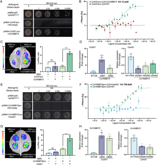
Reciprocally, both Y1H (Figure 6E) and MST (Figure 6F) confirmed that CsAS1 directly binds to the CsYABBY3 promoter (CsYABBY3pro), and Dual‐LUC assays demonstrated activation of CsYABBY3pro by CsAS1(Figure 6G). Moreover, CsYABBY3 expression positively correlated with CsAS1 transcript abundance in CsAS1‐OE hairy roots and VIGS‐mediated CsAS1‐silenced plants, suggesting that CsYABBY3pro is a direct transcriptional target of CsAS1 (Figure 6H). Together, these results indicate that CsYABBY3 and CsAS1 mutually activate each other’s transcription, forming a positive feedback regulatory loop.
CsYABBY3‐CsAS1 Synergistically Regulating Cannabinoid Biosynthesis
Given that CsYABBY3 alone acts as a positive regulator of cannabinoid biosynthesis, we further investigated the role of the CsYABBY3‐CsAS1 module in cannabinoid metabolism in C. sativa. Dual‐LUC assays revealed that co‐expression of CsYABBY and CsAS1 in N. benthamiana significantly activated the full‐length CsPT4pro and CsCBDASpro (Figure 7A), as well as the truncated CsPT4‐P1pro (Figure 7B), CsPT4‐P2pro (Figure 7C), and CsCBDAS‐P1pro (Figure 7D), compared to either factor alone. These results indicate that the CsYABBY3‐CsAS1 module cooperatively enhances transcription of downstream biosynthetic genes.
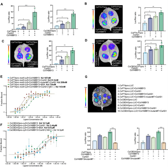
To assess how CsYABBY3 interface mutants affect promoter recognition in complex with CsAS1, we performed MST using complexes of mutants #1‐#8 with CsAS1 and measured binding to the CsPT4pro motif (Figure 7E) and CsCBDASpro (Figure 7F). The CsYABBY3 mutant #7‐CsAS1 complex showed a substantially reduced affinity for both promoters relative to the wild‐type CsYABBY3‐CsAS1, approaching the levels of CsYABBY3 alone or CsYABBY3 paired with inactive CsAS1. Dual‐LUC assays revealed that co‐expression of CsYABBY3 mutant #7 with CsAS1 significantly repressed CsPT4pro and CsCBDASpro activities, whereas other mutants (except #8) showed no significant differences (Figure 7G; Figure S8D). These results demonstrate that substitution at M199 residue disrupts the CsYABBY3‐CsAS1 interaction, weakening promoter binding and impairing transcriptional activation.
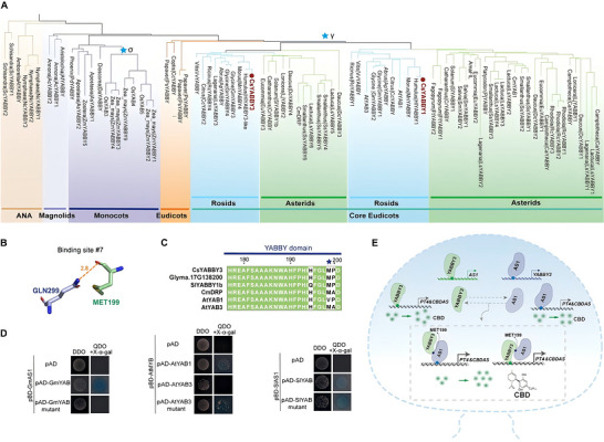
To explore the evolutionary significance of this interaction, we analyzed the phylogeny of the FIL/YAB3 subfamily (Figure 8A; Table S4). In core eudicots, FIL/YAB3 genes diverged into two subclades: Subclade I and Subclade II, likely as the result of their shared whole‐genome triplication‐γ. Sequence alignment revealed that the M199 residue is highly conserved across core eudicots, which structurally serves as a key docking site (Figure 8B,C). Based on the phylogenetic analysis, we cloned AtYAB1 and AtYAB3 from Arabidopsis (subclade I), only GmYAB and SlYABBY1b from soybean and tomato (subclade II), along with their respective AS1 homologs. Y2H assays confirmed that all these species form the FIL/YAB3‐AS1 protein complex. In soybean, as in cannabis, M199 is the critical residue for the GmYAB‐GmAS1 interaction. However, in Arabidopsis, while AtYAB1 interacts with AtAS1, AtYAB3 does not. Introduction of the double substitution (M199V/A200P) into AtYAB3 restored its interaction with AtAS1 (Figure 8D). Similarly, in tomato, the single M199 residue is insufficient to maintain the FIL/YAB‐AS1 interaction. These results suggest that the mechanism by which M199 determines FIL/YAB3‐AS1 interaction is conserved within subclade II of FIL/YAB3 in Rosids. While both subclades in core eudicots exhibit functional redundancy, they have evolved distinct mechanisms due to structural changes.
Discussion
C. sativa, a plant of substantial economic and therapeutic significance, synthesizes specialized cannabinoids primarily within its glandular trichomes [ref. 4]. While the biosynthetic pathways of these compounds are well understood, their regulatory networks, and in relation to trichome development remain inadequately elucidated. Here, we identify a novel transcriptional regulatory module involving FIL/YAB3 member CsYABBY3 and the R2R3‐MYB factor CsAS1 (Figure 8). These transcription factors directly activate the promoters of key biosynthetic genes (CsPT4 and CsCBDAS), thereby synergistically enhancing cannabinoid production and trichome development. The increased density of both trichome types in the CsYABBY3‐OE tomato model suggests a general role in initiation, while the higher proportion of glandular types likely reflects a specialized preference for glandular specification through its synergy with CsAS1. This establishes a reciprocal CsYABBY3‐CsAS1 feedback loop as a core molecular switch for trichome‐specific metabolism, adding a critical regulatory layer to established biosynthetic cascades [ref. 27, ref. 28, ref. 29, ref. 30].
Mechanistically, this feedback loop integrates mutual transcriptional activation with direct protein–protein interaction. DAP‐seq and functional assays confirm that CsYABBY3 and CsAS1 reciprocally bind each other’s promoters (Figure S11A,B; Figure 6). This bidirectional activation ensures the robust co‐accumulation of both regulators, which subsequently physically interact to amplify the expression of shared downstream targets. Similar regulatory architectures are well‐established in plants, such as the SEP3‐AP1/AP3/AG complex in Arabidopsis [ref. 31] and the CaWRKY27b‐CaWRKY40 circuit in pepper [ref. 32]. Regarding their co‐occupancy on shared targets (CsPT4 and CsCBDAS), the physical distance between the distinct CsYABBY3 and CsAS1 motifs is insufficient for a pre‐formed complex to simultaneously bridge both sites. Instead, our structural modeling and molecular docking support a mutual recruitment mechanism (Figure S11C–E). Either TF can independently anchor to its specific motif and subsequently recruit its partner to form a functional heterodimer in situ. Docking simulations indicate that complex formation does not sterically hinder DNA interaction; rather, the entire complex stably co‐binds to a single motif, enhancing overall DNA‐binding affinity without site competition.
The FIL/YAB3 subfamily phylogenetically and functionally diverges across angiosperms. In monocots, they exhibit diversified expression patterns, such as the uniform expression of rice OsYABBY1 in specific cell layers [ref. 33, ref. 34] and the abaxial expression of maize ZYB9 and ZYB14 [ref. 35]. In contrast, eudicot FIL/YAB3 members generally maintain the ancestral adaxial expression pattern, exemplified by Arabidopsis AtYAB1 and AtYAB3, Antirrhinum GRAMINIFOLIA (GRAM), and soybean GmFILa, participating in lateral organ polarity and leaf morphogenesis [ref. 36, ref. 37]. Within the FIL/YAB3 subfamily in core eudicots, subclade II represents a distinct evolutionary lineage. The FIL/YAB3‐AS1 physical interaction is conserved across various core eudicots highlights its fundamental role as an ancestral regulatory scaffold (Figure 8E). We propose that the CsAS1‐CsYABBY3 module represents an evolutionary co‐option, where this conserved developmental switch was recruited to synchronize glandular trichome differentiation with the specialized metabolic demands of cannabinoid biosynthesis. Similar recruitment of YABBY homologs for specialized metabolism has been observed in other aromatic and medicinal species, such as M. spicata and A. annua [ref. 38, ref. 39]. Crucially, our identification of this conserved M199 residues as a requisite docking site further suggests that this biochemical interface is an inherent evolutionary feature, providing a universal molecular framework that lineages can adapt to orchestrate high‐output specialized pathways. Mutations in M199 disrupt complex formation and abolish the transcriptional activation of downstream targets, echoing the well‐known MYB‐bHLH‐WD40 (MBW) pigment model [ref. 40], where specific amino acid interactions are fundamental for functional transcriptional complexes.
The initiation and termination of this feedback loop likely involve the coordination of early hormonal fluctuations and upstream regulators, as well as the physiological maturation and senescence of the glandular trichome head [ref. 6, ref. 28, ref. 41]. A similar mechanism has been observed in other plants [ref. 42, ref. 43], such as in Arabidopsis, where the MYC2‐DOF2.1‐MYC2 circuit has been shown to regulate leaf senescence [ref. 44]. Ultimately, these findings provide significant insights for optimizing cannabinoid production in the C. sativa industry. Future research should focus on expanding the network of regulators that interact with this complex to provide a more comprehensive understanding of the transcriptional landscape in C. sativa. Moreover, while our study establishes a robust transcriptional framework, future endeavors aimed at characterizing the endogenous protein stability and post‐translational dynamics of the CsYABBY3‐CsAS1 complex will further refine the spatiotemporal precision of this regulatory axis. It will also be important to explore how environmental factors, such as light, temperature, and nutrient availability, influence the expression of CsYABBY3 and CsAS1, as these factors are known to affect trichome density and cannabinoid content in plants. Additionally, validating the role of the CsYABBY3‐CsAS1 feedback loop under field conditions will be crucial for translating these findings into practical agricultural applications.
Experimental Section
Plant Materials and Growth Conditions
Previously described cannabis organ transcriptomes from multiple cultivars established by our group were used [ref. 45]. C. sativa, A. thaliana, and N. benthamiana plants were sourced from the greenhouse of Northeast Forestry University. Plants were grown under a 16‐h light/8‐h dark photoperiod at 23°C.
RNA‐seq and Transcriptome Analysis
Total RNA was extracted from flowers, bracts, sugar leaves, leaves, stem, roots, seeds of C. sativa cultivar“Diku” using the Quick RNA extraction kit (Huayueyang, Beijing, China). Samples with RNA integrity number of > 8 were retained for sequencing. Illumina sequencing was performed at BGI Genomics Co. Ltd. (Beijing, China). and RNA‐seq data were processed, assembled, and annotated. All the assembled transcripts were aligned to six databases: NR, Swiss‐Prot, Pfam, COG, GO, and KEGG to obtain annotation information. Gene expression was quantified using Fragments Per Kilobase Million values.
Standards and Chemical Reagents
OA (CAS: 508‐02‐1), CBD (CAS: 13956‐29‐1), CBDA (CAS: 1244‐58‐2), CBG (CAS: 25654‐31‐3); CBGA (CAS: 25555‐57‐1), and GPP (CAS: 104715‐14‐2) standards were obtained from Sigma–Aldrich (USA).
MALDI‐Imaging
Bracts, sugar leaves, and leaves were mounted on indium tin oxide (ITO)‐coated glass slides. To enhance ionization, samples were treated with a matrix solution containing 10 mg/mL DHB in a 1:1 (v/v) mixture of methanol and water. MALDI‐imaging was performed with a spatial resolution of 15 µm using MirionV3 software. Other experimental parameters were based on previous protocols published by our group [ref. 46].
Phylogenetic Analysis and Sequence Alignment
Amino acid sequences of CsYABBY3 protein and its homologs were retrieved from the NCBI database using BLAST (https://blast.ncbi.nlm.nih.gov/). Homology matching was performed using BLASTP searches and the ‘Blast Several Sequences to a Big Database’ function in TBtools, with parameters set to an E‐value < 10− 5 and redundant sequences removed, ultimately identifying all YABBY gene family members in other species. Multiple amino acid sequences were aligned using ClustalW, and phylogenetic trees were constructed using Maximum likelihood method implemented in the MEGAX program with 1000 bootstrap replicates [ref. 47, ref. 48].
Total RNA Isolation and qRT‐PCR
RNA was extracted from flash‐frozen samples using the FastPure Plant Total RNA Isolation Kit (Vazyme, China). cDNA synthesis and qRT‐PCR was performed using ReverTra Ace qPCR RT Master Mix with gDNA Remover (TOYOBO, Japan) and ChamQ Universal SYBR qPCR Master Mix (Vazyme, China) [ref. 49, ref. 50]. Relative gene expression was calculated using the 2−△△CT methods as previously described [ref. 51]. Primers used for qRT‐PCR are listed in Table S3.
Gene and Promoter Cloning
Open reading frames (ORFs) of CsAS1, CsYABBY3, and homologs from other species were amplified using KOD One PCR Master Mix (TOYOBO, Japan) from corresponding cDNA templates using gene‐specific primers (Table S3). Promoter regions of CBDA biosynthetic genes and TFs were amplified from genomic DNA of the cannabis cultivar Diku.
Subcellular Localization
Coding sequences of CsYABBY3 and CsAS1 were fused in‐frame to eGFP in the pEAQ‐HT‐eGFP vector. Constructs were transformed into Agrobacterium tumefaciens strain GV3101 (Psoup) and infiltrated into N. benthamiana leaves. GFP fluorescence was imaged on a Zeiss LSM510 META confocal laster‐scanning microscope after 24 h in darkness followed by 48 h under light.
Prediction of YABBY Transcription Factor Binding Sites
Promoter sequences of CsPT4 and CsCBDAS were scanned for YABBY TF binding sites using the JASPAR 2024 database (http://jaspar.genereg.net) [ref. 52]. Sites with relative score above 0.90 were mapped to genomic positions.
RNA in Situ Hybridization
C. sativa vegetative leaves were fixed in ice‐cold 1 × PBS with 4% paraformaldehyde/4% DMSO, vacuum‐infiltrated for 3 min, and post‐fixed with replaced fixative overnight at 4°C. Samples were dehydrated, cleared, and embedded in Paraplast (Sigma–Aldrich, cat. no. P3683); 8 µm sections were cut using a Leica RM2235 microtome. Gene‐specific 400–1000 bp fragments (Table S3) were cloned bidirectionally into the pEASY‐Blunt Zero cloning vector (TransGen Biotech, CB501) to generate antisense/sense probes. The digoxigenin‐labelled (Roche, 11277073910) antisense and sense probes were transcribed with T7 RNA polymerase (Promega, P4074). Sections were dewaxed, treated with proteinase K, dehydrated, and incubated in probe buffer for 12 h at 50°C in a humidified box. The sections were then incubated with anti‐digoxin antibody (Roche, 11093274910) for 2 h at 25°C, and developed with NBT/BCIP reaction solution (Roche, 11681451001) for 36–48 h at 25°C in a dark humidified box. The images were acquired by bright‐field microscope (Olympus BX‐50).
SEM
The middle part of the first leaf from the third internode was surveyed using a HITACHI UHR FE‐SEM SU8010 Series scanning electron microscope (Hitachi, Tokyo, Japan). Trichome counts were determined using the IMAGEJ software (https://imagej.nih.gov/ij/download.html).
Cannabinoid Extraction and LC‐MS/MS
Lyophilized samples (100 mg) were extracted with 1 mL 95% methanol, sonicated at room temperature for 30 min, and incubated at 4°C overnight. Supernatants after 10 000 g for 15 min centrifugation (twice extractions) were pooled and filtered through 0.22 µm for LC‐MS/MS analysis on an Agilent UPLC 1290II‐G6400 triple quadrupole mass spectrometer. Separation used an MS/MS spectra were obtained in negative ionization mode using a C18 column (Eclipse Plus C18 column (2.1 mm × 100 mm, 1.8 µm) in negative ionization mode. The mobile phases were 0.1% (v/v) formic acid in water (A) and methanol (B) under a linear gradient program: 0/70, 2/70, 10/100, 13/100, 14/95, and 15/95 (min/%B). Cannabinoid content was calculated using th formulas: total CBD = CBD + 0.877 × CBDA, total CBG = CBG + 0.878 × CBGA [ref. 53, ref. 54, ref. 55].
Dual‐Luciferase Reporter Assay
The coding regions of CsYABBY3 and CsAS1 were cloned into pGreenII62‐SK to generate the effectors. The promoter sequence of CsPT4 and CsCBDAS was cloned into the pGreenII 0800‐LUC for reporter constructs. Agrobacterium‐mediated transient expression was performed in N. benthamiana leaves, and LUC activity was visualized using the NightSHADE LB 985 Plant Imaging System (Berthold Technologies, Germany) and subsequently measured using the Dual‐Luciferase Reporter Assay System E1910 (Promega, USA). The LUC/REN ratio was normalized relative to the corresponding controls the experiments were carried out at least three times.
DAP‐seq (DNA Affinity Purification Sequencing) Assay
DAP‐seq was conducted following previously published protocols [ref. 56]. Briefly, C. sativa genomic DNA was isolated and added to an affinity‐purified CsYABBY3 and CsAS1 protein linked to a Halo tag. The binding portion was eluted, amplified, sequenced, and mapped to the reference genome. Genes corresponding to the detected peaks were identified and subjected to GO and KEGG analysis.
Protein Expression and Purification
CsYABBY3 or its mutant (CsYABBY3‐mutant #1∼8) and CsAS1 were cloned into the pCOLD‐TF expression vector, and recombinant plasmids were transferred into the E. coli BL21 (DE3). After induction, proteins were purified using a nickel column and eluted with 200 mm imidazole. Protein concentrations were determined using Bradford reagents (Transgen, Beijing, CN), and SDS–PAGE was performed for confirmation.
MST Assay
The promoter of CsPT4 and CsCBDAS were conjugated with the Cy5 fluorophore. CsYABBY3 and its mutants were fluorescently labeled using the Monolith Protein Labeling Kit RED‐NHS second Generation (Nanotemper Technologies, Germany) according to the manufacturer’s protocol. For DNA‐protein interaction, the Cy5 labeled DNA fragments served as targets, and proteins served as ligands; for protein‐protein interaction, the NHS labeled CsAS1 was used as the targets, and CsYABBY3 and its mutants served as the ligands. Binding assays were performed in PBS buffer, and data were analyzed using MO. Affinity Analysis v2.2.4.
Isothermal Titration Calorimetry (ITC) Assay
ITC was performed using an iTC200 microcalorimeter (MicroCal). Interaction was performed in a buffer (20 mm Tris–HCl pH7.5) at 25°C. CsYABBY3 (200 µm) was titrated into the cell containing 200 µl of CsAS1 (20 µm) or EV (20 µm), respectively. Binding curves were generated by plotting the heat change of the binding reaction, and the data were analyzed using MicroCal Analysis Software.
LCI Assay
Full‐length CDSs of CsAS1 and CsYABBY were fused to N‐terminal and C‐terminal luciferase constructs, respectively. These constructs were co‐expressed in N. benthamiana leaves via Agrobacterium infiltration. For LCI, 1 mm of potassium salt of D‐fluorescein (Promega, USA) in PBS was sprayed onto the leaf surface and incubated for 7 min in the dark. LCI imaging was performed using the NightSHADE LB 985 Plant Imaging System (Berthold Technologies, Germany).
Y1H Assay
Y1H assays were performed using the Machmarker Gold Yeast One‐Hybrid System (Clontech, Japan). Promoters of CsPT4 and CsCBDAS were cloned into pAbAi and the constructs were transformed into the Y1H Gold yeast to generate the bait strains. Full‐length CDSs of CsYABBY3 and CsAS1 were cloned into pGADT7 vector and transformed into the bait yeast strains. Yeast transformations were selected on SD‐Ura/‐Leu agar medium plates containing 100–900 ng/m lAureobasidin A.
Yeast Screening Library and Y2H Assay
Full‐length CDS of CsYABBY3 was cloned into the pGBKT7 vector, which served as the bait to screen potential interacting proteins from pGADT7‐ C. sativa cDNA library constructs. The positive transformants were sequenced and mapped to the reference genome. Y2H assays were performed based on the Matchmaker Gold Yeast Two‐Hybrid System (Clontech) to confirm protein interaction point‐to‐point. The pGADT7‐CsAS1 and pGBKT7‐CsYABBY3 constructs were co‐transformed into Y2H Gold yeast strain. Yeast transformants were selected on QDO (SD/‐Trp/‐Leu/‐His/‐Ade) with X‐α‐Gal.
Co‐IP Assay
Pro35S:CsYABBY3‐GFP and Pro35S:CsAS1‐HA constructs were introduced into A. tumefaciens strain AGL1 and co‐infiltrated into N. benthamiana leaves. Total protein was extracted in ice‐cold buffer containing 50 mm pH 8.0 Tris‐MES, 150 mm NaCl, 1 mm MgCl2, 500 mm sucrose, 10 mm EDTA, 5 mm DTT, 1 mm PMSF, and 1 mm Cocktail. Immunoprecipitation was performed with anti‐GFP Magnetic Beads (share‐bio, Shanghai) to pull down CsYABBY3‐GFP complexes; co‐precipitated CsAS1‐HA was detected by immunoblotting with anti‐HA antibody. Input and immunoprecipitated fractions were probed with anti‐GFP and anti‐HA.
Transient Overexpression and RNAi in Cannabis Leaves
For transient overexpression, full‐length CDS of CsYABBY3 and CsAS1 were cloned into a 35S‐driven overexpression vector carrying the RUBY reporter to generate CsYABBY3‐OE and CsAs1‐OE constructs. For RNAi, ∼300 bp gene‐specific fragments of CsYABBY3 or CsAS1 were inserted into the pHANNIBAL vector. All constructs were introduced into the Agrobacterium tumefaciens strain AGL1 and vacuum‐infiltrated into C. sativa leaves. Infiltrated plants were maintained for 5–7 days prior to sampling for transcript and metabolite analyses.
Stable Transformation via Hairy Roots
Hairy root transformation followed published procedures [ref. 57]. Agrobacterium rhizogenes strain K599 harboring CsYABBY3‐OE or CsAs1‐OE constructs was used to inoculate Cannabis seedlings after removal of approximately two‐thirds of the root system. After ∼4 weeks of cultivation, RUBY‐positive (red) transformed hairy roots were harvested for gene expression and cannabinoid quantification.
VIGS
Gene‐specific ∼300‐bp fragments of CsYABBY3 and CsAS1 were cloned into pTRV2 to generate pTRV2‐CsYABBY3 and pTRV2‐CsAS1. A. tumefaciens AGL1 carrying pTRV1 and respective pTRV2 constructs (or pTRV2‐PDS as a visual indicator) was infiltrated into young cannabis seedlings leaves. At this developmental stage, the leaves primarily bear non‐glandular trichomes and sessile glandular trichomes. Therefore, the analysis was primarily focused on the transcript abundance of CsYABBY3 and the resulting metabolic profile. Plants were maintained for 3–4 weeks before qRT‐PCR and LC‐MS analyses.
AsODN Assay
AsODNs targeting CsYABBY3 and CsAS1, along with corresponding sense ODN controls, were designed using SFold (https://sfold.wadsworth.org/cgibin/soligo.pl). Healthy Cannabis leaves at comparable developmental stages were immersed in 10 µm ODNs diluted in 200 mm sucrose and incubated in the dark at 23°C with 60%–70% humidity for 48 h. Following treatment, the leaves were collected for subsequent analyses.
Tomato Transformation
The full‐length CDS of CsYABBY3 was inserted into 35S‐driven pCAMBIA1300 vector and introduced into A. tumefaciens strain GV3101. Stable transformation of tomato (S. lycopersicum cv. MicroTom) was performed following the published method [ref. 58]. Transgenic lines were selected on hygromycin‐containing medium and verified by PCR. For further functional analysis, T1 transgenic plants at the fruiting stage were selected to observe leaf epidermal trichomes, and leaf and fruit tissues were collected to determine the composition and content of terpenoids.
Determination of Total Terpenoids
Approximately 0.1 g of freeze‐dried and ground transgenic tomato leaf or fruit tissue was accurately weighed and extracted with 10 mL of methanol by ultrasonication for 30 min. The extract was kept at 4°C overnight and centrifuged to obtain the supernatant, which was filtered for analysis. A standard curve was generated using oleanolic acid as the reference compound ranging from 25 to 400 µg. Each tube was treated with 200 µL of 5% vanillin‐acetic acid and 400 µL of concentrated sulfuric acid, followed by incubation in a 60°C water bath for 15 min. Five milliliters of glacial acetic acid was added, and absorbance was measured at 543 nm. The same procedure was applied to 500 µL of the sample extracts, and total terpenoid content was calculated from the standard curve. Samples without oleanolic acid were used as blank controls.
Molecular Docking
Protein structure models of CsYABBY3 and CsAS1 were predicted using Alphafold3 software (https://alphafoldserver.com/). Molecular docking analyses were subsequently performed, and the results were visualized and analyzed by using PyMOL [ref. 59].
Statistical Analysis
All experiments were independently repeated at least three times, and each experiment possessed at least three biological replicates, unless indicated otherwise. Statistical analyses were performed using GraphPad Prism 8.0. Comparisons between two groups were conducted using unpaired two‐tailed student’s t‐test. Statistical significance was indicated as follows: ****p < 0.0001, ***p < 0.001, **p < 0.01, *p < 0.05; ns, no significance (P >0.05).
Accession Numbers
Sequence data from this article can be found in the National Center for Biotechnology Information repository (https://www.ncbi.nlm.nih.gov/bioproject/) under the following accession numbers: CsYABBY1 (Cs_C05H1G183830), CsYABBY2 (Cs_C07H1G284650), CsYABBY3 (Cs_C01H1G013660), CsYABBY4 (Cs_C09H1G358710), CsYABBY5 (Cs_C02H1G050890), CsAS1 (Cs_C09H1G365360), CsPT4 (Cs_C10H1G408210), CsCBDAS (Cs_C06H2G258260), CsAAE (Cs_C04H1G135240), CsGPPS (Cs_C02H1G047885), CsOLS (Cs_C09H1G367230), CsOAC (Cs_C08H2G335610). RNA‐seq data of this study have been deposited in the PRJCA029174.
Author Contributions
Wei Sun, Zhichao Xu, Ming Luo, and Shilin Chen conceived the project and designed the research plan. Xuewen Zhu, Yaolei Mi, Pucheng Fan, and Yiming Zhang performed most of the experiments. Xue Cao, Weiqiang Chen, Wei Yang, Huihua Wan, and Xiangxiao Meng analyzed the data. Jing Wang, Jun Li, Shuo Shen, Mingkun Huang and Xiaoyu Zhang contributed materials and/or analysis tools. Xuewen Zhu, Yaolei Mi, and Wei Sun wrote the paper. All authors read and approved the final manuscript.
Conflicts of Interest
The authors declare no conflicts of interest.
Supplementary Materials
- Supporting File: advs75055‐sup‐0001‐SuppMat.pdf. (PDF)
- Supporting Table 1: advs75055‐sup‐0002‐Supplementary Table 1.pdf. (PDF)
- Supporting Table 2: advs75055‐sup‐0003‐Supplementary Table 2.pdf. (PDF)
- Supporting Table 3: advs75055‐sup‐0004‐Supplementary Table 3.pdf. (PDF)
- Supporting Table 4: advs75055‐sup‐0005‐Supplementary Table 4.pdf. (PDF)
References
- A Chemotaxonomic Analysis of Cannabinoid Variation in Cannabis (Cannabaceae),”. American Journal of Botany, 2004. [DOI | PubMed]
- Bracts, Buds, and Biases: Uncovering Gaps in Trichome Density Quantification and Cannabinoid Concentration in Cannabis sativa L,”. Plants, 2025. [DOI | PubMed]
- Cannabidiol‐Therapeutic and Legal Aspects,”. Die Pharmazie‐An International Journal of Pharmaceutical Sciences, 2020
- Cannabis sativa: Origin and History, Glandular Trichome Development, and Cannabinoid Biosynthesis,”. Horticulture Research, 2023. [PubMed]
- Inflorescence Development in Female Cannabis Plants Is Mediated by Photoperiod and Gibberellin,”. Horticulture Research, 2024. [DOI | PubMed]
- Cannabis Glandular Trichomes Alter Morphology and Metabolite Content During Flower Maturation,”. The Plant Journal, 2020. [DOI | PubMed]
- CsMIKC1 Regulates Inflorescence Development and Grain Production in Cannabis sativa Plants,”. Horticulture Research, 2024. [DOI | PubMed]
- Cannabinoid Synthases and Osmoprotective Metabolites Accumulate in the Exudates of Cannabis sativa L. Glandular Trichomes,”. Plant Science, 2019. [DOI | PubMed]
- Complete Biosynthesis of Cannabinoids and Their Unnatural Analogues in Yeast,”. Nature, 2019. [DOI | PubMed]
- Engineering Yeasts as Platform Organisms for Cannabinoid Biosynthesis,”. Journal of Biotechnology, 2017. [DOI | PubMed]
- Overexpression of CsMIXTA, a Transcription Factor From Cannabis sativa, Increases Glandular Trichome Density in Tobacco Leaves,”. Plants, 2022. [DOI | PubMed]
- Members of the YABBY Gene Family Specify Abaxial Cell Fate in Arabidopsis,”. Development (Cambridge, England), 1999. [DOI | PubMed]
- YABBY Polarity Genes Mediate the Repression of KNOX Homeobox Genes in Arabidopsis,”. The Plant Cell, 2002. [DOI | PubMed]
- Roles of YABBY Transcription Factors in the Modulation of Morphogenesis, Development, and Phytohormone and Stress Responses in Plants,”. Journal of Plant Research, 2020. [DOI | PubMed]
- Evolution of the YABBY Gene Family in Seed Plants,”. Evolution & development, 2016. [DOI | PubMed]
- CRABS CLAW , a Gene That Regulates Carpel and Nectary Development in Arabidopsis , Encodes a Novel Protein With Zinc Finger and Helix‐Loop‐Helix Domains,”. Development (Cambridge, England), 1999. [DOI | PubMed]
- Ancestral Expression Patterns and Evolutionary Diversification of YABBY Genes in Angiosperms,”. The Plant Journal, 2011. [DOI | PubMed]
- Recruitment of CRABS CLAW to Promote Nectary Development Within the Eudicot Clade,”. Development (Cambridge, England), 2005. [DOI | PubMed]
- FILAMENTOUS FLOWER, a Meristem and Organ Identity Gene of Arabidopsis, Encodes a Protein With a Zinc Finger and HMG‐Related Domains,”. Genes & Development, 1999. [DOI | PubMed]
- Molecular Mechanisms of Leaf Morphogenesis,”. Molecular Plant, 2018. [DOI | PubMed]
- YABBYs and the Transcriptional Corepressors LEUNIG and LEUNIG_HOMOLOG Maintain Leaf Polarity and Meristem Activity in Arabidopsis,”. The Plant Cell, 2009. [DOI | PubMed]
- DNA‐Binding Specificities of Plant Transcription Factors and Their Potential to Define Target Genes,”. Proceedings of the National Academy of Sciences of the United States of America, 2014. [DOI | PubMed]
- Asymmetric Leaf Development and Blade Expansion in Arabidopsis Are Mediated by KANADI and YABBY Activities,”. Development (Cambridge, England), 2004. [DOI | PubMed]
- The PHANTASTICA Gene Encodes a MYB Transcription Factor Involved in Growth and Dorsoventrality of Lateral Organs in Antirrhinum,”. Cell, 1998. [DOI | PubMed]
- Transcriptional Regulation of Flavonoid Biosynthesis in Artemisia Annua by AaYABBY5,”. Horticulture Research, 2021. [DOI | PubMed]
- TRICHOMES OF CANNABIS SATIVA L. (CANNABACEAE),”. American Journal of Botany, 1976. [DOI]
- Three Novel Transcription Factors Involved in Cannabinoid Biosynthesis in Cannabis Sativa L,”. Plant Molecular Biology, 2021. [DOI | PubMed]
- Deep Learning‐Based Quantification and Transcriptomic Profiling Reveal a Methyl Jasmonate‐Mediated Glandular Trichome Formation Pathway in Cannabis sativa ,”. The Plant Journal, 2024. [DOI | PubMed]
- Genome‐Wide Analysis of WRKY Gene Family in High‐CBD Hemp (Cannabis sativa L.) and Identification of the WRKY Genes Involved in Abiotic Stress Responses and Regulation Cannabinoid Accumulation,”. Industrial Crops and Products, 2024. [DOI]
- Genome‐Wide Identification, Expression, and Sequence Analysis of CONSTANS‐Like Gene Family in Cannabis Reveals a Potential Role in Plant Flowering Time Regulation,”. BMC Plant Biology, 2021. [DOI | PubMed]
- Target Genes of the MADS Transcription Factor SEPALLATA3: Integration of Developmental and Hormonal Pathways in the Arabidopsis Flower,”. PLoS Biology, 2009. [DOI]
- A CaCDPK29–CaWRKY27b Module Promotes CaWRKY40‐Mediated Thermotolerance and Immunity to Ralstonia Solanacearum in Pepper,”. New Phytologist, 2022. [DOI | PubMed]
- Molecular Characterization the YABBY Gene Family in Oryza Sativa and Expression Analysis of OsYABBY1,”. Molecular Genetics and Genomics, 2007. [DOI | PubMed]
- The YABBY Gene TONGARI‐BOUSHI1 Is Involved in Lateral Organ Development and Maintenance of Meristem Organization in the Rice Spikelet,”. The Plant Cell, 2012. [DOI | PubMed]
- Specification of Adaxial Cell Fate during Maize Leaf Development,”. Development, 2004. [DOI | PubMed]
- GRAMINIFOLIA Promotes Growth and Polarity of Antirrhinum Leaves,”. Development, 2004. [DOI | PubMed]
- Overexpression of a Soybean YABBY Gene, GmFILa, Causes Leaf Curling in Arabidopsis Thaliana,”. BMC Plant Biology, 2019. [DOI | PubMed]
- Metabolic Engineering of Terpene Biosynthesis in Plants Using a Trichome‐Specific Transcription Factor MsYABBY5 From Spearmint (Mentha spicata),”. Plant Biotechnology Journal, 2016. [DOI | PubMed]
- The YABBY Family Transcription Factor AaYABBY5 Directly Targets Cytochrome P450 Monooxygenase (CYP71AV1) and Double‐Bond Reductase 2 (DBR2) Involved in Artemisinin Biosynthesis in Artemisia Annua,”. Frontiers in Plant Science, 2019. [DOI | PubMed]
- Transcriptional Control of Flavonoid Biosynthesis by MYB–bHLH–WDR Complexes,”. Trends in Plant Science, 2015. [DOI | PubMed]
- Functional Diversification of MYB23 and GL1 Genes in Trichome Morphogenesis and Initiation,”. 2005. [DOI]
- Feedback Transcription Regulation Between OsNAC3 and OsDREB1A Promotes Postgermination Growth in Rice,”. Science Advances, 2025. [DOI]
- PIF1 and RVE1 Form a Transcriptional Feedback Loop to Control Light‐Mediated Seed Germination in Arabidopsis,”. Journal of Integrative Plant Biology, 2020. [DOI | PubMed]
- A Jasmonate‐Activated MYC2–Dof2.1–MYC2 Transcriptional Loop Promotes Leaf Senescence in Arabidopsis,”. The Plant Cell, 2020. [DOI | PubMed]
- Genome‐Wide Identification of Key Enzyme‐Encoding Genes and the Catalytic Roles of Two 2‐Oxoglutarate‐Dependent Dioxygenase Involved in Flavonoid Biosynthesis in Cannabis sativa L,”. Microbial Cell Factories, 2022. [DOI | PubMed]
- Unveiling the Catalytical Roles of Three Glycosyltransferases in Triterpenoid Saponins in Quinoa,”. Plant biotechnology Journal, 2025. [PubMed]
- MEGA X: Molecular Evolutionary Genetics Analysis Across Computing Platforms,”. Molecular Biology and Evolution, 2018. [DOI | PubMed]
- Clustal W and Clustal X Version 2.0,”. Bioinformatics, 2007. [DOI | PubMed]
- RNA Integrity and the Effect on the Real‐Time qRT‐PCR Performance,”. Molecular Aspects of Medicine, 2006. [DOI | PubMed]
- A Survey of Tools for the Analysis of Quantitative PCR (qPCR) Data,”. Biomolecular Detection and Quantification, 2014. [DOI | PubMed]
- Characterization and Co‐Expression Analysis of ATP‐Binding Cassette Transporters Provide Insight Into Genes Related to Cannabinoid Transport in Cannabis Sativa L,”. International Journal of Biological Macromolecules, 2023. [DOI | PubMed]
- JASPAR: An Open‐Access Database for Eukaryotic Transcription Factor Binding Profiles,”. Nucleic Acids Research, 2004. [DOI]
- Mechanism and Kinetics of CBDA Decarboxylation Into CBD in Hemp,”. Journal of Plant Biochemistry and Biotechnology, 2023
- Impact of Harvest Time and Pruning Technique on Total CBD Concentration and Yield of Medicinal Cannabis,”. Plants, 2022. [DOI | PubMed]
- Development and Challenges of Mass Spectrometry Database for Traditional Chinese Medicine: A Review,”. Science of Traditional Chinese Medicine, 2025. [DOI]
- Mapping Genome‐Wide Transcription‐Factor Binding Sites Using DAP‐seq,”. Nature Protocols, 2017. [DOI | PubMed]
- A Simple Widely Applicable Hairy Root Transformation Method for Gene Function Studies in Medicinal Plants,”. Acta Pharmaceutica Sinica B, 2025. [PubMed]
- A Highly Efficient Transformation Protocol for Micro‐Tom, a Model Cultivar for Tomato Functional Genomics,”. Plant and Cell Physiology, 2006. [DOI | PubMed]
- Pymol: An Open‐Source Molecular Graphics Tool,”. CCP4 Newsletter on Protein Crystallography, 2002