
Dual RNA sequencing reveals the transcriptomic and cellular response of Cannabis sativa to infection by the fungal pathogen Sclerotinia sclerotiorum
Abstract
Supplementary Information:
The online version contains supplementary material available at 10.1038/s41598-026-47998-2.
Article type: Research Article
Keywords: Microbiology, Molecular biology, Plant sciences
Affiliations: https://ror.org/02gfys938grid.21613.370000 0004 1936 9609Department of Biological Sciences, University of Manitoba, Winnipeg, MB Canada; https://ror.org/051dzs374grid.55614.330000 0001 1302 4958Morden Research and Development Centre, Agriculture and Agri-Food Canada, Morden, MB Canada; https://ror.org/04s5mat29grid.143640.40000 0004 1936 9465Department of Biology, University of Victoria, Victoria, BC Canada
License: © Crown 2026 CC BY 4.0 Open Access This article is licensed under a Creative Commons Attribution 4.0 International License, which permits use, sharing, adaptation, distribution and reproduction in any medium or format, as long as you give appropriate credit to the original author(s) and the source, provide a link to the Creative Commons licence, and indicate if changes were made. The images or other third party material in this article are included in the article’s Creative Commons licence, unless indicated otherwise in a credit line to the material. If material is not included in the article’s Creative Commons licence and your intended use is not permitted by statutory regulation or exceeds the permitted use, you will need to obtain permission directly from the copyright holder. To view a copy of this licence, visit http://creativecommons.org/licenses/by/4.0/.
Article links: DOI: 10.1038/s41598-026-47998-2 | PubMed: 41991617 | PMC: PMC13246782
Relevance: Moderate: mentioned 3+ times in text
Full text: PDF (5.5 MB)
Introduction
Cannabis sativa L. (hereafter referred to as Cannabis or C. sativa) is believed to have originated in Central Asia1,2 and remains one of the most widely cultivated, yet controversial plants grown worldwide3. Grown for its fibre, medicinal, and psychoactive properties, Cannabis quickly spread throughout Asia and Europe, and today is grown and sold internationally both legally and illegally4. C. sativa is a diploid (2n = 20) and dioecious flowering plant species, one of few plants to use an XY chromosomal system of sex differentiation, with a male determining Y 2,5. Female inflorescences, specifically the bracts of the flowers, are densely covered in resin-containing secretory glandular trichomes, the site of cannabinoid and terpene biosynthesis and storage6–8. Δ9-tetrohydrocannabinolic acid (THCA) and cannabidiolic acid (CBDA) are examples of psychoactive and non-psychoactive cannabinoids found within the resin of C. sativa secretory glandular trichomes. Along with high levels of mono- and sesquiterpenes which impart scent and flavour characteristics, the composition of this metabolite-rich resin greatly influences Cannabis consumer preference6,9,10. In other plant species, terpenes are known to serve various roles ranging from attracting beneficial pollinators, to serving as chemical deterrents to herbivores, though how these secondary metabolites specifically benefit C. sativa has yet to be explored11,12. As the female inflorescence is the region of highest glandular trichome density, and thus cannabinoid/terpene-containing resin, it is this structure that is harvested for retail drug sale and is the focus of this study.
In October of 2018, Canada became the second country to legalize C. sativa for non-medical use and retail sale13,14, and as of March 2024, its registered growing area in Canada was reported at 1.39 and 6.24 million m2 for indoor and outdoor growing area, respectively15. Recently, emerging diseases of C. sativa have been reported because of this extensive cultivation. Reports of S. sclerotiorum, the causal agent of white mold, have been described in field and greenhouse settings across North America, and has resulted in the pathogen being deemed an emerging concern for both the medicinal Cannabis and industrial hemp industries16–21. Known to infect more than 600 plant species worldwide including agricultural and horticultural crops, ornamentals, trees/shrubs and weed species, S. sclerotiorum is responsible for devastating yield losses22–25. Although losses in yield vary considerably based on geographic location and species, losses in favourable conditions for infection are often reported at 20–35%, although losses over 50% and up to 80–100% have been documented26,27. S. sclerotiorum infection is difficult to control largely due to its rapid and aggressive disease progression along with its capacity for long-term persistence in the soil in the form of sclerotia. In appropriate conditions, these sclerotia may germinate myceliogenically or carpogenically; resulting in direct host infection via mycelia or by airborne ascospores released by apothecia, respectively22,27. Studies conducted in susceptible crop species such as B. napus and Helianthus annulus (sunflower), among others, have revealed that S. sclerotiorum uses simple and complex appressoria in tandem with a variety of cell wall degrading enzymes, oxalic acid, and other pathogenic effectors to penetrate and degrade host plant tissues, 27–60 –30. Ultimately, this leads to cell death, necrotic lesion formation, and eventually systemic infection and plant death31,32. While the specific lifecycle and interactions between S. sclerotiorum and many crop hosts have been well-documented, the lifecycle and cellular and molecular interactions of the C. sativa – S. sclerotiorum pathosystem has yet to be explored. Infection reports detailing symptoms of S. sclerotiorum infection in C. sativa have highlighted the development of friable tan/brown necrotic cankers and lesions developing on the crown, along the stem, and within the inflorescence of plants16,17,21. Also documented was the presence of white mycelium and sclerotia present at the site of the lesion, as well as within the pith cavity of the stem. Despite the recent publication of these infection reports, the interaction between C. sativa and S. sclerotiorum has yet to be described at the cellular and molecular levels.
Plants have evolved complex defense mechanisms to defend against pathogenic attack. Upon detection of specific pathogen-derived molecules, plants respond through the activation of innate immune pathways33,34. Such molecules include pathogen- or damage-associated molecular patterns (PAMPs and DAMPs, respectively) detected by pattern recognition receptors (PRRs), specifically receptor like protein kinases (RLKs), or through detection of pathogenic elicitors by nucleotide binding leucine rich repeat (NLR) receptors35. While recognition of PAMPs/DAMPs by PRRs initiates pattern triggered immunity (PTI), pathogenic elicitor detection via NLR receptors results in the initiation of effector triggered immunity (ETI). Although PTI is generally regarded to confer immunity against non-adapted pathogens and ETI, through a more robust immune response, against host-adapted pathogens, elaborate crosstalk between pathways has been previously observed with co-induction having led to increased pathogen resistance36,37. Following pathogen recognition and immune activation, early defense responses include cellular calcium import and signal transduction cascades, reactive oxygen species (ROS) burst, and phytohormone signalling that lead to defense-related gene induction and induced resistance responses32,35,38. These defense pathways include systemic acquired resistance (SAR), associated with salicylic acid (SA), and induced systemic resistance (ISR) associated with ethylene (ET) and jasmonic acid (JA)39. Induction of SA-dependent SAR results in increased systemic pathogenesis related (PR) protein expression. Conversely, ISR induction, often activated by beneficial microbe colonization, results in the adoption of a primed defense state allowing for more rapid defense responses upon subsequent pathogen challenge40.
In the present work, we studied the transcriptomic response of the C. sativa cola to infection with S. sclerotiorum across a seven-day period and complemented these experiments with a detailed anatomical study of the infection process. RNA sequencing results revealed large transcriptomic shifts occurring in both the host plant and fungal pathogen. While genes involved in redox buffering and carbohydrate metabolism were enriched in S. sclerotiorum, C. sativa responded to infection through initiating facets of the plant defense response including hormone and cellular signalling cascades, the SAR response, and cell wall reinforcement activities. Host and pathogen transcriptional reprogramming aligned with degradation of host cortical and vascular phloem tissues. Together, these results serve as the first transcriptomic and cellular descriptions of S. sclerotiorum infection of C. sativa.
Results
S. sclerotiorum initiates rapid infection in C. sativa floral tissue
First, we performed S. sclerotiorum infection assays of the C. sativa cola to better understand disease progression over time (Fig. 1). At one day post inoculation (dpi) no external disease symptoms were visible (Fig. 1A, i-iii). At 3 dpi, we first observed floral tissue necrosis at the site of inoculation (Fig. 1A, iv). By 5 dpi, necrosis was observed throughout the inoculated inflorescence and had extended to the inflorescence axis, nearing the main stem of the cola (Fig. 1A, v). Finally, at 7 dpi, necrosis had become widespread, affecting neighbouring inflorescences of the cola and had extended down into the main stem axis (Fig. 1A, vi). Infected necrotized tissues were pale brown in colour, and friable. Necrotic tissue present in the interior of the cola was soft and water-soaked, while necrotic tissue found towards the exterior of the cola was dry and brittle. Alignment of RNA sequencing reads to S. sclerotiorum increased in infected samples as infection time progressed, while the opposite trend was observed for reads aligned to the C. sativa genome (Fig. 1B). This finding was further supported by qPCR results quantifying relative fungal load (Supplementary Figure S1). Targeting S. sclerotiorum 18 S rDNA, data revealed S. sclerotiorum became more abundant in the C. sativa cola during the seven day infection process.
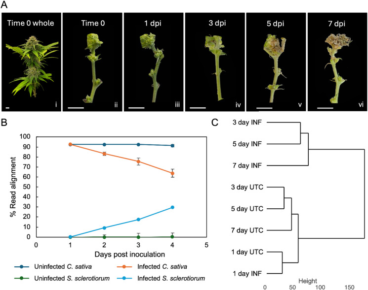
Global shifts in gene expression were observed in both the host plant and fungal pathogen as S. sclerotiorum initiated infection in C. sativa. Hierarchical clustering the top 10,000 most variably expressed C. sativa genes revealed that treatments clustered together based on infection status with the exception of the 1 dpi timepoint which remained clustered with uninfected samples (Fig. 1C). These results were supported by principal component analysis (PCA) of individual samples which revealed that the largest source of variation in our data was attributed to infection status (Supplementary Figure S2). Furthermore, infected samples clustered into distinct groups based on time post inoculation, highlighting large shifts in gene activity as infection progressed across the seven-day infection period. Similarly, hierarchical clustering of the top 1000 most variably expressed S. sclerotiorum genes revealed the 1 dpi timepoint to cluster with in vitro grown S. sclerotiorum, while all other infection timepoints clustered distinctly (Supplementary Figure S3). PCA of individual samples directly supported hierarchical clustering results (Supplementary Figure S4). We validated the RNA sequencing results by comparing the relative expression of the SAR marker gene PATHOGENESIS RELATED PROTEIN 1 (PR-1) using RT-qPCR. Data show PR1 accumulates at similar levels regardless of the method used to evaluate its activity (Supplementary Figure S5).
S. sclerotiorum rapidly infects C. sativa tissues and preferentially infects phloem tissues
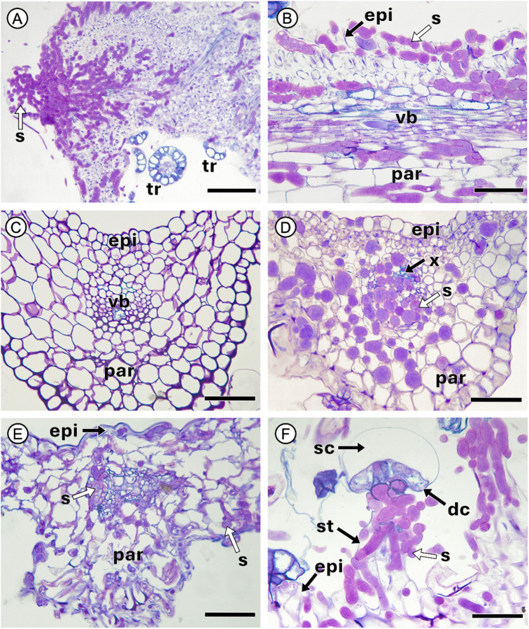
To better understand the interaction between S. sclerotiorum and C. sativa we tracked fungal infection of the cola at the cellular level directly from the site of inoculation (Fig. 2A). At 3 dpi, the inoculation site was clearly visible, as was the extension of fungal hyphae as S. sclerotiorum began to infect host floral tissue. At this timepoint, sectioning of reduced leaves proximal to the inoculation site revealed the presence of fungal hyphae along the surface of the epidermis as well as within epidermal cells, palisade mesophyll and general parenchymatic tissues, and phloem tissue of the vascular bundle (Fig. 2B). Xylem tissues remained relatively untouched whereas the phloem showed extensive colonization by the fungus as compared to uninfected reduced leaves (Fig. 2B-D). While the presence of hyphae was found throughout the reduced leaf, minimal plant cell wall degradation was visible. By 7 dpi, the C. sativa reduced leaf showed severe degradation of all tissue layers apart from the xylem (Figs. 3E). Although still structurally intact, S. sclerotiorum hyphae were visible throughout the xylem at this timepoint. The C. sativa stalked glandular trichomes of the inflorescence were also infected at this timepoint (Fig. 2F).
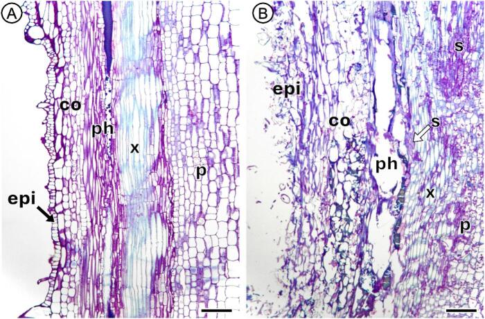
In the floral stem of the plant, S. sclerotiorum infection resulted in extensive host tissue degradation (Fig. 3). The floral stem of C. sativa is divided into distinct cell and tissue layers including the epidermis, cortex, phloem, xylem and pith (Fig. 3A). Extensive tissue degradation was revealed in infected floral stem tissues where S. sclerotiorum hyphae were abundant in the epidermis, cortex, vascular tissues, and pith of the C. sativa stem at 7 dpi (Fig. 3B). Most heavily degraded was the phloem, where cell walls had collapsed entirely, leading to the occurrence of regions of open space in areas where the fungus had used the phloem to travel further into the main stem of the plant. Although hyphae were also present within the xylem tracheids; degradation of this tissue layer was limited.
Differential gene expression analysis reveals the induction of C. sativa defense responses and altered terpenoid production at the mRNA level by S. sclerotiorum
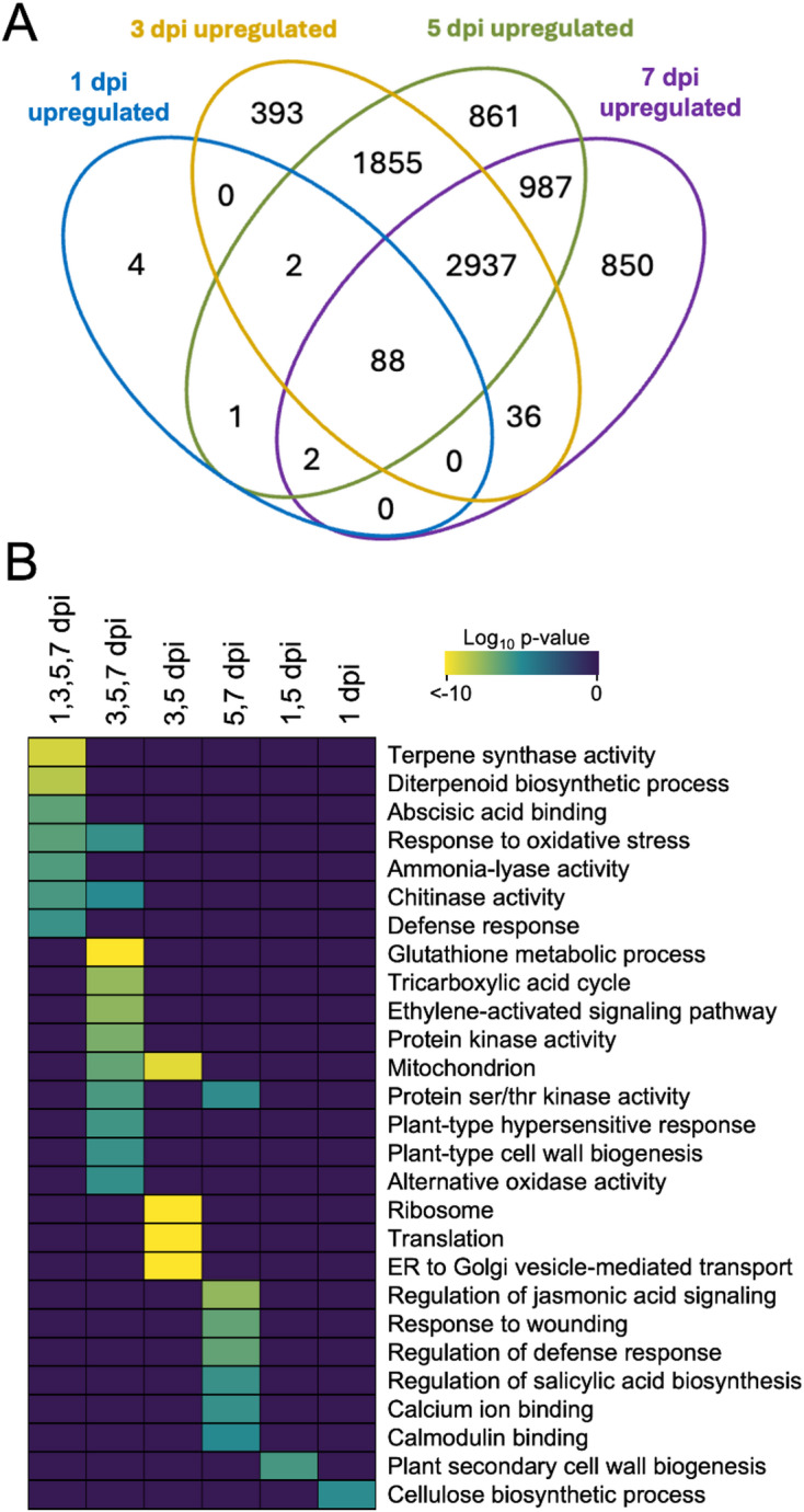
Next, we carried out differential gene expression analysis to better understand how C. sativa responds to S. sclerotiorum at the mRNA level (Fig. 4). The largest number of up-regulated differentially expressed genes (DEGs) in C. sativa were found at the intersection of 3-, 5- and 7 dpi, and of 3- and 5 dpi, with 2937 and 1855 genes, respectively (Fig. 4A). Specific to each timepoint, 4 genes were upregulated at 1 dpi, 393 genes at 3 dpi, 861 genes at 5 dpi, and 850 genes at 7 dpi. To better understand the biological and molecular processes associated with these gene sets, we conducted a GO enrichment analysis (Fig. 4B). Enriched at all timepoints were GO terms associated with terpene biosynthesis (terpene synthase activity and diterpenoid biosynthetic process), plant stress responses (abscisic acid binding and response to oxidative stress), plant defense, and chitinase activity. Specific to 3- and 5 dpi, we observed enrichment of GO terms associated with protein synthesis/transport (translation and endoplasmic reticulum to Golgi vesicle-mediated transport). Shared between 5- and 7 dpi, were GO terms involved in hormone signalling (regulation of jasmonic acid signalling and regulation of SA biosynthesis), response to wounding, and calcium/calmodulin signalling. More generally, shared between 3-, 5-, and 7 dpi were terms pertaining to oxidative stress responses (glutathione metabolic process and hypersensitive response), ethylene signalling, and protein kinase and protein ser/thr kinase activity. In our data, we identified a larger number of enriched GO terms shared between infection time points while few GO terms were enriched at specific timepoints. All GO enrichment and differential gene expression analysis results and associated P-values are provided in the supporting information (Supplementary Material 1).
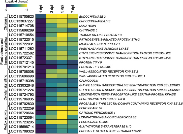
In response to infection, we uncovered genes encoding chitinases and endochitinases to be upregulated in C. sativa (Fig. 5). Those with highest fold changes in response to infection at 5 dpi included ENDOCHINTINASE 2 (LOC115705823; 1982-fold) and ENDOCHITINASE-LIKE (LOC133037227; 1074-fold). Also seen to be highly upregulated at 5 dpi were genes involved the plant SAR response, including PATHOGENESIS RELATED (PR) genes. These genes included THAUMATIN-LIKE PROTEIN 1B (PR-5; LOC115710654; 3487-fold), PATHOGENESIS-RELATED PROTEIN STH-2 (PR-10a; LOC115722015; 917-fold) and MAJOR ALLERGEN PRU AV 1 (PR-10; LOC115722031; 899-fold). Additionally, we observed the induction of genes involved in JA/ET hormone signalling. JASMONATE-ZIM DOMAIN (JAZ) protein genes JAZ5 and JAZ8 in addition to ETHYLENE RESPONSIVE FACTORs (ERF) ERF096 and ERF098 were also significantly differentially expressed. Furthermore, we uncovered the notable upregulation of numerous RLK genes that included WALL ASSOCIATED RECEPTOR KINASE 2 (WAK2; LOC115708008) and WALL ASSOCIATED RECEPTOR KINASE-LIKE 1 (WAKL1; LOC115696698) which both exhibited a 500-fold increase in expression, in addition to various other serine/threonine RLKs that include G-TYPE LECTIN S-RECEPTOR-LIKE SERINE/THREONINE-PROTEIN KINASE 3 (LECRK3; LOC113032648) and LECRK4 (LOC115721224). Finally, a number of peroxidases in C. sativa were also found to be highly upregulated in response to S. sclerotiorum. These peroxidases included PEROXIDASE 57 (PER57; LOC115722259), CATIONIC PEROXIDASE 1 (LOC115720664), LIGNIN-FORMING ANIONIC PEROXIDASE (LOC115723064), and PEROXIDASE 5-LIKE (LOC115723295) with fold changes at 5 dpi of 9710, 2868, 2375, and 1470, respectively.
We were next interested in understanding the genes and biological processes that were downregulated in C. sativa in response to infection with S. sclerotiorum (Supplementary Figure S6). The largest number of down-regulated genes were shared between 3-, 5- and 7 dpi (2866 genes; Supplementary Figure S6A). Large numbers of shared DEGs were also observed between 3- and 5 dpi (1458 genes) and 5- and 7 dpi (1179 genes). Specific to each timepoint, we identified 850 DEGs at 7 dpi, 1540 DEGs at 5 dpi, 512 at 3 dpi, and interestingly, 0 at 1 dpi. GO enrichment revealed enrichment of terms associated with photosynthesis, cellular development, and terpene synthesis (diterpenoid biosynthetic process and terpene synthase activity) were shared between 3-, 5-, and 7 dpi (Supplementary Figure S6B). Abscisic acid biosynthesis was enriched in gene sets shared between 3- and 5- dpi while biological processes associated with hormone activity like jasmonic acid biosynthesis and cytokinin signalling were specific to gene sets at 3 dpi.
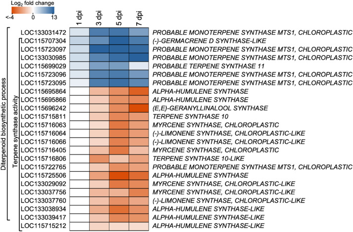
While terpene/diterpenoid biosynthesis GO terms were enriched in both up and downregulated gene sets, the specific genes involved in either GO term were unique (Fig. 6). Upregulated in response to S. sclerotiorum infection were C. sativa terpene synthases that included MONOTERPENE SYNTHASE MTS1 (LOC133031472, LOC115723097, LOC133030985, LOC115723096, LOC115723095) and (-)-GERMACRENE D SYNTHASE-LIKE (LOC115707304). Both genes were very highly upregulated with fold changes seen as high as 6361 at 7dpi (LOC13303985) and 6400 at 5dpi (LOC115707304). Conversely, genes downregulated in response to infection included ALPHA-HUMULENE SYNTHASE and –SYNTHASE-LIKE (LOC115695864, LOC115695866, LOC115725506 and LOC133038934, LOC133039417, LOC115715212), (E-E)-GERANYLLINALOOL SYNTHASE (LOC115696242), (-)-LIMONENE SYNTHASE (LOC115716064, LOC115716066, LOC133037760) and MYRCENE SYNTHASE (LOC115716405, LOC133029092, LOC133037756). Some of the most downregulated genes included (E-E)-GERANYLLINALOOL SYNTHASE (14.8-fold compared to uninfected plants at 7 dpi) and ALPHA-HUMULENE SYNTHASE (LOC115725506; 14.6-fold compared to uninfected plants at 5 dpi).
Differential gene expression analysis of S. sclerotiorum infecting C. sativa identified biological processes associated with carbohydrate metabolic activity and redox processing
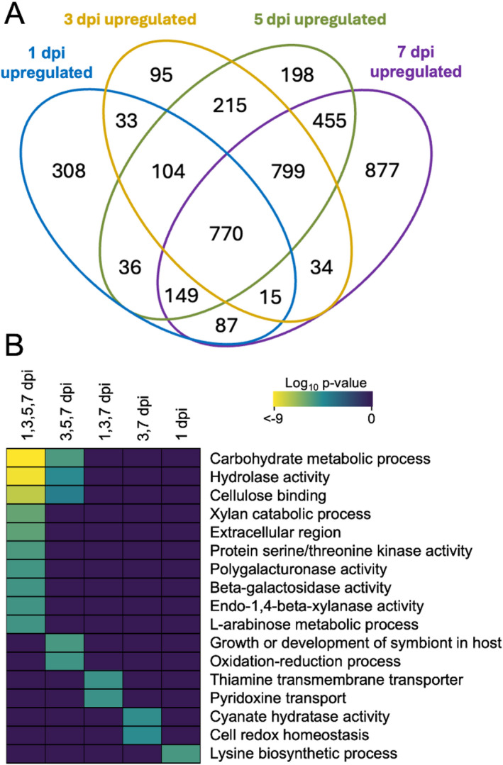
To investigate changes in S. sclerotiorum gene activity during C. sativa infection, we carried out differential gene expression and gene ontology (GO) term enrichment analysis (Fig. 7). Differential expression analysis revealed a high degree of shared differentially expressed genes at all sample timepoints (770 upregulated DEGs; Fig. 7A). Further, we observed the specific upregulation of 308 DEGs at 1 dpi, 95 at 3 dpi, 198 at 5 dpi, and 877 at 7 dpi. GO terms associated with host plant cell wall breakdown and carbohydrate metabolism (carbohydrate metabolic process, cellulose binding, xylan catabolic process and polygalacturonase activity) in addition to protein serine/threonine kinase activity were shared across all time points. GO terms associated with carbohydrate and cell wall breakdown, together with fungal growth and development within the host were enriched in gene sets shared between 3-, 5-, and 7- dpi. Redox processes and homeostasis were enriched in both the 3-, 5-, 7 dpi shared group, and the 3-, 7 dpi shared group (Fig. 7B).
Differential gene expression analysis of downregulated genes identified large numbers of shared DEGs between all sampled time points (610 genes) and shared between 3-, 5- and 7 dpi (710 genes; Supplementary Figure S7). Gene ontology terms enriched across all sampled timepoints identified enriched GO terms associated with translation and protein folding (ribosome, translation, unfolded protein binding, and protein folding) in addition to mitochondrial activity (mitochondrion and tricarboxylic acid cycle).
Validation of RNA Sequencing Results with RT-qPCR
To validate the findings of our RNA sequencing experiments, we studied the relative abundance of four C. sativa defense markers including PR-1, PR-10a, PR-5, and ENDOCHITINASE-2 using reverse-transcriptase quantitative PCR (RT-qPCR). Data show similar levels of gene activity of the four selected transcripts in both the RNA sequencing counts data and relative mRNA abundance observed using RT-qPCR (Figure S8).
Discussion
Known as both a stem and bud rot pathogen, S. sclerotiorum is regarded as an emerging fungal pathogen of C. sativa19. This study serves as the first description of the Cannabis-Sclerotinia pathosystem at the mRNA level where we show the rapid initiation of infection in the C. sativa inflorescence. Within one week of inoculation, severe disease symptoms were widespread in the plant. Global gene activity underpinning this interaction revealed C. sativa responded to infection through the elicitation of lignin deposition, redox buffering and generalized plant defense/immune hormone signalling cascades. Anatomical investigation at the site of inoculation showed the rapid colonization and degradation of host plant tissues by S. sclerotiorum starting in the epidermis and mesophyll before targeting the vascular system of the plant.
Detection of plant pathogens ties together the concepts of PTI and ETI, where unique molecular responses are initiated resulting from the recognition of pathogenic elicitors, PAMPs or DAMPs37,41. In response to S. sclerotiorum infection, we uncovered the upregulation of genes whose products are involved in pathogen perception and early defense responses such as ser/thr RLKs, NLRs, wall associated kinases (WAK) and WAK-like proteins (WAKL)41,42. WAKs and WAKLs have long been known to play a role in the plant defense response through binding pectin and oligosaccharides which act as DAMPs during biotic stress responses43–45. Expression of WAKs have previously been seen to be upregulated in Arabidopsis thaliana in response to SA and wounding, and have been associated with resistance against both hemibiotrophic and necrotrophic pathogens through pathogen- or host-derived elicitor detection, and subsequent cell wall restructuring44,46. In addition to A. thaliana, immunity-related WAKs/WAKLs have been documented in various crop species that include Triticum aestivum (wheat), Oryza sativa (rice), Hordeum vulgare (barley), Zea mays (maize), Sesamum indicum (sesame), Solanum lycopersicum (tomato), Gossypium hirsutum (cotton), and Brassica napus (canola) thereby suggesting WAKs/WAKLs as an evolutionarily conserved feature of the plant defense response against fungal necrotrophs44,45,47. Notable upregulation of C. sativa WAK2 and WAKL1 midway through the seven-day infection period begs the questions of whether earlier induction of these genes would result in greater host resistance, leaving room for further study.
The plant cell wall is the first line of defencse serving as a barrier to restrict attacking pathogens. Fungal necrotrophic pathogens make use of CWDEs to impair cell wall integrity and ultimately degrade host plant tissues48. Despite the presence of S. sclerotiorum throughout the epidermis, mesophyll and vasculature of the C. sativa leaf at 3 dpi, cell wall degradation at this timepoint was not yet observed. In comparison, previous studies have detailed extensive cellular degradation resulting from S. sclerotiorum infection as early as 2 dpi in leaves of B. napus28 and A. thaliana33, and 3 dpi in Glycine max49(soybean). Our data show that S. sclerotiorum progressed into C. sativa leaf tissues more slowly, however it should be noted that this delay could be the result of the complex three-dimensional structure of the cola, versus the direct inoculation of the leaf as was used in the above studies. Work carried out by Wytinck et al. (2022) in the B. napus stem showed similar findings where S. sclerotiorum infection resulted in extensive colonization and degradation of the host epidermis, cortex, and phloem, while xylem tissues remained largely intact. These results suggest a common infection strategy by S. sclerotiorum across diverse plant species, despite the structural and complex metabolic differences underpinning C. sativa.
Our GO enrichment analysis revealed induction of cell wall degrading activities in S. sclerotiorum as infection was initiated. Hydrolase activity, xylan catabolic process, polygalacturonase activity, beta-galactosidase activity, and 1,4-beta-xylanase activity were found across all infection time points. Production of CWDEs by S. sclerotiorum facilitates tissue penetration and maceration, through cell wall weakening characteristic of necrotrophic fungal infection22. Polygalacturonases (PGs) are a class of fungal pectinases that target unesterified pectate polymers of the middle lamella and primary cell wall of the host plant22. As plant cell walls are weakened and degraded by PGs, the secreted oligogalacturonides have been shown to elicit ROS burst, including H2O2 and O2−, as the plant attempts to restrict pathogen attack22,50. ROS production to develop localized cell death occurring as a result of pathogen infection is termed as the plant hypersensitive response (HR), and is regarded as one of the most important factors in impeding the growth of biotrophic pathogens51. Although the HR is generally effective against biotrophs, necrotrophic pathogen virulence, including that of S. sclerotiorum and B. cinerea, has been suggested to be strengthened as a result of HR elicitation29,52. Studies in Nicotiana tabacum (tobacco) and A. thaliana have also revealed that plants unable to initiate the HR demonstrated increased resistance to S. sclerotiorum53,54. While we uncovered rapid upregulation of S. sclerotiorum PG activity that continued throughout the seven-day infection period, the C. sativa HR was not induced until 3 dpi, likely revealing one facet of the S. sclerotiorum coordinated and timed control of host ROS activation to favour fungal proliferation in host tissues.
Similarly, the major S. sclerotiorum pathogenicity factor oxalic acid, has also been previously found to manipulate host ROS production to initially suppresses ROS signalling, before eliciting ROS production, leading to cell death28,55. Recent studies have challenged the classical view of the S. sclerotiorum necrotrophic lifestyle, suggesting the possibility of a brief biotrophic phase early in the infection process. These studies suggest that S. sclerotiorum is capable of suppressing SA-mediated SAR early in infection, and thus the HR, before the true necrotrophic portion of the lifestyle occurs in which initiation of ROS production leads to localized cell death and subsequent widespread infection56,57. Our results support the initial suppression of SA-dependent SAR as PHENYLALANINE AMMONIA-LYASE (PAL), a major enzyme involved in SA biosynthesis, was not upregulated until 3 dpi, with highest upregulation occurring two days later, at 5 dpi. Similarly, we found GLUTATHIONE S-TRANSFERASE U10 and GLUTATHIONE S-TRANSFERASE were not upregulated until 3 dpi. Glutathione transferases are cellular protectant enzymes involved in redox homeostasis and ROS detoxification that are rapidly induced by H2O258. Previous work in B. napus revealed plants partially resistant to S. sclerotiorum demonstrated increased redox buffering capacity as early as 1 dpi28. These results suggest that a more rapid induction of redox buffering by C. sativa may result in an increased resistance or tolerant phenotype.
In addition to ROS accumulation, S. sclerotiorum infection has previously been found to initiate lignin biosynthesis within the host plant59. Lignins are biopolymers important for plant cell wall structural support, and have previously been reported to be deposited in Brassica species in response to S. sclerotiorum infection60. Specific to our study, we found that in response to S. sclerotiorum infection, C. sativa highly upregulated PAL in addition to various class III peroxidases that include PEROXIDASE 57, CATIONIC PEROXIDASE 1, LIGNIN-FORMING ANIONIC PEROXIDASE, and PEROXIDASE 5-LIKE. Belonging to the PR-9 subfamily of PR proteins, class III plant peroxidase gene expression has previously been found to increase in plants challenged with fungi, in addition to bacteria, viruses and viroids61–63. Plant peroxidases are also capable of creating physical barriers to limit pathogen invasion in response to stimuli such as wounding, pathogen presence, or hormone accumulation61. The upregulation of peroxidase activity uncovered in this study serves as the first description of genes involved in H2O2-dependent lignin deposition by C. sativa to restrict further incursion of S. sclerotiorum.
While PAL is known to be involved in lignification, it is also involved in SA biosynthesis64,65. SA biosynthesis is initiated during both ETI and PTI in response to recognition of PAMPs or pathogenic effectors66. Increased SA levels are required for plant SAR initiation, which is accompanied and characterized by increased systemic PR gene expression39. Belonging to the PR families PR-3, -4, -8, and − 11, chitinases are among the most abundant PR proteins67. Our dataset revealed the upregulation of chitinase genes across the S. sclerotiorum infection process. Additionally, we also observed increases in gene activity of the PR-5 and PR-10 subfamilies. It is thought that PR-5 proteins exhibit antifungal activity by inserting themselves into fungal membranes to create a transmembrane pore, later leading to influx of water and subsequent fungal osmotic rupture68. Unlike the PR-5 proteins however, the function of PR-10 proteins remains largely unclear, which may be attributed to the large multi-gene families they code for68,69. As our study serves as one of the first transcriptome-level investigations of the infection of C. sativa with any fungal pathogen, future studies that explore PR protein activity in other fungal interactions with C. sativa may reveal how PR gene expression may be engineered or selected to develop more resistant germplasm.
As complex specialized secondary plant metabolites, terpenes serve various purposes to plants that include attracting pollinators and insect predators of feeding herbivores, and creating both chemical and physical barriers to herbivorous insects, as well as invading pathogens11,12. Specific to our study, we uncovered differential expression activity of various C. sativa terpene synthases. While terpene synthase activity of C. sativa in response to biotic stressors has not previously been investigated, our results suggest that the largescale transcriptional reprogramming that occurs as a result of coordinating a defense response against S. sclerotiorum may impact the terpenoid profile exhibited by infected plants. As the terpene composition of the resin produced by glandular trichomes of the female inflorescence is largely responsible for the scent and flavour characteristics of harvested Cannabis products, terpenes greatly impact consumer preferences6. Consequently, terpenoid profiles often serve as a basis for modern selective breeding. With varying terpenoid profiles attributed to unique C. sativa cultivars, the question arises of how different cultivars may respond to pathogen attack based on terpenoid profile composition; and whether selective breeding may allow for the production of cultivars that exhibit an increased resistance phenotype.
Taken together, this study provides a comprehensive investigation into the transcriptional and anatomical changes that occur as S. sclerotiorum initiates infection in the C. sativa inflorescence. Our data reveal large shifts in host gene activity in response to infection with S. sclerotiorum that largely peak at 5 dpi. Gene categories identified in C. sativa show complex shifts in defense hormone signalling and redox buffering associated with the plant immune responses across time post inoculation. Anatomical study revealed extensive degradation of host cortical and vascular phloem tissues associated with the production of fungal toxins and CWDEs. Additional studies conducting molecular and biochemical validation of the various gene products and metabolites identified herein will allow for increased understanding of this pathosystem and can help to direct future crop improvement studies.
Materials and methods
Cannabis sativa growth conditions
Female C. sativa plants, cultivar ‘Kona’, were sourced from Rogue Botanical, a licensed grower in southern Manitoba, Canada. Plants were obtained in vegetative growth, 20 days after being clonally propagated. At 22 days old, plants were transplanted from 4-inch pots to 6-inch pots in Sunshine growing mix #4 (Sungro, Agawan, MA, USA). Plants were grown in a controlled environment chamber under long day conditions (18 h light, 6 h dark), 500 µmol/m2/s− 1, 23 °C and 50% relative humidity. After 30 days in vegetative growth, the photoperiod was adjusted to 12 h light, 12 h dark to promote flowering. Plants were fertilized using Advanced Nutrients Sensi Grow/Bloom nutrient packages, as per manufacturer’s instructions (Advanced Nutrients, West Hollywood, CA, USA).
Experimental research on C. sativa, including the collection of plant material, were conducted in accordance with relevant institutional, national, and international guidelines and legislation.
Sclerotinia sclerotiorum inoculation of the Cannabis sativa cola
S. sclerotiorum was grown in vitro on potato dextrose agar (BD Difco) plates supplemented with 15 µg/mL tetracycline HCl. S. sclerotiorum mycelial plugs were taken from the leading edge of a 3-day-old actively growing plate using a P1000 pipette tip. Mycelial plugs were carefully placed at the inflorescence node of the third-most distal inflorescence of the C. sativa cola using forceps. Infection took place over a seven-day period with colas being harvested after 1-, 3-, 5- and 7- days post inoculation (dpi). Both infected and untreated control (UTC) colas were harvested at each timepoint. Twelve C. sativa plants were used for this experiment with four biological replicates sampled across each timepoint.
Sample collection, RNA isolation, library preparation and RNA sequencing
Harvested colas were immediately trimmed down to the main floral stem, while maintaining ~ 1 cm3 of floral tissue of the inoculated inflorescence and immediately flash frozen using liquid nitrogen. Tissue was ground to a fine powder using a mortar and pestle with liquid nitrogen prior to RNA extraction.
RNA was extracted using the Purelink Plant RNA Reagent (Invitrogen, Waltham, MA, USA) as per manufacturer’s protocol. Following RNA extraction, Qiagen’s RNeasy Plant Minikit and RNase-Free DNase Set was used for DNAse treatment following the “RNA Cleanup” protocol available in Qiagen’s RNeasy Mini Handbook (Qiagen, Toronto, ON, Canada). As sample purity was often compromised as a result of the DNAse treatment procedure, samples then underwent a sodium acetate precipitation. This precipitation used 3M C2H3NaO2 (pH 5.2) and subsequent ethanol washes (100% followed by 75%) before resuspension in molecular grade water to yield RNA of increased purity.
cDNA libraries were constructed by Genome Québec following their polyA Enriched RNA Library Preparation protocol. Paired-end 100 bp reads were sequenced for a minimum of 25 million reads per library on the Illumina NovaSeq sequencing system at Genome Québec (Montréal, Québec, Canada). All sequencing data can be found at the Gene Expression Omnibus, under accession GSE284432.
Fungal load qPCR and RT-qPCR
Expression of PATHOGENESIS RELATED PROTEIN 1 (PR-1; LOC115704466), PATHOGENESIS-RELATED PROTEIN STH-2 (PR-10a; LOC115722015), THAUMATIN-LIKE PROTEIN 1B (PR-5; LOC115710654), and ENDOCHITINASE 2 (LOC115705823) were assessed using RT-qPCR. cDNA was synthesized using qScript™ cDNA SuperMix according to manufacturer’s instructions (Quantabio, Beverly, MA, USA). To quantify defense gene transcript abundance, RT-qPCR was run on PR1, PR-10a, PR-5, and ENDOCHITINASE 2 with the housekeeping genes TIP41-LIKE PROTEIN (TIP41; LOC115703022) and ADENINE PHOSPHO-RIOSYLTRANSFERASE 1 (APT1; LOC115713640) used as internal controls70. Primer sequence information is found in Table S1.
We also used qPCR to determine the relative fungal load between samples. Genomic DNA was extracted from ground tissue using a modified cetyltrimethylammonium bromide (CTAB) method71. As a target for S. sclerotiorum, we used 18 S rDNA as described in Wytinck et al. (2022). Primer sequences and target loci for both RT-qPCR and fungal load qPCR can be found in Supplementary Table S1. SsoFast EvaGreen Supermix was used as per manufacturer’s instructions for both RT-qPCR and qPCR (Bio-Rad Laboratories, Hercules, CA, USA).
RNA sequencing analysis
Raw reads were processed using computing clusters available through Compute Canada and the Digital Research Alliance of Canada (https://www.alliancecan.ca/en). Prior to read alignment, sequence read quality was first assessed using FastQC (v0.12.1; https://www.bioinformatics.babraham.ac.uk/projects/fastqc/)72. Paired-end read alignment was carried out using the C. sativa cultivar ‘Pink Pepper’ reference genome (NCBI RefSeq assembly GCF_029168945.1) and the S. sclerotiorum reference genome73,74 (NCBI RefSeq assembly GCF_000146945.2; ) using HISAT2 (v2.2.1; https://daehwankimlab.github.io/hisat2/)75. Transcript abundance was determined using featureCounts (v2.0.3; https://subread.sourceforge.net/)76. With one of the barriers to working with the C. sativa transcriptome being the level of genome annotation, we used predicted protein orthologs publicly available for the loci of the C. sativa cultivar ‘Pink Pepper’ genome through the NCBI Genomes database (RefSeq accession GCF_029168945.1; Lim, 2023). Differential gene expression analysis, low counts filtering, library normalization, principle component analysis and further data visualization was done using libraries DESeq2 (v1.42.1; https://bioconductor.org/packages/release/bioc/html/DESeq2.html)77, ashr (v2.2.63; https://cran.r-project.org/web/packages/ashr/index.html)78, and ggplot2 (v3.5.2; https://cran.r-project.org/web/packages/ggplot2/index.html)79 in R (v4.3.1; https://www.r-project.org/)80. Genes with counts lower than 10 across all samples were filtered prior to normalization and differential gene expression analysis. Raw sequenced read counts were normalized using the median of ratios method in DESeq281. Differentially expressed genes were called with a p-value < 0.01 when adjusted for false discovery rate (FDR) by the Benjamini-Hochberg method82. GO term enrichment was carried out on differentially expressed gene sets using SeqEnrich (v2.0)83,84. GO and DEG lists (Supplementary Material 1), raw counts aligned to C. sativa (Supplementary Material 3), and raw counts aligned to S. sclerotiorum (Supplementary Material 4) are provided as supplemental datasets.
Sample preparation for light microscopy
Sample preparation, sectioning and staining followed the methods previously described by Chan and Belmonte (2013) with slight modifications. Harvested colas were trimmed to the above-mentioned region of interest before being fixed in a solution of 2.5% glutaraldehyde and 1.6% paraformaldehyde in 1x phosphate-buffered saline. Tissue was added to fixative solution before being vacuum infiltrated for 30 min to ensure adequate penetration of the fixative into the C. sativa tissues. Tissue samples were fixed for 24 h at 4 °C. Tissue was decoloured in methyl cellosolve for 24 h, followed by daily 100% ethanol changes for three days at 4 °C. Historesin (Leica Microsystems, Wetzlar, Germany) was gradually infiltrated into processed tissue using a 30%, 50%, 75% and 100% ethanol: historesin mixture. Pure Historesin was exchanged three times over the course of a week, while vacuum infiltrating the tissue in for 30 min halfway through this period. Tissue was then embedded in round molds using an embedding medium composed of 91.5% Historesin, 2.4% polyethylene glycol 400, and 6.1% Historesin Hardener (Leica Microsystems, Wetzlar, Germany; Chan & Belmonte, 2013).
Sectioning and staining for light microscopy
Hardened Historesin blocks were sectioned at 3 μm using disposable Epredia Edge-Rite steel blades (Epredia, Kalamazoo, MI, USA) mounted on a Leica RM2245 microtome (Leica Microsystems, Wetzlar, Germany). Sections were placed on glass slides for staining.
Sections were first stained with periodic acid-Schiff stain (15 min in 0.1% periodic acid, followed by 15 min in Schiff’s reagent) before being stained with 0.1% toluidine blue O suspended in distilled water for 30 s. Following staining, coverslips were mounted on the slides using Cytoseal 60 (Richard-Allen Scientific, Kalamazoo, MI, USA). Slides were viewed using a brightfield light microscope and micrographs were taken using the Leica Application Suite software version 4.6.0 (Leica Microsystems, Wetzlar, Germany). Image cropping and the addition of scale bars was carried out in Adobe Photoshop version 25.7.0 (Adobe Systems Inc., San Jose, CA, USA).
Supplementary Materials
References
- T Long, M Wagner, D Demske, C Leipe, PE Tarasov. Cannabis in Eurasia: origin of human use and Bronze Age trans-continental connections. Veg. Hist. Archaeobotany, 2017. [DOI]
- G Ren. Large-scale whole-genome resequencing unravels the domestication history of Cannabis sativa. Sci. Adv., 2021. [DOI | PubMed]
- S Braich, RC Baillie, LS Jewell, GC Spangenberg, N Cogan. O. I. Generation of a Comprehensive Transcriptome Atlas and Transcriptome Dynamics in Medicinal Cannabis. Sci. Rep., 2019. [DOI | PubMed]
- H van Bakel. The draft genome and transcriptome of Cannabis sativa. Genome Biol., 2011. [DOI | PubMed]
- MG Divashuk, OS Alexandrov, OV Razumova, IV Kirov, G Karlov. I. Molecular Cytogenetic Characterization of the Dioecious Cannabis sativa with an XY Chromosome Sex Determination System. PLOS ONE, 2014. [DOI | PubMed]
- JK Booth, JE Page, J Bohlmann. Terpene synthases from Cannabis sativa. PLoS ONE, 2017. [DOI | PubMed]
- P Rodziewicz, S Loroch, Ł Marczak, A Sickmann, O Kayser. Cannabinoid synthases and osmoprotective metabolites accumulate in the exudates of Cannabis sativa L. glandular trichomes. Plant. Sci., 2019. [DOI | PubMed]
- Z Xie. Cannabis sativa: origin and history, glandular trichome development, and cannabinoid biosynthesis. Hortic. Res., 2023. [DOI | PubMed]
- JK Booth, J Bohlmann. Terpenes in Cannabis sativa – From plant genome to humans. Plant. Sci., 2019. [DOI | PubMed]
- JT Fischedick. Identification of Terpenoid Chemotypes Among High (–)-trans-∆9- Tetrahydrocannabinol-Producing Cannabis sativa L. Cultivars. Cannabis Cannabinoid Res., 2017. [DOI | PubMed]
- F Chen, D Tholl, J Bohlmann, E Pichersky. The family of terpene synthases in plants: a mid-size family of genes for specialized metabolism that is highly diversified throughout the kingdom. Plant. J., 2011. [DOI | PubMed]
- CI Keeling, J Bohlmann. Genes, enzymes and chemicals of terpenoid diversity in the constitutive and induced defence of conifers against insects and pathogens. New. Phytol, 2006. [DOI | PubMed]
- 13.Government of Canada. Consolidated federal laws of Canada, Cannabis Regulations. (2018). https://laws-lois.justice.gc.ca/eng/regulations/SOR-2018-144/FullText.html
- D Hammond. Evaluating the impacts of cannabis legalization: The International Cannabis Policy Study. Int. J. Drug Policy, 2020. [DOI | PubMed]
- 15.Government of Canada. Licensed area market data. (2024). https://www.canada.ca/en/health-canada/services/drugs-medication/cannabis/research-data/market/licensed-area.html
- PS Bains, HS Bennypaul, SF Blade, C Weeks. First Report of Hemp Canker Caused by Sclerotinia sclerotiorum in Alberta, Canada. Plant. Dis., 2000. [DOI | PubMed]
- AR Garfinkel. First Report of Sclerotinia sclerotiorum Causing Stem Canker on Cannabis sativa in Oregon. Plant. Dis., 2021. [DOI]
- ZK Punja. Emerging diseases of Cannabis sativa and sustainable management. Pest Manag Sci., 2021. [DOI | PubMed]
- ZK Punja, L Ni. The bud rot pathogens infecting cannabis (Cannabis sativa L., marijuana) inflorescences: symptomology, species identification, pathogenicity and biological control. Can. J. Plant. Pathol., 2021. [DOI]
- LD Thiessen, T Schappe, S Cochran, K Hicks, AR Post. Surveying for Potential Diseases and Abiotic Disorders of Industrial Hemp (Cannabis sativa) Production. Plant. Health Prog, 2020. [DOI]
- X Yang, A Justice, C Colburn. First report of Sclerotinia sclerotiorum causing stem canker on industrial hemp in South Carolina, USA. Plant. Dis., 2023. [DOI | PubMed]
- MD Bolton, BPHJ Thomma, BD Nelson. Sclerotinia sclerotiorum (Lib.) de Bary: biology and molecular traits of a cosmopolitan pathogen. Mol. Plant. Pathol., 2006. [DOI | PubMed]
- X Liang, JA Rollins. Mechanisms of Broad Host Range Necrotrophic Pathogenesis in Sclerotinia sclerotiorum. Phytopathology®, 2018. [DOI | PubMed]
- 24.Saharan, G. S. & Mehta, N. Sclerotinia Diseases of Crop Plants: Biology, Ecology and Disease Management (Springer Science & Business Media, 2008).
- J Willbur, M McCaghey, M Kabbage, DL Smith. An overview of the Sclerotinia sclerotiorum pathosystem in soybean: impact, fungal biology, and current management strategies. Trop. Plant. Pathol., 2019. [DOI]
- JT Alkooranee. Detecting the Hormonal Pathways in Oilseed Rape behind Induced Systemic Resistance by Trichoderma harzianum TH12 to Sclerotinia sclerotiorum. PLOS ONE, 2017. [DOI | PubMed]
- MM Hossain, F Sultana, W Li, LSP Tran, MG Mostofa. Sclerotinia sclerotiorum (Lib.) de Bary: Insights into the Pathogenomic Features of a Global Pathogen. Cells, 2023. [DOI | PubMed]
- IJ Girard. RNA sequencing of Brassica napus reveals cellular redox control of Sclerotinia infection. J. Exp. Bot., 2017. [DOI | PubMed]
- DD Hegedus, SR Rimmer. Sclerotinia sclerotiorum: When to be or not to be a pathogen?. FEMS Microbiol. Lett., 2005. [DOI | PubMed]
- R Horbach, AR Navarro-Quesada, W Knogge, HB Deising. When and how to kill a plant cell: Infection strategies of plant pathogenic fungi. J. Plant. Physiol., 2011. [DOI | PubMed]
- MC Derbyshire, M Denton-Giles. The control of sclerotinia stem rot on oilseed rape (Brassica napus): current practices and future opportunities. Plant. Pathol., 2016. [DOI]
- N Wytinck. Host induced gene silencing of the Sclerotinia sclerotiorum ABHYDROLASE-3 gene reduces disease severity in Brassica napus. PLOS ONE, 2022. [DOI | PubMed]
- PL Walker. Control of white mold (Sclerotinia sclerotiorum) through plant-mediated RNA interference. Sci. Rep., 2023. [DOI | PubMed]
- MH Ghozlan, E EL-Argawy, S Tokgöz, DK Lakshman, A Mitra. Plant Defense against Necrotrophic Pathogens. Am. J. Plant. Sci., 2020. [DOI]
- W Song, A Forderer, D Yu, J Chai. Structural biology of plant defence. New. Phytol, 2021. [DOI | PubMed]
- PL Walker. Tissue-specific mRNA profiling of the Brassica napus–Sclerotinia sclerotiorum interaction uncovers novel regulators of plant immunity. J. Exp. Bot., 2022. [DOI | PubMed]
- RN Pruitt, AA Gust, T Nürnberger. Plant immunity unified. Nat. Plants, 2021. [DOI | PubMed]
- 38.Cale, N. L. et al. Global mRNA profiling reveals the effect of boron as a crop protection tool against Sclerotinia sclerotiorum. AoB PLANTS. plae05610.1093/aobpla/plae056 (2024).
- CMJ Pieterse. Induced Systemic Resistance by Beneficial Microbes. Annu. Rev. Phytopathol., 2014. [DOI | PubMed]
- Y Yu. Induced Systemic Resistance for Improving Plant Immunity by Beneficial Microbes. Plants, 2022. [DOI | PubMed]
- 41.Dodds, P. N., Chen, J. & Outram, M. A. Pathogen perception and signaling in plant immunity. Plant. Cell.koae02010.1093/plcell/koae020 (2024).
- 42.Kohorn, B. D. & Kohorn, S. L. The cell wall-associated kinases, WAKs, as pectin receptors. Front Plant. Sci.3, 88 (2012).
- 43.Harvey, A., van den Berg, N. & Swart, V. Describing and characterizing the WAK/WAKL gene family across plant species: a systematic review. Front Plant. Sci.15, 1467148 (2024).
- C Stephens, KE Hammond-Kosack, K Kanyuka. WAKsing plant immunity, waning diseases. J. Exp. Bot., 2022. [DOI | PubMed]
- W Yan. Genome-wide characterization of the wall-associated kinase-like (WAKL) family in sesame (Sesamum indicum) identifies a SiWAKL6 gene involved in resistance to Macrophomina Phaseolina. BMC Plant. Biol., 2023. [DOI | PubMed]
- ZH He, D He, BD Kohorn. Requirement for the induced expression of a cell wall associated receptor kinase for survival during the pathogen response. Plant. J., 1998. [DOI | PubMed]
- J Wu. Comparative transcriptomic analysis uncovers the complex genetic network for resistance to Sclerotinia sclerotiorum in Brassica napus. Sci. Rep., 2016. [DOI | PubMed]
- 48.Bellincampi, D., Cervone, F. & Lionetti, V. Plant cell wall dynamics and wall-related susceptibility in plant–pathogen interactions. Front Plant. Sci.5, 228 (2014).
- AL Davidson. Histopathology of Sclerotinia sclerotiorum infection and oxalic acid function in susceptible and resistant soybean. Plant. Pathol., 2016. [DOI]
- P Wojtaszek. Oxidative burst: an early plant response to pathogen infection. Biochem. J., 1997. [DOI | PubMed]
- EM Govrin, A Levine. The hypersensitive response facilitates plant infection by the necrotrophic pathogen Botrytis cinerea. Curr. Biol., 2000. [DOI | PubMed]
- FR Rossi. Reactive oxygen species generated in chloroplasts contribute to tobacco leaf infection by the necrotrophic fungus Botrytis cinerea. Plant. J., 2017. [DOI | PubMed]
- 53.Dickman, M. B. et al. Abrogation of disease development in plants expressing animal antiapoptotic genes. Proc. Natl. Acad. Sci. 98, 6957–6962 (2001).
- BP Thomma, IA Penninckx, BP Cammue, WF Broekaert. The complexity of disease signaling in Arabidopsis. Curr. Opin. Immunol., 2001. [DOI | PubMed]
- SG Cessna, VE Sears, MB Dickman, PS Low. Oxalic Acid, a Pathogenicity Factor for Sclerotinia sclerotiorum, Suppresses the Oxidative Burst of the Host Plant. Plant. Cell., 2000. [DOI | PubMed]
- M Kabbage, O Yarden, MB Dickman. Pathogenic attributes of Sclerotinia sclerotiorum: Switching from a biotrophic to necrotrophic lifestyle. Plant. Sci., 2015. [DOI | PubMed]
- S Seifbarghi. Changes in the Sclerotinia sclerotiorum transcriptome during infection of Brassica napus. BMC Genom., 2017. [DOI]
- C Lamb, RA Dixon. THE OXIDATIVE BURST IN PLANT DISEASE RESISTANCE. Annu. Rev. Plant. Biol., 1997. [DOI]
- B Calla. Genomic evaluation of oxalate-degrading transgenic soybean in response to Sclerotinia sclerotiorum infection. Mol. Plant. Pathol., 2014. [DOI | PubMed]
- MB Uloth, PL Clode, MP You, MJ Barbetti. Attack modes and defence reactions in pathosystems involving Sclerotinia sclerotiorum, Brassica carinata, B. juncea and B. napus. Ann. Bot., 2016. [DOI | PubMed]
- L Almagro. Class III peroxidases in plant defence reactions. J. Exp. Bot., 2009. [DOI | PubMed]
- LC van Loon, M Rep, CMJ Pieterse. Significance of Inducible Defense-related Proteins in Infected Plants. Annu. Rev. Phytopathol., 2006. [DOI | PubMed]
- K Sasaki. Ten Rice Peroxidases Redundantly Respond to Multiple Stresses Including Infection with Rice Blast Fungus. Plant. Cell. Physiol., 2004. [DOI | PubMed]
- DS Kim, BK Hwang. An important role of the pepper phenylalanine ammonia-lyase gene (PAL1) in salicylic acid-dependent signalling of the defence response to microbial pathogens. J. Exp. Bot., 2014. [DOI | PubMed]
- C Ullah, YH Chen, MA Ortega, CJ Tsai. The diversity of salicylic acid biosynthesis and defense signaling in plants: Knowledge gaps and future opportunities. Curr. Opin. Plant. Biol., 2023. [DOI | PubMed]
- TE Mishina, J Zeier. Pathogen-associated molecular pattern recognition rather than development of tissue necrosis contributes to bacterial induction of systemic acquired resistance in Arabidopsis. Plant. J., 2007. [DOI | PubMed]
- R Chouhan, S Ahmed, SG Gandhi. Over-expression of PR proteins with chitinase activity in transgenic plants for alleviation of fungal pathogenesis. J. Plant. Pathol., 2023. [DOI]
- 68.Sinha, M. et al. Current Overview of Allergens of Plant Pathogenesis Related Protein Families. Sci. World J. 543195 (2014). (2014).
- H Fernandes, K Michalska, M Sikorski, M Jaskolski. Structural and functional aspects of PR-10 proteins. FEBS J., 2013. [DOI | PubMed]
- 70.Balthazar, C., Cantin, G., Novinscak, A., Joly, D. L. & Filion, M. Expression of Putative Defense Responses in Cannabis Primed by Pseudomonas and/or Bacillus Strains and Infected by Botrytis cinerea. Front Plant. Sci.11, 572112 (2020).
- S Porebski, LG Bailey, BR Baum. Modification of a CTAB DNA extraction protocol for plants containing high polysaccharide and polyphenol components. Plant. Mol. Biol. Rep., 1997. [DOI]
- 72.Andrews, S. & FastQC A Quality Control tool for High Throughput Sequence Data. (2010). https://www.bioinformatics.babraham.ac.uk/projects/fastqc/
- 73.Lim, J. D. Cannabis sativa Isolate KNU-18-1 (Cultivar: Pink Pepper), Whole Genome Sequencing Project. Genbank (2023).
- J Amselem. Genomic Analysis of the Necrotrophic Fungal Pathogens Sclerotinia sclerotiorum and Botrytis cinerea. PLOS Genet., 2011. [DOI | PubMed]
- D Kim, JM Paggi, C Park, C Bennett, SL Salzberg. Graph-based genome alignment and genotyping with HISAT2 and HISAT-genotype. Nat. Biotechnol., 2019. [DOI | PubMed]
- Y Liao, GK Smyth, W Shi. featureCounts: an efficient general purpose program for assigning sequence reads to genomic features. Bioinformatics, 2014. [DOI | PubMed]
- MI Love, W Huber, S Anders. Moderated estimation of fold change and dispersion for RNA-seq data with DESeq2. Genome Biol., 2014. [DOI | PubMed]
- M Stephens. False discovery rates: a new deal. Biostatistics, 2017. [PubMed]
- H Wickham. ggplot2. WIREs Comput. Stat., 2011. [DOI]
- 80.R Core Team. R: A language and environment for statistical computing. (2021).
- S Anders, W Huber. Differential expression analysis for sequence count data. Genome Biol., 2010. [DOI | PubMed]
- Y Benjamini, Y Hochberg. Controlling the False Discovery Rate: A Practical and Powerful Approach to Multiple Testing. J. R Stat. Soc. Ser. B Methodol., 1995. [DOI]
- MG Becker, PL Walker, NC Pulgar-Vidal, MF Belmonte, SeqEnrich. A tool to predict transcription factor networks from co-expressed Arabidopsis and Brassica napus gene sets. PLOS ONE, 2017. [DOI | PubMed]
- A Chan, MF Belmonte. Histological and ultrastructural changes in canola (Brassica napus) funicular anatomy during the seed lifecycle. Botany, 2013. [DOI]