
Sarcopenia and cachexia: molecular mechanisms and therapeutic interventions
Abstract
Sarcopenia is defined as a muscle‐wasting syndrome that occurs with accelerated aging, while cachexia is a severe wasting syndrome associated with conditions such as cancer and immunodeficiency disorders, which cannot be fully addressed through conventional nutritional supplementation. Sarcopenia can be considered a component of cachexia, with the bidirectional interplay between adipose tissue and skeletal muscle potentially serving as a molecular mechanism for both conditions. However, the underlying mechanisms differ. Recognizing the interplay and distinctions between these disorders is essential for advancing both basic and translational research in this area, enhancing diagnostic accuracy and ultimately achieving effective therapeutic solutions for affected patients. This review discusses the muscle microenvironment’s changes contributing to these conditions, recent therapeutic approaches like lifestyle modifications, small molecules, and nutritional interventions, and emerging strategies such as gene editing, stem cell therapy, and gut microbiome modulation. We also address the challenges and opportunities of multimodal interventions, aiming to provide insights into the pathogenesis and molecular mechanisms of sarcopenia and cachexia, ultimately aiding in innovative strategy development and improved treatments.
Article type: Review Article
Keywords: cachexia, molecules, pathogenesis, sarcopenia, targeted therapy
Affiliations: Department of Neurology West China Hospital of Sichuan University Chengdu Sichuan China; Institute of Brain Science and Brain‐Inspired Technology of West China Hospital Sichuan University Chengdu Sichuan China; Department of Neurology Chengdu Shangjin Nanfu Hospital Chengdu Sichuan China
License: © 2025 The Author(s). MedComm published by Sichuan International Medical Exchange & Promotion Association (SCIMEA) and John Wiley & Sons Australia, Ltd. CC BY 4.0 This is an open access article under the terms of the http://creativecommons.org/licenses/by/4.0/ License, which permits use, distribution and reproduction in any medium, provided the original work is properly cited.
Article links: DOI: 10.1002/mco2.70030 | PubMed: 39764565 | PMC: PMC11702502
Relevance: Moderate: mentioned 3+ times in text
Full text: PDF (3.2 MB)
INTRODUCTION
Rosenberg first defined sarcopenia in 1989 as a “muscle‐wasting geriatric syndrome” characterized by the progressive loss of skeletal muscle mass and strength.ref. mco270030-bib-0001, ref. mco270030-bib-0002 Studies indicate that sarcopenia affects 5–13% of adults aged 60–70 years, with prevalence rising to between 11 and 50% in individuals over 80 years old. Sarcopenia is associated with a range of conditions, including cardiac disease, respiratory disease, and cognitive impairment, leading to mobility challenges and a diminished overall quality of life (QOL). It also contributes to a decline in independence, often necessitating long‐term care placement.ref. mco270030-bib-0003 Patients face various physical challenges, such as mobility disorders, an increased risk of falls and fractures, reduced capacity to perform daily tasks, disabilities, diminished QOL, loss of independence, and a higher risk of mortality.ref. mco270030-bib-0004 Sarcopenia imposes significant social, health, and economic burdens, especially as the elderly population continues to grow. The rising incidence of sarcopenia presents a pressing challenge for the healthcare system. Research has extensively examined hormones, including insulin signaling, inflammatory pathways, and impaired proteostasis, as potential risk factors for sarcopenia.ref. mco270030-bib-0005 However, the molecular mechanisms and triggers of sarcopenia remain incompletely understood, which hampers the development of effective therapies. Therefore, investigating the underlying molecular mechanisms and identifying potential therapeutic targets are essential for advancing the clinical management of sarcopenia.
Cachexia is widely recognized as a severe wasting syndrome associated with diseases such as cancer and immunodeficiency disorders, which cannot be fully alleviated through conventional nutritional supplementation.ref. mco270030-bib-0006, ref. mco270030-bib-0007 With an estimated annual death rate of 2 million people worldwide, cachexia is a significant contributor to human morbidity and mortality.ref. mco270030-bib-0008 Patients with cachexia share a common factor: an unresolved underlying disease process—a wound that does not heal. They experience involuntary weight loss, specifically muscle and fat loss, which may extend to other tissues (e.g., heart and bone), often accompanied by anorexia, fatigue, and sickness behavior such as anhedonia. The diversity of diseases leading to cachexia suggests that either multiple processes converge into an advanced state or various early diseases follow a common pathway of progression.ref. mco270030-bib-0009 The uncertainty surrounding the mechanisms driving cachexia necessitates further research to identify the initiators and to understand how they disrupt communication and function across multiple organ systems. Cancer cachexia, commonly observed in older patients, frequently coexists with sarcopenia. It affects approximately 70% of cancer patients and is responsible for up to 22% of cancer‐related deaths.ref. mco270030-bib-0010, ref. mco270030-bib-0011, ref. mco270030-bib-0012 In fact, these two syndromes overlap significantly, particularly among the elderly.ref. mco270030-bib-0010, ref. mco270030-bib-0013 Most individuals with cachexia are also sarcopenic; however, most sarcopenic individuals are not classified as cachectic, suggesting that sarcopenia can be considered a component of cachexia.ref. mco270030-bib-0010, ref. mco270030-bib-0013, ref. mco270030-bib-0014 The mechanisms underlying muscle loss in cachexia and sarcopenia differ.ref. mco270030-bib-0015 Sarcopenia is often linked to metabolic disorders, while cachexia is characterized by increased total energy expenditure and basal metabolic rates. Sarcopenia does not typically involve weight loss, in contrast to cachexia, which is associated with significant weight reduction and a decrease in both fat and fat‐free mass.ref. mco270030-bib-0013, ref. mco270030-bib-0016, ref. mco270030-bib-0017 Moreover, although both conditions are related to systemic inflammation, cachexia is marked by more intense inflammatory responses, while sarcopenic patients often exhibit mild or undetectable systemic inflammation.ref. mco270030-bib-0017, ref. mco270030-bib-0018 Sarcopenia is influenced by various factors, whereas in cachexia, the activation of proinflammatory cytokines plays a major and direct role in disrupting muscle metabolism.ref. mco270030-bib-0013 Several questions emerge: should we differentiate our approach to age‐related sarcopenia from that of sarcopenia associated with diseases in older cancer patients? Should we advocate for physical activity and pharmacotherapy for all cancer patients, even those experiencing a negative energy balance? A thorough understanding of these pathological conditions—including their definitions, screening tools, and diagnostic criteria—is crucial for tackling these questions effectively.
Currently, several emerging treatment strategies for sarcopenia and cachexia are being developed. For instance, anti‐inflammatory cytokines have shown promise in alleviating both conditions in rodent models. Additionally, physical activity is recognized as a crucial component in the management of sarcopenia and cancer cachexia.ref. mco270030-bib-0019, ref. mco270030-bib-0020 Pharmacotherapy options effective for these conditions include β‐blockers, which can reduce body energy expenditure and enhance substrate utilization efficiency, with some also exhibiting beneficial effects for cachexia.ref. mco270030-bib-0021 Notably, GLP‐1 receptor (GLP‐1R) agonists and β3‐adrenergic receptors (β3‐ARs) are particularly promising, as their stimulation has been associated with improvements in muscle wasting, reduction of aging‐related inflammation, enhanced insulin sensitivity in adipocytes.ref. mco270030-bib-0022 Given the current limitations of therapies aimed at reversing sarcopenia and cachexia, these strategies may provide potential benefits. Due to the multifaceted nature of both conditions, multimodal therapeutic approaches are preferred, as a single intervention is unlikely to be effective. These approaches typically combine pharmacological treatments, nutritional supplementation, and structured moderate physical exercise programs.
In this review, we first summarize the pathogenesis of sarcopenia and cachexia, followed by an exploration of the common molecular mechanisms underlying both conditions. We emphasize key signaling pathways shared by the two diseases, including myostatin and activin signaling, NF‐κB and signal transducer and activator of transcription 3 (STAT3) pathways, the insulin‐like growth factor 1(IGF‐1)/protein kinase B (Akt)/the mammalian target of rapamycin (mTOR) pathway, mitochondrial dysfunction, and the roles of cytokines (myokines and adipokines). Additionally, we examine the involvement of microRNAs (miRNAs) and long noncoding RNAs (lncRNAs) in the interaction between muscle and adipose tissue, which contributes to the development of sarcopenia and cachexia. The review also evaluates current diagnostic tools, biomarkers, and therapeutic strategies, including emerging treatments. Finally, we discuss future research directions, highlighting the need for innovative, multitargeted approaches to effectively manage these conditions. By integrating the latest research findings and identifying critical gaps, this review aims to bridge academic research with clinical practice, advancing the understanding and treatment of sarcopenia and cachexia.
PATHOGENESIS OF SARCOPENIA
Sarcopenia arises from a combination of factors, including muscle atrophy, imbalance in muscle regeneration, neuromuscular junction (NMJ) impairments, cellular senescence, low‐grade inflammation, insulin resistance (IR), aged adipose tissue and hormonal changes. Together, these factors lead to significant alterations in body composition, notably a reduction in muscle mass and strength alongside an increase in fat mass. While each factor individually influences muscle and fat quality and quantity, their complex interactions further intensify these changes, compounding their impact on the body.
Muscle atrophy and regeneration imbalance
Satellite cell dysfunction
Sarcopenia is a multifactorial condition, that results in a progressive age‐related loss of muscular size and strength. Decline of muscle regenerative potential in sarcopenia is attribute to decrease in satellite cell number, also known as muscle stem cells (MuSCs).ref. mco270030-bib-0023, ref. mco270030-bib-0024 It is conceivable that if satellite cell proliferative capacity could be maintained during aging, sarcopenia would possibly be delayed.ref. mco270030-bib-0025, ref. mco270030-bib-0026 However, contrasting findings have also been reported. Victor et al.ref. mco270030-bib-0027 suggested that while older mice (20–24 months of age) exhibited a significant decrease in MuSCs and accompanying fiber atrophy, geriatric mice (28–32 months of age) did not show any further loss of MuSCs despite the presence of multiple sarcopenic symptoms. Given the conflicting evidence, the direct causative relationship between the loss of MuSCs and sarcopenia remains controversial. There is a suggestion that sarcopenia and MuSC dysfunction may be separate entities, as sarcopenia is observed to occur prior to any significant impairment in MuSC function.ref. mco270030-bib-0028
During the aging process, various internal changes significantly affect the function of MuSCs, including the accumulation of reactive oxygen species (ROS), DNA damage, oxidative stress, as well as altered expressions of signaling molecules such as transforming growth factor‐β (TGF‐β), FGF2, p38, p16Ink4, and the Wnt signaling pathway.ref. mco270030-bib-0029, ref. mco270030-bib-0030, ref. mco270030-bib-0031 In addition, reduced autophagy, disruptions in protein homeostasis, altered expressions of molecules like Sprouty1, p27Kip1, p53, and the notch signaling pathway could also be observed in MuSCs as they age.ref. mco270030-bib-0030 These aging‐related changes collectively lead to a decline in the function of MuSCs, which in turn affects skeletal muscle health and its ability to regenerate. Normally, in healthy muscles, the generation or differentiation of satellite cells into adipocytes is minimal. However, the number of satellite cells per single myofiber may vary,ref. mco270030-bib-0032, ref. mco270030-bib-0033 their proliferative capacity,ref. mco270030-bib-0033, ref. mco270030-bib-0034 and the adipogenic potentialref. mco270030-bib-0035 might change. Taylor‐Jones et al.ref. mco270030-bib-0035 found that myoblasts from 23‐month‐old adult mice had an increased adipogenic potential than those from 8‐month‐old adult mice. When proliferating, satellite cells from aged mice are capable of transitioning from a myogenic to a fibrogenic phenotype.ref. mco270030-bib-0036 Human skeletal muscle satellite cells obtained from older individuals secreted elevated amount of myokines, including IL1a, IL‐6, IL‐8, and TNF‐α compared with the younger group.ref. mco270030-bib-0037 Alteration of these secreted factors can potentially affect multiple tissues and influence metabolic homeostasis. Apart from internal changes, MuSCs could also be influenced by environmental signals, resulting in a reduction in their population and a decline in their functionality. This negatively impacts muscle regeneration and exacerbates sarcopenia (described below). During aging, the environment in which stem cells reside is known to be more proinflammatory, a condition referred to as “inflammaging,” and aged MuSCs undergo cellular senescence, which can lead to the release of senescence‐associated secretory phenotype (SASP) factors that contribute to inflammation, creating vicious cycle.ref. mco270030-bib-0027, ref. mco270030-bib-0038 Overall, the MuSCs and muscle microenvironment is believed to play a crucial role in the process of muscle wasting, suggesting its prominent influence.
Impaired muscle protein synthesis and increased degradation
The main factor in maintaining the quality of skeletal muscle is the balance between muscle protein synthesis and degradation. With advanced age, the balance will be broken. When the decomposition rate exceeds the synthesis rate, muscle protein will be lost, causing impaired muscle mass or activity.ref. mco270030-bib-0039
Protein synthesis in skeletal muscle is primarily induced during normal growth, such as through growth factors and growth hormone, or by conditions like resistance exercise. The main anabolic signal in skeletal muscle is IGF‐1.ref. mco270030-bib-0040 When IGF‐1 binds to its transmembrane tyrosine kinase receptor on myocyte membranes, it triggers intracellular trans‐phosphorylation and creates a docking site for insulin receptor substrate 1 (IRS‐1). Activation of this receptor phosphorylates IRS‐1, initiating the phosphatidylinositide 3‐kinases (PI3K)/Akt pathway. This pathway activation leads to increased protein synthesis and muscle hypertrophy by inhibiting glycogen synthase kinase‐3 (GSK3), activating mTOR complex‐1 (mTORC‐1), promoting mTORC‐1‐mediated phosphorylation of p70S6 kinase, and inhibiting IF‐4E‐binding protein.ref. mco270030-bib-0041 Furthermore, PI3K/Akt activation prevents protein degradation in skeletal muscle by phosphorylating and inactivating O‐type forkhead box (FOXO) transcription factors. FOXO1, FOXO3a, and FOXO4 are expressed in skeletal muscle and, when phosphorylated, are unable to translocate into the nucleus, thereby preventing the upregulation of proteolysis‐related genes like muscle RING‐finger protein‐1 (MuRF‐1) and muscle atrophy F‐Box (MAFbx, Atrogin‐1).ref. mco270030-bib-0042, ref. mco270030-bib-0043 In summary, IGF‐1 stimulates protein synthesis and muscle hypertrophy while also suppressing protein degradation in skeletal muscle, thereby promoting muscle growth and maintenance in response to growth stimuli and exercise.ref. mco270030-bib-0005
Proteolysis in skeletal muscle is primarily activated by two signaling pathways: the p38 mitogen‐activated protein kinase (MAPK) pathway and the NF‐κB pathway. Additionally, skeletal muscle mass loss can be induced by the myokine myostatin, which binds to the activin receptor on myocytes. This binding leads to the phosphorylation and activation of SMAD2 and SMAD3 transcription factors, resulting in the upregulation of Atrogin‐1 and MuRF‐1, and the suppression of PI3K/AKT activity. This activity allows FOXO transcription factors to stimulate autophagy, promote the expression of Atrogin‐1 and MuRF‐1, and upregulate other mediators of muscle catabolism.ref. mco270030-bib-0005, ref. mco270030-bib-0044 Atrogin‐1 and MuRF‐1 function as E3 ubiquitin ligases and play pivotal roles in the progression of muscle atrophy. Knockout mouse models for each gene have demonstrated resistance to muscle atrophy induced by denervation.ref. mco270030-bib-0045 The equilibrium between protein synthesis and degradation in skeletal muscles is crucial for regulating muscle mass and strength.
Role of inflammation and oxidative stress
Chronic low‐grade inflammation (inflammaging)
As the above studies have summarized, chronic inflammation along with aging, termed “inflammaging,” is a common ground for various aging‐related diseases, including osteoporosis, osteoarthritis (OA), and sarcopenia.ref. mco270030-bib-0046, ref. mco270030-bib-0047, ref. mco270030-bib-0048, ref. mco270030-bib-0049, ref. mco270030-bib-0050, ref. mco270030-bib-0051, ref. mco270030-bib-0052 When kept under a certain threshold, chronic inflammatory stimulation should not always be harmful due to its secondary adaptive activation of anti‐inflammatory networks.ref. mco270030-bib-0053, ref. mco270030-bib-0054, ref. mco270030-bib-0055, ref. mco270030-bib-0056 Adaptive responses influence aging trajectories and outcomes, resulting in unsuccessful aging and age‐associated diseases rather than normal aging and longevity. Active and dynamic mechanisms are responsible for activating anti‐inflammatory responses, especially for preventing and treating harmful inflammation.ref. mco270030-bib-0057
However, during muscle atrophy, both proinflammatory and anti‐inflammatory cytokine expression can be altered, leading to increased catabolism and suppression of protein synthesis in skeletal muscle cells, resulting in sarcopenia.ref. mco270030-bib-0049, ref. mco270030-bib-0058 Thus, inflammatory cytokines play an important role in sarcopenia.
Oxidative damage to muscle cells
ROS are continuously produced in cells under normal conditions by various enzymes, such as xanthine oxidase and NAD(P)H oxidase, but primarily as by‐products of mitochondrial oxidative phosphorylation. Cells employ several detoxifying mechanisms to maintain redox balance, including the antioxidant enzyme network comprising superoxide dismutase, catalase, and glutathione peroxidase. During aging, this carefully regulated balance between prooxidants and antioxidants is disrupted due to a decline in antioxidant enzymes, leading to increased ROS levels and oxidative damage to mitochondrial DNA (mtDNA).ref. mco270030-bib-0059 The heightened production of ROS is believed to contribute to sarcopenia by destabilizing mitochondria in skeletal muscle fibers, thereby making them more susceptible to apoptotic stimuli and downregulating pathways related to mitochondrial biogenesis.ref. mco270030-bib-0060, ref. mco270030-bib-0061 Additionally, oxidative stress may drive other processes associated with sarcopenia, including proteolysis, upregulation of TNF‐α levels, and inhibition of muscle cell differentiation.ref. mco270030-bib-0062, ref. mco270030-bib-0063 Overall, these findings suggest that redox homeostasis—both in skeletal muscle and motor neurons—may play a critical role in the development of sarcopenia.
Hormonal regulation and metabolic factors
Decline in anabolic hormones
The progressive decline in testosterone levels, averaging 1–2% per year after the age of 30 years,ref. mco270030-bib-0064 may be linked to clinical symptoms of hypogonadism, including reductions in muscle mass, strength, and physical performance.ref. mco270030-bib-0065 Testosterone is regarded as a key factor in maintaining muscle mass and function during aging.ref. mco270030-bib-0066 However, no studies have specifically investigated testosterone supplementation in the context of sarcopenia or among sarcopenic patients. Thus, the efficacy and safety of testosterone supplementation for sarcopenia remain undetermined. Similarly, there have been no randomized controlled trials (RCTs) examining the effects of estrogens in sarcopenic patients. While several studies have evaluated the impact of estrogens on muscle mass, strength, and physical performance in the context of osteoporosis in postmenopausal women—who are typically younger than the usual sarcopenic population—the benefits of estrogens in preventing muscle loss and strength decline are not consistently confirmed in most RCTs, as highlighted by various reviews and meta‐analyses.ref. mco270030-bib-0067, ref. mco270030-bib-0068, ref. mco270030-bib-0069
IR and its impact on muscle metabolism
Skeletal muscle plays a crucial role in postprandial glucose uptake, accounting for 60–80% of glucose removal from the bloodstream in response to insulin.ref. mco270030-bib-0070 Consequently, skeletal muscle is vital for regulating systemic glucose homeostasis. Impairments in skeletal muscle function, are characterized by reduced glucose uptake and disrupted insulin signaling pathways. These dysfunctions can result in impaired mitochondrial biogenesis and further dysfunction within skeletal muscle.ref. mco270030-bib-0071 Moreover, disruption of skeletal muscle has been linked to obesity and IR in mouse models.ref. mco270030-bib-0072, ref. mco270030-bib-0073, ref. mco270030-bib-0074
Neuromuscular junction
Sarcopenia is characterized by various changes in the NMJ, including motor endplate dispersion, a decrease in the number of presynaptic vesicles and acetylcholine receptors (AChRs), as well as reduced binding affinity to AChRs. Severe motor neuron diseases associated with muscle, such as amyotrophic lateral sclerosis,ref. mco270030-bib-0075 multiple sclerosis,ref. mco270030-bib-0076 and duchenne muscular dystrophyref. mco270030-bib-0077 display NMJ defects. This suggests that preserving the integrity of the NMJ may play a crucial role in the treatment of sarcopenia and various neuronal disorders.
The relationship between aging and NMJ dysfunction in muscle remains unclear, but factors such as inflammation‐aging, oxidative stress, and mitochondrial dysfunction are believed to be associated with NMJ functional impairment.ref. mco270030-bib-0078 Although alterations in the NMJ are recognized as significant features of sarcopenia,ref. mco270030-bib-0079, ref. mco270030-bib-0080 there remains debate regarding whether these pathological events originate within sarcopenia itself or contribute to its development.
Cellular senescence
The accumulation of senescent cells in skeletal muscle is recognized as a significant contributor to the development of sarcopenia.ref. mco270030-bib-0081 Cellular senescence represents a state of irreversible cell cycle arrest induced by various stresses and pathophysiological processes such as DNA damage, oxidative stress, proteome instability, telomere shortening, and impaired autophagy.ref. mco270030-bib-0050, ref. mco270030-bib-0082, ref. mco270030-bib-0083, ref. mco270030-bib-0084 Senescent cells can be identified by specific markers including senescence‐associated beta‐galactosidase (SA‐β‐gal), and cell cycle inhibitory proteins like p16INK4a and p21Cip1.ref. mco270030-bib-0084 These cells are characterized by significant chromatin alterations and the development of SASPs, which includes cytokines, chemokines, matrix‐remodeling proteins, and growth factors that can negatively impact tissue function.ref. mco270030-bib-0082, ref. mco270030-bib-0083, ref. mco270030-bib-0084 Initially, senescent cells can trigger cellular senescence in nearby healthy cells through the secretion of SASP factors and via gap junction‐mediated cell–cell interactions.ref. mco270030-bib-0085, ref. mco270030-bib-0086 Moreover, SASP can enter the bloodstream, worsening inflammaging.
Recent research indicates varying susceptibility to senescence among different cell populations within skeletal muscle with age. Research has demonstrated that aged MuSCs are more prone to undergoing senescence or apoptosis compared with their younger counterparts.ref. mco270030-bib-0087 Besides, fibro/adipogenic progenitors (FAPs) have been found to exhibit high expression levels of p16INK4a, suggesting these cells are prone to entering a senescent state. On the other hand, myofibers in the skeletal muscle of aged mice show elevated expression of p21.ref. mco270030-bib-0088 Further, in this study, the analysis of human skeletal muscle has revealed significant inverse associations between the enrichment of SASP‐related pathways and leg strength in older compared with younger skeletal muscle. SIRT1 is a key antisenescence gene, as well as other genes involved in aging and the MAPK signaling pathway. Rathbone et al.ref. mco270030-bib-0089 reported that overexpression of SIRT1 increases satellite cell proliferation, indicating that elevated SIRT1 expression might rescue an aging‐induced reduction in satellite, cell numbers and regenerative capacity in older muscle models. In support of this, combined resistance exercise with an activator of SIRT1 (resveratrol) showed elevated satellite cell proliferation and increased muscle fiber size and function of older humans to a greater extent than resistance exercise alone.ref. mco270030-bib-0090 Moreover, resveratrol can prevent cell death and stimulate the differentiation of myotubes, while silencing SIRT1 evokes greater cell death and decreased differentiation.ref. mco270030-bib-0091
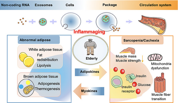
Aged adipose tissue
As previously summarized, aged adipose tissue may be a risk factor for sarcopeniaref. mco270030-bib-0022, ref. mco270030-bib-0049, ref. mco270030-bib-0050 (Figure 1). Fat tissue serves as a significant endocrine organ and could potentially be the largest organ in aging or obese individuals in certain instances.ref. mco270030-bib-0092 The primary components of adipose tissue include white adipose tissue (WAT), along with brown adipose tissue (BAT) and the intermediate beige fat.ref. mco270030-bib-0093, ref. mco270030-bib-0094 WAT is also present within muscles and impacts muscle functionality.ref. mco270030-bib-0095, ref. mco270030-bib-0096, ref. mco270030-bib-0097, ref. mco270030-bib-0098, ref. mco270030-bib-0099, ref. mco270030-bib-0100 BAT and WAT have contrasting roles in energy metabolism.ref. mco270030-bib-0101
BAT
BAT generates small, numerous fat droplets dispersed in the cytoplasm. Abundant mitochondria within BAT facilitate the burning of fat to generate heat. BAT expresses high levels of uncoupling protein‐1 (UCP‐1), crucial for heat production. Importantly, BAT can metabolize WAT, aiding in obesity alleviation by producing heat energy.ref. mco270030-bib-0102, ref. mco270030-bib-0103, ref. mco270030-bib-0104 Thus, BAT holds considerable importance in promoting human well‐being and combating conditions like diabetes, obesity, and other metabolic ailments.ref. mco270030-bib-0105, ref. mco270030-bib-0106, ref. mco270030-bib-0107, ref. mco270030-bib-0108 Indeed, brown fat adipocytes originate from skeletal muscle precursor cells, specifically Myf5‐positive cells, whereas beige adipocytes differentiate from Myf5‐negative progenitor cells, despite exhibiting similar morphology and function to brown adipocytes.ref. mco270030-bib-0109, ref. mco270030-bib-0110
As individuals age, there is a decline in BAT mass and adipogenic activity, coupled with reduced BAT thermogenesis. This decline may exhibit sex‐based differences.ref. mco270030-bib-0111 For example, Ouellet’s studyref. mco270030-bib-0112 demonstrated a significant decline in BAT mass among the elderly compared with the young cohort. Additionally, this decline was more pronounced in males across all age groups than in females.ref. mco270030-bib-0112 Valle’s researchref. mco270030-bib-0113 indicated that the capacity of BAT to sustain reduced heat production is poorer in elderly male rats compared with females. These findings may help explain the increase in visceral fat accumulation with age and why women tend to be more resilient to cold conditions compared with men.ref. mco270030-bib-0114 Indeed, the underlying cause of decreased BAT activity during aging remains elusive. Such changes could potentially exacerbate the development of obesity and IR, both hallmarks of sarcopenia. However, the interplay between BAT and sarcopenia warrants further investigation for clarity. Furthermore, BAT possesses endocrine capabilities that influence metabolism in distant organs, releasing factors that function in an autocrine manner. For example, BAT can secrete not only traditional adipokines such as leptin and adiponectin, but also FGF21 and 12,13‐HOME. Aging may affect the signaling of these molecules, which in turn could influence skeletal muscle physiology.ref. mco270030-bib-0101, ref. mco270030-bib-0115, ref. mco270030-bib-0116 We will describe this insight in the following section.
WAT
WAT is associated with insulation and cushioning, lipid storage and release, and endocrine signaling. It is extensively present in subcutaneous tissue (sWAT) and surrounding viscera (vWAT). The varying morphological and molecular mechanisms are fundamental to these two depots, directly impacting distinct metabolic health and diseases.ref. mco270030-bib-0117, ref. mco270030-bib-0118 vWAT is characterized by numerous large adipocytes, whereas sWAT contains a higher proportion of small adipocytes.ref. mco270030-bib-0119 vWAT contains a large number of blood vessels, and nerves are more susceptible to lipolysis in response to signaling from the nervous system, with a rich blood supply, while sWAT is preferentially for long‐term lipid storage.ref. mco270030-bib-0120 As individuals age, there is a tendency for the ratio of vWAT to sWAT to rise, reflecting the accumulation of vWAT.ref. mco270030-bib-0121 The substantial release of free fatty acids (FFAs) directly into the portal vein circulation results in their dissemination to the liver and peripheral tissues, contributing to triglyceride (TG) accumulation in these nonadipose tissues and reducing insulin sensitivity. This process might explain why increased vWAT is related to metabolic diseases, including diabetes and obesity.ref. mco270030-bib-0122, ref. mco270030-bib-0123
Furthermore, the maintenance of WAT functionality relies on precise regulation to produce lipid‐storing adipocytes. Adipose progenitor and stem cells (APSCs) play a pivotal role in metabolic regulation by generating mature adipocytes. These APSCs are proposed to be central to systemic metabolism disruption. Specifically, their capacity for proliferation and differentiation sustains the renewal, enlargement, and adaptive functionality of adipose tissue.ref. mco270030-bib-0124 However, the decreased proliferation and differentiation capacity of APSCs mainly occur due to aging.ref. mco270030-bib-0092, ref. mco270030-bib-0125 The dysfunction of APSCs compromises the plasticity of adipose tissue and is correlated with an increased susceptibility to IR.ref. mco270030-bib-0126 Additionally, preadipocytes’ ability to manage fatty acids diminishes with age, as they are less capable of differentiating and effectively storing lipids. This impairment can result in a higher vulnerability to lipotoxicity and reduced efficiency in fatty acid‐induced adipogenesis.ref. mco270030-bib-0127 Certainly, these factors could contribute to age‐related metabolic alterations in older individuals. The mechanism underlying impaired preadipocyte differentiation involves the downregulation of C/EBPα and peroxisome proliferator‐activated receptor (PPAR)γ expression in adipose tissue.ref. mco270030-bib-0128, ref. mco270030-bib-0129 Indeed, beyond differentiation, the self‐renewal capacity of preadipocytes is also compromised during aging or degenerative diseases.ref. mco270030-bib-0126
Moreover, aging induces senescence in both rodent and human WAT, leading to a reduction in adipogenic potential.ref. mco270030-bib-0130 For instance, markers like p16INK4a and SA‐β‐gal are heightened throughout WAT depots, and the ability of preadipocytes to differentiate is diminished in older mice.ref. mco270030-bib-0131 Clearance of senescent cells from 18‐month‐old mice by Xu alleviated the senescent cell‐mediated inhibition of adipogenesis and mitigated age‐related fat loss.ref. mco270030-bib-0132 This suggests that senolytic drugs could serve as a promising treatment to diminish the burden of senescent cells and enhance therapeutic potential in adipose tissue.ref. mco270030-bib-0133 Senescent cells, including adipocytes and preadipocytes, can secrete SASP factors such as proinflammatory cytokines, which can induce inflammation in tissues and impact other organs. When adipose progenitor cells are cocultured with aging cells, only around 20% of these progenitor cells accumulate fat. In contrast, when cocultured with nonaging cells, the proportion of progenitor cells with fat accumulation increases to over 50%. This suggests that the presence of senescent cells can impair the adipogenic potential of progenitor cells.ref. mco270030-bib-0132 Indeed, immune cells within adipose tissue can release proinflammatory cytokines under the influence of aging or obesity. The aging process is characterized by an increase in the number of M1 phenotype macrophages and a decrease in M2 phenotype macrophages in WAT. For example, animal studies have demonstrated that as animals age, the ratio of M1 to M2 macrophages rises, signifying a decrease in M2 alternatively activated macrophages and fostering a proinflammatory environment within the tissue.ref. mco270030-bib-0134, ref. mco270030-bib-0135
Overall, age‐related factors, such as the redistribution of WAT, senescence, and inflammation, can compromise the normal functions of adipose tissue. These disruptions can lead to abnormal lipid accumulation, metabolic dysfunction, and IR in WAT. This review categorizes the process of fat infiltration and delineates the events occurring at each stage, in conditions during aging. However, it is noteworthy that while certain cells or molecules play a pivotal role in a particular phase, they may also contribute to the overall process of muscle homeostasis. In addition, lean and youthful WAT produces various molecules like leptin and adiponectin that can affect other organs or the adipose tissue itself. As inflammation and reduced adipogenesis become more common with aging, they might also impact the endocrine role of adipose tissue. The production and secretion of these adipokines are influenced by aging, which has been explored in depth in the literature.ref. mco270030-bib-0118, ref. mco270030-bib-0136 We will discuss the roles of adipokines in sarcopenia with aging in the following section.
Accumulation of lipid derivatives in muscle cells leads to the condition known as myosteatosis
At first, it is hypothesized that an excess of lipids can “spill over” into various tissues, leading to ectopic fat infiltration, notably in skeletal muscles, a phenomenon termed myosteatosis. These lipids accumulate in the form of intermuscular adipose tissue (IMAT), intramuscular adipose tissue, and intramyocellular lipid droplets (IMCLs).ref. mco270030-bib-0049, ref. mco270030-bib-0137 Evidence indicates that myosteatosis is linked to preceding muscle atrophy, contributing to the development of sarcopenia.ref. mco270030-bib-0138 Although the precise biological mechanisms driving the escalation of myosteatosis or the infiltration of fat into skeletal muscles remain uncertain, recent findings propose a significant involvement of FAPs. FAPs, constitute a subset within the muscle with the capacity for differentiation into adipocytes and fibrocytes.ref. mco270030-bib-0137 The process of differentiation hinges on various stimuli, potentially leading to either excessive fat infiltration or fibrosis, both commonly observed in various pathological conditions.ref. mco270030-bib-0137 FAPs have a direct role in regulating muscle regeneration. For example, depletion of FAPs in muscles has been demonstrated to cause muscle weakness and atrophy, alterations in fiber type composition, and denervation at NMJ, ultimately leading to a failure to maintain muscle mass.ref. mco270030-bib-0139 With aging, aberrant signaling in FAPs can trigger their profibrotic behavior, leading to skeletal muscle fibrosis. Additionally, this process can compromise satellite cell function.ref. mco270030-bib-0140 Transplantation of young FAPs into aged mice has shown the ability to restore the myogenic commitment of MuSCs.ref. mco270030-bib-0140, ref. mco270030-bib-0141, ref. mco270030-bib-0142 Interestingly, transplanting senescent FAPs intraperitoneally into mice is sufficient to induce a sarcopenia‐like functional phenotype, as evidenced by decreased grip strength and walking speed.ref. mco270030-bib-0139, ref. mco270030-bib-0143
FAPs exert many of their functional effects on muscle regeneration and repair by secreting paracrine factors within their local microenvironment.ref. mco270030-bib-0144, ref. mco270030-bib-0145 Aging could potentially impair the secretion of a soluble myogenic support signal from FAPs. For instance, FAPs may lose the ability to stimulate the production of the matricellular protein Wnt1‐inducible signaling pathway protein 1 (WISP1), crucial for satellite cell expansion and commitment. This impairment could further contribute to the decline in muscle function with age.ref. mco270030-bib-0140 In addition, FAPs serve as the main mononuclear cell source of IL‐33, a cytokine linked to type 2 immunity. Nonetheless, with aging, there is a decline in IL‐33 production by FAPs, resulting in reduced accumulation of regulatory T cells (Tregs) and impaired muscle repair.ref. mco270030-bib-0146 Studying the mechanisms that lead to these alterations in IL‐33 production by FAPs is crucial for comprehending the broader effects of FAPs on muscle regeneration and repair as one ages.ref. mco270030-bib-0140, ref. mco270030-bib-0146 Modulating the cytokines released by FAPs could serve as a potential therapeutic approach to alleviate age‐related muscle decline and promote muscle regeneration. Additionally, changes in the microenvironment or niche where FAPs reside during aging contribute to the dysfunctional state of FAPs, disrupting the cellular niche necessary for proper skeletal muscle regeneration.ref. mco270030-bib-0147, ref. mco270030-bib-0148 The differential effects of conditioned medium from myogenic cells isolated from aged versus young donors on FAPs proliferation and adipogenic differentiation highlight the impact of age‐related changes in the muscle microenvironment.ref. mco270030-bib-0147 The disruption described can indeed lead to the development of muscle fibrosis. Furthermore, in the presence of chronic inflammatory conditions associated with various metabolic disorders, the signals that typically regulate FAP apoptosis and inhibit proliferation are disturbed. This disturbance results in a chronic state of tissue remodeling, ultimately leading to the accumulation of fibrotic and fatty tissues. This aberrant tissue accumulation further impairs skeletal muscle function and regeneration, exacerbating the effects of aging and metabolic disorders on muscle health. Understanding and addressing these mechanisms are crucial for developing effective therapeutic interventions.ref. mco270030-bib-0145
Aged FAPs not only contribute to the decline in muscle repair and regeneration but are also the main stem cell group involved in the formation of IMAT in sarcopenia.ref. mco270030-bib-0137, ref. mco270030-bib-0139, ref. mco270030-bib-0149 Numerous signaling pathways are implicated in this process. With aging, there is a diminished response in the Notch signaling pathway, resulting in compromised satellite cell proliferation and hindrance in muscle regeneration.ref. mco270030-bib-0150 The reduced activity of the Notch signaling pathway may be implicated in the development of IMAT driven by FAPs during aging.ref. mco270030-bib-0150 In specific, Notch signaling has been observed to inhibit the differentiation of FAPs into adipocytes both in laboratory settings and in living organisms. Treatment with 5 µM DAPT, an activator of the Notch signaling pathway, has been shown to reduce the accumulation of adipose tissue.ref. mco270030-bib-0150 Moreover, during aging, changes in Wnt10b signaling lead to the increased expression of critical adipogenic genes, thereby contributing to the accumulation of IMAT.ref. mco270030-bib-0151 Aging is also linked to a decline in the synthesis of nitric oxide (NO) in skeletal muscle. NO plays a role in regulating the fate of FAPs by inhibiting their differentiation into adipocytes.ref. mco270030-bib-0152, ref. mco270030-bib-0153 Moreover, aging is associated with comorbidities that can influence the behavior of FAPs. Buras et al.ref. mco270030-bib-0154 found that prolonged consumption of a high‐fat diet (HFD) leading to obesity can promote the proliferation of FAPs, as well as their differentiation into adipocytes and collagen‐depositing fibroblasts. This observation is reinforced by the discovery that adipogenic progenitors can be isolated from obese human skeletal muscle using the CD56negCD15pos cellular fraction, which corresponds to PDGFRαpos FAPs that give rise to intramuscular adipose tissue.ref. mco270030-bib-0155, ref. mco270030-bib-0156 In obese mice, the proliferation of FAPs is facilitated by elevated levels of circulating adipokines such as Thrombospondin 1 (THBS1) and TGF‐β1, which are secreted by the hypertrophic adipose tissue.ref. mco270030-bib-0154, ref. mco270030-bib-0157 The presence of various factors derived from adipocytes enhances the adipogenic differentiation of FAPs, suggesting a direct mechanism by which adipose tissue expansion in obesity contributes to the accumulation of IMAT. Moreover, diabetes could also influence the behavior of FAPs. A study utilizing various genetic and diet‐induced mouse models of diabetes demonstrated that the ectopic deposition of adipocytes in skeletal muscle originated from FAPs.ref. mco270030-bib-0158
The adipocytes derived from FAPs may display reduced insulin sensitivity compared with typical adipocytes, as indicated by decreased insulin receptor phosphorylation. This suggests that the accumulation of adipocytes originating from FAPs could contribute to compromised peripheral insulin sensitivity.ref. mco270030-bib-0156 Indeed, these findings indicate that FAPs directly contribute to the buildup of IMAT in skeletal muscle during aging and metabolic disorders, which are pivotal to the adverse alterations observed in skeletal muscle under metabolic stress. However, since IMAT represents only a small portion of the overall adipose depots in the body, its adverse effects on glucose disposal are likely mediated through secondary mechanisms that hinder myofiber’s capacity to uptake glucose (as described below).
As previously mentioned, FAPs can develop into IMAT during aging, which refers to adipose tissue interspersed between and around skeletal muscle groups. Several studies highlight a relationship between IMAT levels and muscle function. Indeed, increased accumulation of IMAT can lead to a decrease in muscle mass, muscle strength, and insulin sensitivity.ref. mco270030-bib-0100, ref. mco270030-bib-0159, ref. mco270030-bib-0160 These research findings indicate that individuals aged 70–79 years with elevated baseline levels of IMAT are more prone to experiencing mobility limitations compared with those with lower baseline levels of IMAT.ref. mco270030-bib-0160 Studies indicate that higher levels of IMAT in the quadriceps upon admission are more strongly correlated with poorer recovery of activities of daily living (ADL) than lower muscle mass in older hospitalized patients.ref. mco270030-bib-0161 These studies illustrate that IMAT serves as a potential predictor of sarcopenia, a condition observed in diverse populations, including obese, diabetic, dystrophic, sarcopenic, and aging animals and patients.ref. mco270030-bib-0158, ref. mco270030-bib-0162, ref. mco270030-bib-0163 IMAT is linked to metabolic health and the onset of IR and other metabolic disorders.ref. mco270030-bib-0164, ref. mco270030-bib-0165 The proximity of IMAT to muscle fibers enables it to release various proinflammatory cytokines such as IL‐6 directly onto the muscle fibers, triggering local inflammation. This inflammatory reaction can contribute to the onset of IR and other metabolic disturbances.ref. mco270030-bib-0099, ref. mco270030-bib-0100, ref. mco270030-bib-0166 Moreover, muscle dysfunction can contribute to a detrimental cycle where reduced physical activity leads to increased levels of IMAT, which in turn exacerbates muscle dysfunction.ref. mco270030-bib-0167, ref. mco270030-bib-0168, ref. mco270030-bib-0169 Further exploration is necessary to comprehensively grasp the precise mechanisms by which IMAT impacts skeletal muscle.
Given the possible mechanistic connection between the adverse effects of IMAT accumulation on muscle and mobility function, addressing IMAT and its related inflammation could prove pivotal in rehabilitation settings addressing age‐related diseases and disabilities.ref. mco270030-bib-0170, ref. mco270030-bib-0171, ref. mco270030-bib-0172 Promising evidence indicates that physical therapy may have beneficial effects on levels of IMAT and intramuscular inflammation.ref. mco270030-bib-0173, ref. mco270030-bib-0174 Other researchers examined the impact of 12 weeks of eccentric exercise training on older individuals and observed an 11% reduction in the area of IMAT in the thigh.ref. mco270030-bib-0175 A study has shown that a 14‐week periodized conventional strength training protocol can have positive effects on intermuscular fat and muscle quality in patients with knee OA.ref. mco270030-bib-0176 In a captivating study conducted by Wroblewski et al.,ref. mco270030-bib-0177 attention was directed toward elderly elite athletes who participated in fitness and sports competitions at least four or five times per week. The results unveiled that these individuals did not undergo any decline in lean muscle mass or a rise in IMAT accumulation with age.ref. mco270030-bib-0177
Aside from IMAT, fat infiltration into muscle can manifest as IMCLs. IMCLs, a smaller subset of lipids, are stored as lipid droplets within muscle cells.ref. mco270030-bib-0098 Predominantly containing TGs, diacylglycerols (DAGs), and ceramides, these lipids serve as an energy source during exercise. However, their excessive accumulation can induce lipotoxic and inflammatory effects, heightening the risk of certain pathological conditions, particularly metabolic diseases.ref. mco270030-bib-0178, ref. mco270030-bib-0179 The primary consequence of muscle‐associated LDs is the degradation of mitochondrial function in muscle, leading to reduced lipid β‐oxidation and lipolysis, along with increased ROS production. These changes foster IR and lipotoxicity, as well as activate inflammatory signaling pathways, ultimately resulting in skeletal muscle inflammation, metabolic abnormalities, and contributing to the onset of sarcopenia.ref. mco270030-bib-0049, ref. mco270030-bib-0098, ref. mco270030-bib-0180, ref. mco270030-bib-0181, ref. mco270030-bib-0182, ref. mco270030-bib-0183, ref. mco270030-bib-0184 Over time, these alterations negatively impact the contractile function and metabolic characteristics of skeletal muscle, significantly affecting human health and exacerbating IR.ref. mco270030-bib-0081
Myosteatosis induced by IMAT and IMCL can result in local muscle inflammation and alters myocyte insulin sensitivity through paracrine or autocrine effects. Inflammatory signals can activate pattern recognition receptors in myocytes, such as Toll‐like receptor 4 (TLR4), leading to direct metabolic effects.ref. mco270030-bib-0119 Besides, MuSCs gradually undergo senescent, releasing SASP factors.ref. mco270030-bib-0027, ref. mco270030-bib-0038 Therefore, muscle tissues can trigger metabolic dysfunction and inflammaging by transplanting proinflammatory cytokines into blood‐storm, contributing to IR and inflammation.ref. mco270030-bib-0049 Collectively, this evidence contributes to the recognition of myosteatosis as a potential significant predictor in sarcopenia.
Aging‐induced myosteatosis can exacerbate systemic inflammaging and IR by recruiting immune cells
As described earlier, age‐induced myosteatosis plays a significant role in contributing to IR and inflammation.ref. mco270030-bib-0185 The elevated levels of proinflammatory cytokines are recognized for inducing the infiltration of immune cells, such as T cells, B cells, and macrophages, into adipose tissue, muscle, and other tissues.ref. mco270030-bib-0051, ref. mco270030-bib-0052, ref. mco270030-bib-0186 Furthermore, age‐related immune system dysregulation, known as immune aging or immunosenescence, can disturb the immune environment. This disruption alters interactions between immune cells and signaling pathways crucial for skeletal muscle function.ref. mco270030-bib-0187, ref. mco270030-bib-0188 For instance, Wang et al.ref. mco270030-bib-0189 conducted bone marrow transplantations in mice with disparate ages and observed that immune system aging led to a reduction in MuSC numbers. This process promoted their transformation into a fibrogenic phenotype, ultimately impacting the onset of sarcopenia.ref. mco270030-bib-0189
The lymphocyte compartment encompasses the primary circulating subpopulations of immune cells.ref. mco270030-bib-0190 Within lymphocytes, T cells are pivotal in the processes of skeletal muscle repair, regeneration, and differentiation. Specifically, CD8+ T cells contribute to skeletal muscle regeneration through the secretion of MCP‐1, which recruits Gr1(high) macrophages. These macrophages, in turn, support myoblast proliferation, aiding in the regeneration process.ref. mco270030-bib-0191 During immune aging, the decline and alteration in the phenotype of T lymphocytes, transitioning from CD8+ to CD4+, could be associated with a decrease in muscle mass.ref. mco270030-bib-0192 Research has indicated that a specific subset of CD4+ T helper 1 cells, characterized by the absence of CD28 (CD4+CD28null T cells), showed a negative correlation with muscle mass index in patients with sarcopenia.ref. mco270030-bib-0192, ref. mco270030-bib-0193 Furthermore, CD28null T cells have been found to release cytotoxic particles containing perforin and granzyme B. Additionally, they can produce cytokines like TNF‐α and interferon gamma (IFN‐γ), thereby activating inflammatory pathways.ref. mco270030-bib-0194 The cytokines produced by CD28null T cells, are believed to contribute to the onset of sarcopenia. Additionally, studies have indicated that the secretome of activated T cells from younger individuals can stimulate the proliferation and movement of immortalized murine satellite cells. Conversely, the secretome from activated T cells of older individuals has been observed to induce premature differentiation, while leaving satellite cell proliferation and migration unaffected. This suggests a modified interaction between immune cells and satellite cells during aging, potentially impacting skeletal muscle regeneration and repair processes in sarcopenia.ref. mco270030-bib-0195 These observations imply that proteins released by adaptive immune cells in younger individuals promote the proliferation and movement of satellite cells, whereas proteins secreted by adaptive immune cells in older individuals hinder satellite cell proliferation and migration by triggering premature differentiation. Consequently, the diminished proliferation and migration of satellite cells in older individuals are associated with age‐related deficiencies in T cells. In vitro investigations have additionally revealed that aged muscle cells exhibit reduced responsiveness to immune secretions.ref. mco270030-bib-0196 Dumke’s studyref. mco270030-bib-0196 discovered that a medium conditioned by T cells stimulated the proliferation and migration of satellite cells taken from the muscles of young rats but did not yield the same outcomes with satellite cells obtained from the muscles of older rats. This suggests that both the reaction of satellite cells to immune cells and the immune response to muscle cells alter with age, fostering a vicious cycle that disrupts muscle regeneration as individuals age.ref. mco270030-bib-0196
Additionally, another significant subset of T cells that infiltrate skeletal muscle are known as immune response Tregs, denoted as the CD4 + Foxp3 + sub‐phenotype.ref. mco270030-bib-0197 Age‐related impairments to Treg signaling are also believed to limit muscle regenerative capacity. During muscle regeneration, as described above, the recruitment of Tregs via IL‐33 signaling is decreased in old mice due to ineffective production of it by FAP‐like cells, and these defects compromise muscle regenerative capacity.ref. mco270030-bib-0146 However, the regenerative capacity of aged skeletal muscle can be improved through intramuscular or systemic supplementation with IL‐33, which re‐establishes the recruitment of Tregs into injured muscles.ref. mco270030-bib-0146 The depletion of Tregs, which is characterized by an increased IFN‐γ response and activation of M1 macrophages, leads to an increase in muscle inflammation. It also involves abnormal inflammatory morphology and fibrosis of regenerated muscle fibers.ref. mco270030-bib-0198 Studies have found that accumulation of Treg cells in injured skeletal muscle profoundly declines with age, paralleling a degradation of repair and regeneration processes.ref. mco270030-bib-0146
Indeed, macrophages play a crucial role in tissue repair following damage. They swiftly infiltrate damaged tissue, aiding in debris removal and promoting satellite cell myogenesis, which contributes to tissue regeneration.ref. mco270030-bib-0199, ref. mco270030-bib-0200 Proinflammatory macrophages indeed play a pivotal role in promoting the proliferation and migration potential of satellite cells. The signaling molecules released by these macrophages enhance the regenerative capacity of satellite cells, crucial for muscle tissue repair. However, it is vital to acknowledge that prolonged presence or inhibition of proinflammatory macrophage activity can have adverse effects, potentially exacerbating tissue damage and causing delays in the muscle repair process.ref. mco270030-bib-0201 Macrophages possessing anti‐inflammatory properties release substances that guide the differentiation of satellite cells and aid in the reconstruction, remodeling, and maturation of the extracellular matrix (ECM). However, akin to the negative impacts of prolonged proinflammatory activity on muscle health, an excessive or premature shift toward an anti‐inflammatory phenotype can also have detrimental effects. Balancing the inflammatory response is critical for optimal muscle repair and regeneration.ref. mco270030-bib-0202 Therefore, achieving a timely and accurate shift to anti‐inflammatory macrophage phenotype replacement is essential for promoting tissue growth and restoring homeostasis. With aging, changes in monocyte/macrophage phagocytosis and cytokine secretion have been noted,ref. mco270030-bib-0203 although there are still inconsistencies among research findings. Research on aging has shown mixed results regarding monocyte behavior: some studies indicate an increase in the production of proinflammatory cytokines by human monocytes as people age, while other studies have observed a decrease in this production.ref. mco270030-bib-0204, ref. mco270030-bib-0205, ref. mco270030-bib-0206 Given the vital role of macrophages in governing the proliferation and differentiation of satellite cells, it is reasonable to suggest that any changes in macrophage functions occurring during aging could impair the skeletal muscle’s capacity for regeneration.
In summary, excess lipids can overflow and redistribute to various tissues, including skeletal muscles (IMAT and IMCLs). A specific population of skeletal muscle mesenchymal stem cells called FAPs is responsible for the development of IMAT with aging, which may serve as a potential predictor of sarcopenia. IMCLs can potentially lead to mitochondrial dysfunction, impair the β‐oxidation process of fatty acids, and increase ROS production, contributing to lipotoxicity, IR, and inflammation. Proinflammatory factors attract T cells, macrophages, and other immune cells into adipose and muscle tissues. Immunosenescent cells not only directly impede muscle regeneration but also release numerous proinflammatory cytokines and chemokines, inducing local chronic inflammation that extends into a systemic inflammaging state and promotes IR in adipose and muscle tissues. Thus, local inflammation, lipotoxicity, and IR initiate systemic inflammaging, worsening lipid metabolism dysfunction in a perpetuating cycle.
The interaction between adipose tissue and skeletal muscle plays a significant role in the development of sarcopenic obesity
Given the close relationship and interaction between muscle and adipose tissue, it is reasonable to hypothesize that inflamed adipose tissue and inflamed skeletal muscle create a harmful cycle, contributing to age‐related sarcopenia and sarcopenic obesity. Sarcopenic obesity represents a clinical and functional state marked by the simultaneous presence of obesity and sarcopenia.ref. mco270030-bib-0207 Indeed, obese adipocytes have the potential to trigger inflammation and muscle cell atrophy, thereby contributing to muscle wasting commonly associated with metabolic disorders.ref. mco270030-bib-0208 The consensus is that obesity is typified by a state of low‐level chronic inflammation.ref. mco270030-bib-0209 Several immune cell types, such as macrophages, T cells, B cells, and neutrophils, associated with adipose tissue.ref. mco270030-bib-0210, ref. mco270030-bib-0211 Growing adipose tissue alters the composition and abundance of both local and systemic immune cells, promoting a transition toward a more proinflammatory profile.ref. mco270030-bib-0211 In adipose tissue, this transition involves a shift from anti‐inflammatory M2 macrophages in lean individuals to proinflammatory M1 macrophages in obese individuals.ref. mco270030-bib-0212 In obese tissue, a notable feature called “crown‐like” structures can be observed, wherein macrophages surround deceased or dying fat cells.ref. mco270030-bib-0213, ref. mco270030-bib-0214 Obese individuals who lack crown‐like structures exhibit improved metabolic control and reduced expression of inflammatory genes.ref. mco270030-bib-0215 In obese mice, there is a heightened presence of MCP‐1 in both WAT and plasma.ref. mco270030-bib-0216 Indeed, MCP‐1 is recognized as a potent chemotactic factor for monocytes.ref. mco270030-bib-0217 Obese mice lacking MCP‐1 exhibited decreased macrophage presence and a reduction in the inflammatory state of adipose tissue. There is an interplay between macrophages and adipocytes, where in obesity, adipocytes release surplus saturated FFA, activating macrophages via the TLR4 signaling pathway. Consequently, macrophages produce TNF‐α, which interacts with tumor necrosis factor receptor 1 on adipocytes, activating the NF‐κB pathway and instigating an inflammatory cascade, resulting in further FFA release.ref. mco270030-bib-0218 There are reports proposing that FFA could act on fat cells in an autocrine manner, triggering an inflammatory reaction and elevating adipokine production. This mechanism is thought to occur, in part, via the TLR4 pathway.ref. mco270030-bib-0219 Obesity can influence the composition of T‐cell subsets within adipose tissue, where they are thought to play a role in modulating macrophage phenotypes. Thin mice were observed to have elevated levels of CD4+ Tregs and Th2 polarized cells in adipose tissue. These cells contribute to preserving adipose tissue function and insulin sensitivity by promoting an anti‐inflammatory shift in macrophage activation.ref. mco270030-bib-0220, ref. mco270030-bib-0221 In obesity, the accumulation of CD8+ effector T cells and CD4+ Th1 cells in adipose tissue can result in the generation of Th1 signals. These signals can initiate the recruitment and activation of macrophages, thus perpetuating the proinflammatory cascade linked to IR.ref. mco270030-bib-0222 Thus, the alterations in the signaling equilibrium of Th1‐ and Th2‐types induced by obesity can impact macrophage recruitment and phenotype in adipose tissue, leading to either pathogenesis or environmental protection. Furthermore, B cells have been implicated in obesity‐induced adipose tissue inflammation by promoting T cell and macrophage activation.ref. mco270030-bib-0223 Indeed, the secretion of soluble factors by proinflammatory macrophages can reshape tissue composition, fostering a microenvironment conducive to tissue remodeling and reconstruction.
There is evidence suggesting an upregulation of inflammatory cytokine production and heightened inflammation in skeletal muscle among individuals with obesity.ref. mco270030-bib-0224 In sarcopenic obesity, adipose tissue‐resident macrophages can foster a sterile inflammatory milieu, potentially dampening insulin signaling. This reinforces the concept of “immune metabolism,” acknowledging macrophages’ dual roles—both detrimental and beneficial—in sarcopenic obesity.ref. mco270030-bib-0098, ref. mco270030-bib-0225 In obese individuals, muscles exhibit an accumulation of M1 macrophages compared with lean subjects, and this accumulation has been found to correlate with body mass index (BMI). Similarly, studies with mice fed a HFD have reported comparable findings. Furthermore, research indicates that a short‐term high‐fat, high‐calorie diet, or overfeeding, which can induce IR, leads to an increase in macrophage markers in the skeletal muscle of healthy individuals.ref. mco270030-bib-0226, ref. mco270030-bib-0227 Histologically, macrophages and T lymphocytes are primarily located in the adipose tissue surrounding skeletal muscle, known as intermyocellular/IMAT. These immune cells are situated between muscle fibers or in close proximity to the muscle.ref. mco270030-bib-0228 In obesity, there is a significant increase in the number of macrophages and T cells within these adipose depots (IMAT).ref. mco270030-bib-0228 Immune cells in skeletal muscle, akin to those in visceral adipose tissue, also tend to undergo polarization into proinflammatory phenotypes during obesity.ref. mco270030-bib-0228, ref. mco270030-bib-0229 Research indicates that in obese mice, there is an increase in both CD4+ and CD8+ T cells within skeletal muscle tissue. Moreover, the proportion of Th1 cells expressing IFN‐γ is elevated, whereas the proportion of Tregs is reduced in the skeletal muscle tissue of mice with obesity.ref. mco270030-bib-0228 Macrophages and T lymphocytes are present in skeletal muscle between myofibers, although they occur at a lower frequency compared with the adipose tissue surrounding the muscle.ref. mco270030-bib-0228 Indeed, while proinflammatory markers may be more abundant in adipose tissue depots surrounding skeletal muscle (IMAT), infiltrating immune cells can still contribute to the release of proinflammatory molecules and metabolic dysfunction in skeletal muscle. Additionally, under conditions of inflammation or in the presence of inflammatory molecules like FFAs, both adipocytes and myocytes have been demonstrated to secrete increased levels of chemokines,ref. mco270030-bib-0230 which induce immune cell migration,ref. mco270030-bib-0231, ref. mco270030-bib-0232 including MCP‐1, to recruited infiltration of leukocytes from the circulation into tissues requires. Absolutely, as obesity progresses, recruited immune cells like macrophages and T lymphocytes can indeed secrete chemokines, exacerbating inflammation in both skeletal muscle and adipose tissue, as described. This sets up a feedback loop where the initial inflammatory response triggers the recruitment of more immune cells, which then secrete additional chemokines, amplifying the inflammatory response further. This cycle contributes significantly to the chronic low‐grade inflammation observed in obesity.
Studying changes in fat deposition and distribution during advanced aging is essential, as dysregulated lipid metabolism in adipose tissue can lead to local inflammation and exacerbate the process of inflammaging. These factors are likely contributors to metabolic‐related diseases. The metabolic effects of ectopic fat accumulation present potential therapeutic targets for age‐related conditions, such as sarcopenia. There is evidence supporting the hypothesis that aged adipose tissue serves as a risk factor for sarcopenia, with the bidirectional interaction between adipose tissue and muscle potentially worsening this cycle. However, in older adults, sarcopenic obesity has been associated with better survival outcomes compared with sarcopenia alone.ref. mco270030-bib-0022 As the prevalence of sarcopenic obesity increases globally, it is crucial to investigate the underlying mechanisms of this condition.
PATHOPHYSIOLOGT OF CACHEXIA
The etiology of cachexia involves a complex interplay of mechanisms. A thorough understanding of its pathogenesis is essential for developing targeted prevention and management strategies for this debilitating condition. This section explores the multifaceted etiological factors, cellular and molecular mechanisms, inflammatory processes, and the role of adipose tissue in cachexia.
Systemic inflammation and cytokine dysregulation
Systemic inflammation is a well‐documented feature of cancer cachexia, with increased circulating levels of C‐reactive protein (CRP) associated with weight loss in cancer patients.ref. mco270030-bib-0233, ref. mco270030-bib-0234, ref. mco270030-bib-0235 In 1985, Cerami’s groupref. mco270030-bib-0236, ref. mco270030-bib-0237 demonstrated that circulating mediators could induce cachexia, identifying TNF‐α, initially termed “cachectin.” In cancer cachexia, proinflammatory cytokines produced by immune and tumor cells—particularly TNF‐α, interleukin‐1, ‐6, and ‐8 (IL‐1, IL‐6, IL‐8), and IFNγ—play significant roles in driving the wasting phenotype associated with this syndrome and are classified as procachetic factors.ref. mco270030-bib-0238 For instance, serum concentrations of IL‐1 increase in cachectic patients, although its role in tissue wasting remains debated.ref. mco270030-bib-0239, ref. mco270030-bib-0240 On one hand, IL‐1 is thought to induce anorexia by increasing tryptophan plasma concentrations, leading to elevated serotonin levels that cause early satiety and suppress appetite.ref. mco270030-bib-0241 Inversely, other studies have shown that high circulating IL‐1 does not significantly impact food intake or weight loss, suggesting it may exert local tissue‐specific effects or require high pharmacologic doses to elicit a cachectic response.ref. mco270030-bib-0242, ref. mco270030-bib-0243 The role of IFNγ in cachexia is also not fully understood; however, it has been shown to synergize with TNF‐α to promote muscle wasting.ref. mco270030-bib-0244, ref. mco270030-bib-0245 IFNγ inhibits myosin mRNA in skeletal muscle cells and activates ubiquitin gene expression.ref. mco270030-bib-0241, ref. mco270030-bib-0244 The subsequent sections will delve into the roles of TNF‐α, IL‐6, and IL‐8 in cachexia. Additionally, the decreased expression of anti‐inflammatory cytokines such as IL‐4, IL‐10, and IL‐12 accompanies the upregulation of proinflammatory cytokines, further disrupting the balance between pro‐ and anti‐inflammatory stimuli.ref. mco270030-bib-0239 This systemic inflammatory response primarily affects skeletal muscle, indicating that while nonmuscle tissues (such as the host immune system and tumor cells) mainly contribute to skeletal muscle inflammation, increased cytokine production by skeletal muscle itself may also play a role. Therefore, the contribution of skeletal muscle fibers and resident or recruited mononucleated cells in cytokine production warrants further evaluation.
Protein degradation pathways
Activation of the ubiquitin–proteasome system
The ubiquitin–proteasome system (UPS) is a crucial pathway responsible for the degradation of ubiquitinated proteins within cells, playing a significant role in skeletal muscle protein turnover.ref. mco270030-bib-0246 Increased UPS activation is particularly important in driving muscle wasting associated with cachexia, as evidenced by numerous studies utilizing animal models of cancer, heart failure, and sepsis to explore the underlying molecular mechanisms.ref. mco270030-bib-0247, ref. mco270030-bib-0248, ref. mco270030-bib-0249, ref. mco270030-bib-0250, ref. mco270030-bib-0251
Skeletal muscle is highly susceptible to cachectic factors, such as proinflammatory cytokines, leading to selective degradation of specific muscle proteins rather than a generalized loss. For instance, research involving muscle biopsies from cancer cachexia patients has demonstrated upregulation of ubiquitin mRNA and 20S proteasome subunits, alongside increased proteasome activity compared with healthy controls.ref. mco270030-bib-0252, ref. mco270030-bib-0253 Furthermore, in mouse models with colon‐26 tumors, a marked reduction in myosin heavy chain was observed, correlating with muscle wasting.ref. mco270030-bib-0244 The enhanced activity of the proteasome pathway in cachexia appears to be mediated by the activation of key transcription factors, including FOXO and NF‐κB, which promote the expression of atrogenes such as MuRF‐1 and MAFbx. These atrogenes contribute to elevated proteasome activity and increased catabolism. Additionally, FOXO transcription factors further exacerbate this catabolic signaling by inhibiting the PI3K/Akt pathway, which is critical for protein synthesis. Importantly, prior to the UPS degrading monomeric actin and myosin, the activation of caspase‐3 through the PI3K pathway is necessary for dissociating actomyosin complexes, thereby facilitating the degradation process. This intricate interplay underscores the complexity of muscle wasting in cachectic conditions.ref. mco270030-bib-0254
Involvement of autophagy–lysosomal pathways
The autophagy–lysosome system plays a crucial role in eliminating long‐lived proteins and large supramolecular structures, including dysfunctional mitochondria. During the formation of a double‐membrane structure known as the autophagosome, proteins and organelles destined for degradation are engulfed. The autophagosome then fuses with lysosomes, enabling acidic proteolytic degradation of its contents by cathepsins. There is increasing interest in the role of autophagy in skeletal muscle wasting and the progression of cachexia.ref. mco270030-bib-0255, ref. mco270030-bib-0256 Evidence suggests that autophagy is significantly upregulated during cancer cachexia, with elevated levels of mediators such as BNIP3A mRNA and LC3B protein observed in a small cohort of lung cancer patients.ref. mco270030-bib-0257, ref. mco270030-bib-0258 In the skeletal muscle of cachectic cancer patients, the protein levels of autophagy‐related genes—such as ATG5, ATG7, Beclin1, and LC3B—are also increased, along with a rise in the number of autophagosomes.ref. mco270030-bib-0259, ref. mco270030-bib-0260, ref. mco270030-bib-0261 Additionally, transcription factors like NF‐κB, STAT3, and CCAAT/enhancer‐binding protein‐β (C/EBPβ) contribute to the regulation of E3 ubiquitin ligases and autophagy genes.ref. mco270030-bib-0262, ref. mco270030-bib-0263 Since animal models may not fully replicate the complex events of cancer cachexia in humans, it is essential to validate the significance of these transcription factors by assessing their activity in the skeletal muscle of patients with cancer‐associated cachexia.ref. mco270030-bib-0264 While autophagy is constantly active in removing damaged proteins and organelles, defects in this process can lead to muscle functional impairment, and excessive autophagy can contribute to muscle mass loss.ref. mco270030-bib-0265 Therefore, tight regulation of the autophagy–lysosome system is vital for maintaining skeletal muscle homeostasis. However, in cachexia, the upregulation of autophagy genes results in excessive activation of autophagy pathways, leading to increased breakdown of skeletal muscle.
Metabolic abnormalities and hypercatabolism
One consequence of the systemic metabolic alterations in cancer‐associated cachexia is decreased energy efficiency, primarily due to energy‐wasting mechanisms such as futile metabolic cycles, as demonstrated in preclinical studies.ref. mco270030-bib-0266 These changes result in energetic inefficiency, shifting the energy balance toward weight loss through increased resting energy expenditure (REE).ref. mco270030-bib-0267 Clinical studies indicate that not all cancer patients exhibit hypermetabolism; however, those with elevated REE experience a higher incidence of treatment‐related toxicities. Furthermore, advanced‐stage cancer patients with high REE have shorter median overall survival durations.ref. mco270030-bib-0267, ref. mco270030-bib-0268
Cancer cachexia is often characterized by hypermetabolism, which is frequently accompanied by mitochondrial dysfunction in skeletal muscle, leading to muscle wasting.ref. mco270030-bib-0269 For instance, breast cancer patients show dysregulation of pathways governing oxidative phosphorylation, resulting in mitochondrial dysfunction. Additionally, reduced PPAR signaling, which regulates energy metabolism, contributes to this dysfunction by decreasing β‐oxidation. In Lewis lung carcinoma (LLC) mice, tumor progression correlates negatively with mitochondrial ATP synthesis while promoting mitochondrial ROS production.ref. mco270030-bib-0270 In patients with gastrointestinal cancer‐associated cachexia, studies have identified disrupted mitochondrial morphology.ref. mco270030-bib-0259 Evidence suggests that dysregulated mitochondrial metabolism is critical for muscle wasting in cancer cachexia.ref. mco270030-bib-0271 For example, in older gastric cancer patients, muscle loss is associated with diminished mitochondrial protein content and increased mitophagy.ref. mco270030-bib-0272
Adipose and muscle wasting
WAT: dysfunctional lipid storage and remodeling
Fat is lost more rapidly than lean tissue in cancer cachexia, highlighting the growing recognition that adipose tissue wasting is a significant component of cancer‐associated weight loss and that fat mass can serve as a predictor of survival in these patientsref. mco270030-bib-0273 (Figure 1). Adipose tissue undergoes extensive remodeling during cachexia. In addition to lipid depletion, ECM remodeling occurs, as indicated by transcriptomic profiling of adipose tissue from patients with gastrointestinal cancer.ref. mco270030-bib-0274 Enhanced collagen accumulation and fibrosis have also been observed in adipose tissue specimens from patients experiencing cancer cachexia.ref. mco270030-bib-0275, ref. mco270030-bib-0276 This may contribute to increased recruitment of inflammatory cells into adipose tissue, causing local inflammation and potentially exacerbating the systemic inflammation commonly seen in cachexia.ref. mco270030-bib-0277
Even more strikingly, transcriptomic profiling of adipose tissue from gastrointestinal cancer patients suggests that both energy turnover and fatty acid degradation are significantly elevated in individuals with cachexia.ref. mco270030-bib-0274 Specifically, lipolysis— the hydrolysis of TGs into FFAs and glycerol— is linked to this wasting syndrome, with increased plasma levels of FFAs and glycerol often noted in cachectic patients.ref. mco270030-bib-0278 Lipolysis and palmitate oxidation are heightened in weight‐losing cancer patients,ref. mco270030-bib-0279 and elevated lipolysis in those with gastrointestinal adenocarcinoma correlates with increased expression of hormone‐sensitive lipase, a key lipolytic enzyme, in adipose tissue.ref. mco270030-bib-0280 Indeed, the activities of both hormone‐sensitive lipase and adipocyte TG lipase are elevated in the adipose tissue of cachectic patients, and knockout studies in mice have confirmed the central role of these lipases in the progression of cachexia.ref. mco270030-bib-0281 A futile substrate cycle between lipolysis and lipogenesis further contributes to the negative energy balance observed in murine models of cachexia.ref. mco270030-bib-0016, ref. mco270030-bib-0282
Dysfunction of adipose tissue in cachexia can have additional systemic effects. The infiltration of adipose tissue cells into skeletal muscle appears to contribute to muscle wasting. One study reported a correlation between the increased presence of IMCLs—indicative of adipose tissue cell infiltration in muscle—and body weight loss in patients with cancer.ref. mco270030-bib-0283 Insufficient lipid storage can lead to lipotoxicity, particularly detrimental effects arising from lipid accumulation in muscle, heart, and liver. Altered adipose tissue function may also result in changes to the secretion of lipokines and reactive lipid metabolites (such as lysophosphatidic acid, ceramide, and DAG), as well as adipokines (such as adiponectin and leptin). These alterations can contribute to local or systemic inflammation, ultimately leading to metabolic dysfunction.ref. mco270030-bib-0284, ref. mco270030-bib-0285 Although research in these areas is still in its early stages, the potential for developing multitargeting therapies centered on adipose tissue in future cachexia treatment remains promising.
BAT: brown and cachexia
Recent studies have highlighted a different aspect of adipose tissue biology in the progression of cachexia. Evidence shows enhanced BAT activity in cachectic mice, indicating a shift from energy storage to energy expenditure.ref. mco270030-bib-0286 Research led by Petruzzelli et al. demonstrated that the browning of sWAT occurs in the early stages of cachexia, prior to muscle wasting.ref. mco270030-bib-0245 This phenotypic transformation involves the conversion of white adipocytes to beige fat, characterized by increased UCP1 content and elevated expression of browning markers such as PGC‐1α, Cidea, and Prdm16 across multiple mouse models of cachexia (including syngeneic, genetically engineered, and xenograft human tumors). Increased energy expenditure was observed in cachectic mice, along with higher mitochondrial content and oxygen consumption in sWAT.ref. mco270030-bib-0245 Notably, inflammatory signaling was present in sWAT from these mice. Treatments using neutralizing antibodies targeting IL‐6, IL‐6 receptor‐deficient mice, and the anti‐inflammatory drug sulindac were effective in ameliorating fat depletion and reducing UCP1 expression. Additionally, increased expression of thermogenic markers UCP1 and PGC‐1α was noted in BAT in response to central delivery of TNF‐α.ref. mco270030-bib-0287, ref. mco270030-bib-0288 The study also identified that β‐adrenergic signaling, typically known for inducing beige fat in cold conditions, plays a role in cancer cachexia development. While treatment with a β3‐AR antagonist improved cachexia and reduced UCP1 levels, nonsteroidal anti‐inflammatory treatments were generally more effective.ref. mco270030-bib-0245 Beyond proinflammatory cytokines, other molecules, such as parathyroid hormone‐related peptide (PTHrP), have been implicated in cancer cachexia progression. PTHrP, secreted by LLC, can induce markers of WAT browning, thermogenesis, and energy metabolism in primary adipocytes and in vivo.ref. mco270030-bib-0289 Treatment of cachectic LLC mice with a PTHrP neutralizing antibody preserved body weight and adipose tissue, partially protecting muscle from wasting. Inhibition of PTHrP blocked increases in UCP1, PGC‐1α, Dio2, and Atgl expression in sWAT, normalizing energy expenditure in LLC tumor‐bearing mice. Although the metabolic function of PTHrP is not fully understood, it is expressed in human WAT and may be associated with IR.ref. mco270030-bib-0289, ref. mco270030-bib-0290 In vitro studies indicate that PTHrP stimulates cellular respiration through PKA signaling, suggesting potential interactions with the β‐adrenergic pathway.ref. mco270030-bib-0291, ref. mco270030-bib-0292
Overall, these findings reveal the detrimental metabolic effects of various cachectic mediators on adipose tissue function. Targeting the activation of BAT and the browning of WAT presents promising pharmacological strategies to maintain energy balance and alleviate cachexia in cancer patients.
Tumor and disease‐specific factors (in cancer cachexia)
Tumors can produce factors that induce anorexia, exacerbating the impact of direct muscle‐acting factors. Borner and colleaguesref. mco270030-bib-0293 described a rat hepatoma model where increasing tumor burden led to progressive anorexia, resulting in significant weight loss and muscle wasting. This process is associated with elevated levels of macrophage inhibitory cytokine‐1, also known as growth differentiation factor 15.ref. mco270030-bib-0293
Hogan et al.ref. mco270030-bib-0294 reported that lung cancer cell lines secrete various factors that inhibit myoblast differentiation, including IGFBP‐3, C‐X‐C motif chemokine ligand 1 (CXCL1), and C‐C motif chemokine ligand 2 (CCL2). Other studies have confirmed the tumor‐derived secretion of CXCL1, which has multiple effects within the tumor microenvironment.ref. mco270030-bib-0295, ref. mco270030-bib-0296 Additionally, leukemia inhibitory factor (LIF) has been identified as another significant tumor‐derived cytokine.ref. mco270030-bib-0297 In a murine model of C26 colon carcinoma, LIF was shown to induce atrophy in myotubules, leading to reductions in muscle mass and myofiber size, while fat mass remained unaffected.ref. mco270030-bib-0298
Much research has focused on the tumor’s ability to promote inflammation, a primary contributor to cancer‐induced muscle wasting.ref. mco270030-bib-0299, ref. mco270030-bib-0300 The question arises as to whether specific tumor characteristics trigger the release of proinflammatory mediators. Scientists demonstrated that in various cancers—including breast, head and neck, lung, colorectal, and stomach—there is a positive correlation between the expression of proinflammatory cytokines (such as IL‐1α, IL‐1β, IL‐6, IL‐8, and TNF‐α) and Fn14, the receptor for TWEAK.ref. mco270030-bib-0002, ref. mco270030-bib-0301, ref. mco270030-bib-0302 In mice with Fn14‐expressing tumors, targeting Fn14 with antibodies significantly mitigated tumor‐induced weight loss and extended lifespan. Several human tumor cell lines also secrete activin A and myostatin.ref. mco270030-bib-0303, ref. mco270030-bib-0304 Activin A plays a crucial role in stimulating an inflammatory response at the tumor level. In patients with lung and colorectal cancers, activin A levels correlate with inflammatory markers like CRP,ref. mco270030-bib-0305 which is known to associate with IL‐6—a procachectic cytokine produced by some tumor cells.ref. mco270030-bib-0306, ref. mco270030-bib-0307, ref. mco270030-bib-0308
Last, as previously summarized, in a LLC model of cancer cachexia, tumor‐derived PTHrP has been shown to drive thermogenesis (“browning”) in adipose tissue.ref. mco270030-bib-0289, ref. mco270030-bib-0302 While PTHrP does not directly affect muscle mass, it significantly exacerbates skeletal muscle wasting and dysfunction in the presence of tumors.
MOLECULAR MECHANISMS
Sarcopenia and cachexia are recognized as multifactorial syndromes that share overlapping mechanisms. While most individuals experiencing cachexia are also sarcopenic, the majority of those with sarcopenia do not meet the criteria for cachexia, indicating that sarcopenia may be viewed as a component of cachexia.ref. mco270030-bib-0010, ref. mco270030-bib-0013, ref. mco270030-bib-0014 In this discussion, we will explore the common pathways that link sarcopenia and cachexia, as well as the bidirectional relationship between adipose tissue and muscle, which serves as a molecular mechanism for both conditions.
Common pathways in sarcopenia and cachexia
Myostatin and activin signaling
Myostatin is a key pathway involved in muscle atrophy, particularly in certain cachexia models and sarcopenia. As a member of the TGF‐β family, myostatin is secreted by muscle cells and circulates in the bloodstream.ref. mco270030-bib-0309, ref. mco270030-bib-0310 It negatively regulates skeletal muscle growth, as evidenced by the significantly larger muscle size observed in myostatin knockout mice compared with wild‐type animals, which exhibit hyperplastic and hypertrophic muscle cell activation.ref. mco270030-bib-0311
Myostatin and activin A share the same receptor, activin type 2 receptor B (ActR2B).ref. mco270030-bib-0312 Research has shown that the expression of a dominant‐negative ActR2B in transgenic mice results in skeletal muscle hypertrophy.ref. mco270030-bib-0313 Both myostatin and activin A upregulate FOXO expression, leading to protein breakdown through MuRF1 and MAFbx/Atrogin1, while inhibiting protein synthesis by repressing the Akt/mTOR signaling pathway via SMAD3 activation.ref. mco270030-bib-0309, ref. mco270030-bib-0313, ref. mco270030-bib-0314, ref. mco270030-bib-0315 In skeletal muscle, the binding of myostatin and activin A to ActR2B activates the transcription factors SMAD2 and SMAD3, resulting in the expression of atrogin‐1.ref. mco270030-bib-0012, ref. mco270030-bib-0251
Circulating levels of activin A are consistently elevated in cachectic cancer patients and in cachectic cancer mice.ref. mco270030-bib-0305, ref. mco270030-bib-0316, ref. mco270030-bib-0317, ref. mco270030-bib-0318, ref. mco270030-bib-0319 However, studies indicate that the mRNA levels of activin A in skeletal muscle are either decreased or unchanged, while protein levels are increased.ref. mco270030-bib-0314, ref. mco270030-bib-0316, ref. mco270030-bib-0320, ref. mco270030-bib-0321 This suggests that circulating activin A may originate from sources other than skeletal muscle, particularly tumor cells.ref. mco270030-bib-0303, ref. mco270030-bib-0319 Conversely, while myostatin signaling is commonly activated during cancer cachexia in rodents, it does not appear to be activated in the skeletal muscle of cachectic cancer patients. However, this conclusion must be nuanced, as investigations have shown that genes associated with TGF‐β signaling are upregulated in the skeletal muscle of cachectic cancer patients, and the transcript levels of ActRIIB negatively correlate with muscle mass in these individuals.ref. mco270030-bib-0322, ref. mco270030-bib-0323 One possible explanation for these findings is that myostatin, produced by skeletal muscle, may have decreased circulating levels due to reductions in muscle mass during cancer cachexia. Additionally, strict temporal regulation of myostatin expression has been demonstrated in mouse models of atrophy, indicating that myostatin levels may have increased earlier in the disease process, before significant muscle loss occurred.
In conclusion, targeting myostatin and activin A presents a promising avenue for developing therapies to prevent muscle wasting associated with cachexia and sarcopenia. Nonetheless, further investigation into the roles of myostatin and activin A in muscle physiology is essential for understanding their full implications.
NF‐κB and STAT3 pathways in inflammation
Both the proinflammatory pathways of NF‐κB and STAT3 are implicated in sarcopenia and cachexia. The NF‐κB transcription factor transmits signals from various cytokines, primarily IL‐1β and TNF‐α. For instance, in aging mouse models, the activation of NF‐κB signaling reduces the number of MuSCs, hindering muscle repair and increasing inflammatory gene expression.ref. mco270030-bib-0324 Inhibiting NF‐κB can enhance MyoD expression, a key regulator of myogenesis, while also suppressing proinflammatory mediators and facilitating muscle cell differentiation.ref. mco270030-bib-0325, ref. mco270030-bib-0326 In the context of cancer cachexia, NF‐κB inhibits MyoD expression at the transcriptional level following TNF‐α activation.ref. mco270030-bib-0262, ref. mco270030-bib-0327 Notably, clinical studies have revealed elevated NF‐κB levels in patients with cancer cachexia, particularly those with advanced non‐small‐cell lung cancer, compared with healthy individuals.ref. mco270030-bib-0328
STAT3 plays an essential role in sarcopenia driven by various factors, including aging, physical inactivity, poor nutrition, chronic diseases, hormonal changes, inflammation, oxidative stress, neurodegenerative processes, genetics, and medications. However, opinions on STAT3’s role in muscle biology are conflicting. Some studies suggest that STAT3 promotes muscle regeneration by activating satellite cells and enhancing mitochondrial function in skeletal MuSCs.ref. mco270030-bib-0329 Conversely, other research indicates that activation of the JAK2/STAT3 pathway, mediated by IL‐6, can lead to muscle atrophy, with STAT3 inhibition mitigating muscle wasting caused by cancer cachexia.ref. mco270030-bib-0263 In cachectic cancer mice, both STAT3 activation and the expression of its target genes are elevated alongside increased IL‐6 levels.ref. mco270030-bib-0330, ref. mco270030-bib-0331, ref. mco270030-bib-0332 Inhibiting STAT3 using a mutated construct to induce dominant negative activity can partially reverse skeletal muscle wasting downstream of IL‐6 by inhibiting the UPS, both in vitro and in vivo.ref. mco270030-bib-0333 Transcriptomic analyses also highlight STAT3’s involvement in muscle atrophy during cancer cachexia. Furthermore, excessive STAT3 activation in cancer models exacerbates weight and muscle mass loss compared with controls.ref. mco270030-bib-0265
These findings suggest that there is an optimal range of STAT3 expression; both excessively high and low levels can disrupt muscle function. This duality implies that STAT3 may exert different effects depending on the muscle’s condition. Under stable conditions, STAT3 serves as a vital signaling molecule involved in muscle synthesis and repair. However, during unstable states—such as chronic inflammation or IR—excessive STAT3 activation can contribute to muscle atrophy.ref. mco270030-bib-0334
IGF‐1/AKT/mTOR pathway and its role in muscle maintenance
As we have summarized above, IGF1/AKT/mTOR pathway is positively regulate muscle synesis. IGF‐1 participates in a variety of cellular functional activities. IGF‐1 and its related receptors and pathways have also become therapeutic targets for aging and metabolic diseases.ref. mco270030-bib-0335 For example, IGF‐1 has been shown to play a physiological role in muscle by regulating skeletal muscle proteostasis and hypertrophy as well as protecting against age‐related muscle mass loss.ref. mco270030-bib-0336 Low IGF‐1 secretion is related to poor muscle mass, muscle strength and slow walking speed.ref. mco270030-bib-0337 It has been reported that the age‐induced decrease in IGF‐1 secretion is associated with sarcopenia, muscle frailty, and upper extremity obesity.ref. mco270030-bib-0338, ref. mco270030-bib-0339, ref. mco270030-bib-0340 Furthermore, PI3K/Akt activation prevents protein degradation in skeletal muscle by phosphorylating and inactivating FOXO transcription factors. mTOR is also a cellular energy sensing molecule that can sense the energy state and regulate its downstream signaling pathway, thereby regulating cell metabolism, cell cycle processes and cell growth.ref. mco270030-bib-0341 mTOR signaling plays a fundamental role in adipogenesis, interacting with several proteins to form two distinct complexes named mTORC‐1 and mTORC‐2 reviewed previously,ref. mco270030-bib-0342 which has become a new target for the treatment of aging diseases.ref. mco270030-bib-0343 Furthermore, mTORC1 activation leads to severe muscle atrophy and low body mass,ref. mco270030-bib-0344, ref. mco270030-bib-0345 blocking mTORC1 signaling, inhibiting adipogenesis and impairing the maintenance of fat cells.ref. mco270030-bib-0346, ref. mco270030-bib-0347 Blocking mTOR signaling might extend life span and prevent the onset of age‐related diseases in mammals (e.g., sarcopenia), suggesting that mTOR inhibitors exert beneficial effects on human longevity.ref. mco270030-bib-0345 Agarwal reported that inhibition of the mTOR pathway could alleviate muscle fibrosis and improve myo‐fiber regeneration.ref. mco270030-bib-0348 Specifically, the mTOR pathway participates in skeletal muscle generation and muscle protein synthesis by regulating 4E‐BP1 and S6K1.ref. mco270030-bib-0349 Overall, impairment in anabolic signaling in sarcopenia, as evidenced by suppressed IGF‐1/AKT/mTOR pathway.
Similarly, global transcriptomic and biochemical analyses indicate that the activity of the insulin/IGF‐1–AKT–mTOR pathway is diminished in the cachectic muscle of cancer patients and animal models. For instance, AKT protein levels, along with the phosphorylation of GSK3, mTOR, and S6K, are decreased in the skeletal muscle of cachectic cancer patients compared with their noncachectic counterparts. This is also reflected in the phosphorylated inactive form of the translational repressor 4EBP1.ref. mco270030-bib-0328, ref. mco270030-bib-0350 These changes are associated with weight loss,ref. mco270030-bib-0351 suggesting that greater inhibition of the pathway correlates with more severe cachexia. While some discrepancies have been noted between studies in humans and animals,ref. mco270030-bib-0260, ref. mco270030-bib-0352 impairment of the IGF‐1/AKT/mTOR pathway may be a risk factor for muscle dysfunction in cachexia.
Role of mitochondrial dysfunction in muscle wasting
Mitochondria within skeletal muscle are incredibly dynamic organelles that demonstrate substantial adaptability in their composition, structure, metabolism, and role, responding to diverse stresses such as exercise, muscle disuse, aging, cachexia, and sarcopenia.ref. mco270030-bib-0353, ref. mco270030-bib-0354, ref. mco270030-bib-0355 Mitochondrial decay is one of the main factors that promotes muscle aging.ref. mco270030-bib-0356 Research has shown that the mitochondria of both mice and humans during aging may undergo various changes, such as reduced mitochondrial protein synthesis, mitochondrial enlargement, impairment of mitochondrial permeability transition pore function, alterations in mitochondrial enzyme concentrations, greater generation of ROS, and increased mtDNA mutations.ref. mco270030-bib-0005 PGC‐1α is known to be involved in physiological processes, including mitochondrial biogenesis, thermogenesis, glucose homeostasis, and oxidative stress responses.ref. mco270030-bib-0357, ref. mco270030-bib-0358, ref. mco270030-bib-0359 During aging, mitochondrial biogenesis is decreased in muscle cells, driven by a reduction in PGC‐1α expression and acetylation.ref. mco270030-bib-0360, ref. mco270030-bib-0361 Apoptosis mediated by mitochondria has been shown to contribute to the pathogenesis of sarcopenia due to the vital role of mitochondria in apoptotic signaling integration and the decline in mitochondrial function in skeletal muscle with age.ref. mco270030-bib-0356
Mitochondrial abnormalities resulting from changes in biogenesis and dynamics are significant in cachexia models.ref. mco270030-bib-0362 For instance, FIS1, a protein that regulates mitochondrial morphology, is elevated in the skeletal muscle of patients with cancer‐associated cachexia.ref. mco270030-bib-0259 In mouse models of this condition, key indicators of mitochondrial dynamics—such as mitofusin 2, optic atrophy 1 protein, FIS1, and density‐regulated protein—are downregulated in skeletal muscle, indicating compromised mitochondrial function.ref. mco270030-bib-0363, ref. mco270030-bib-0364 Additionally, mitophagy, the process of removing dysfunctional mitochondria, appears to be heightened in skeletal muscle during cancer‐associated cachexia. This is evidenced by increased levels of mitophagy markers like BNIP3 and microtubule‐associated protein 1B, which are linked to mitochondrial dysfunction and apoptosis.ref. mco270030-bib-0365 These mitochondrial alterations contribute to impaired respiration in skeletal muscle,ref. mco270030-bib-0365, ref. mco270030-bib-0366, ref. mco270030-bib-0367 leading to enhanced production of ROS and oxidative stress, which further exacerbate mitochondrial dysfunction.
Molecules play a role in sarcopenia and cachexia by mediating the crosstalk between adipose and skeletal muscle
As summarized, there is significant crosstalk between adipose tissue and skeletal muscle in the pathogenesis of both sarcopenia and cachexia. Both tissues function as endocrine organs, releasing adipokines and myokines that facilitate a wide range of interorgan communication.ref. mco270030-bib-0368, ref. mco270030-bib-0369, ref. mco270030-bib-0370 This communication can occur through direct cell–cell contact via gap junctions or through the release of extracellular vesicles (EVs) into neighboring tissues or the circulatory system. EVs, including exosomes and microvesicles, are cell‐derived particles that play a crucial role in regulating both local and distant target cells. They carry a diverse array of cargo, such as cytokines, proteins, lipids, organelle components, and noncoding RNAs, including miRNAs and other small regulatory RNAs.ref. mco270030-bib-0371, ref. mco270030-bib-0372 Exosomes derived from adipose and skeletal muscle tissues can exert reciprocal regulatory functions.ref. mco270030-bib-0373, ref. mco270030-bib-0374 Aging and cancer induce changes in cytokines and noncoding RNAs, leading to unfavorable fat deposition and accumulation, persistent low‐grade inflammation, and impairments in lipolysis, glucose uptake, and insulin sensitivity. These alterations contribute to the pathophysiology of sarcopenia and cachexia (see Tables 1 and 2). Below, we will provide a conceptual framework to explain how adipose tissue communicates with skeletal muscle to influence the development of sarcopenia and cachexia (Figure 2).
TABLE 1: Cytokines bridge the crosstalk between muscle and adipose.
TABLE 2: Noncoding RNAs bridge the crosstalk fat mass and muscle.
| Potential molecules | Suggested role in muscle | Suggested role in adipose | References |
|---|---|---|---|
| miR130b | miR130b decreases the expression of PGC‐1α | The expression of miR‐130b in WAT increases with obesity. MiR130b level is positively correlated with BMI and predictive of the metabolic syndrome | ref. mco270030-bib-0634 |
| miR‐33a | miR‐33a impairs myoblasts proliferation | Mice deficient in miR‐33a became obese with increased adipose tissue expansion, increased lipid uptake, and impaired lipolysis and IR. | ref. mco270030-bib-0635, ref. mco270030-bib-0636, ref. mco270030-bib-0637 |
| miR‐133 | Controversial.miR‐133 stimulates myoblast proliferation while hindering myoblast differentiation;miR‐133 promotes myogenic differentiation. | Unknown | ref. mco270030-bib-0638, ref. mco270030-bib-0639, ref. mco270030-bib-0640, ref. mco270030-bib-0641, ref. mco270030-bib-0642, ref. mco270030-bib-0643, ref. mco270030-bib-0644, ref. mco270030-bib-0645, ref. mco270030-bib-0646 |
| miR‐206 | miR‐206 downregulates Notch3 and allow myoblast differentiation, regulates myogenesis by inhibiting connexin 43 expression, participates in skeletal muscle growth and regeneration by repressing the expression of Pax7. | miR‐206 decreases adipocytes proliferation and triglyceride accumulation. | ref. mco270030-bib-0647, ref. mco270030-bib-0648, ref. mco270030-bib-0649, ref. mco270030-bib-0650, ref. mco270030-bib-0651, ref. mco270030-bib-0652, ref. mco270030-bib-0653, ref. mco270030-bib-0654, ref. mco270030-bib-0655, ref. mco270030-bib-0656, ref. mco270030-bib-0657, ref. mco270030-bib-0658 |
| miR‐424 ‐5p (miR‐322) | miR‐424 ‐5p inhibits protein synthesis in muscle cells, causes fiber atrophy, plays an important role in muscle wasting. | Unknown | ref. mco270030-bib-0659, ref. mco270030-bib-0660, ref. mco270030-bib-0661, ref. mco270030-bib-0662, ref. mco270030-bib-0663 |
| H19 | H19 facilitates muscle regeneration, improves glucose metabolism. Deficiency of H19 impairs muscle differentiation. | Unknown | ref. mco270030-bib-0664, ref. mco270030-bib-0665, ref. mco270030-bib-0666, ref. mco270030-bib-0667, ref. mco270030-bib-0668, ref. mco270030-bib-0669, ref. mco270030-bib-0670, ref. mco270030-bib-0671 |
| MALAT1 | MALAT1 is involved in the proliferation and differentiation of skeletal cells | Knocking down MALAT1 significantly inhibited the process of adipogenesis | ref. mco270030-bib-0672, ref. mco270030-bib-0673, ref. mco270030-bib-0674, ref. mco270030-bib-0675, ref. mco270030-bib-0676, ref. mco270030-bib-0677 |
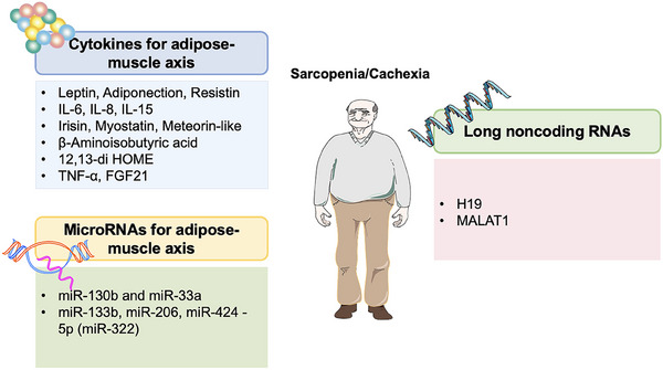
Dysregulation of cytokines
Leptin
Leptin is an adipokine produced and secreted by WAT and BAT, the periosteum, the placenta, and other tissues, and regulates both muscle and adipose tissue. Leptin has a significant role in maintaining energy balance and metabolism. Leptin can also regulate most peripheral organs, such as adipose tissue and muscle.ref. mco270030-bib-0375
Although adipocytes are a main source of leptin, they also express leptin receptor (LepR),ref. mco270030-bib-0376, ref. mco270030-bib-0377 which is an important target of leptin. Leptin decreases insulin‐induced glucose uptake and lipogenesis in white adipocytes by blunting the insulin response.ref. mco270030-bib-0378, ref. mco270030-bib-0379 Moreover, leptin also has a thermogenic effect, promoting energy expenditure in rodents.ref. mco270030-bib-0380, ref. mco270030-bib-0381 Whether leptin can also affect BAT development in humans remains to be determined.
The relationship between skeletal muscle and leptin is complex. On the one hand, leptin exerts beneficial effects on muscle, as reduced skeletal muscle atrophy and mass in leptin‐deficient mice (ob/ob) mice can be corrected by intraperitoneal administration of leptin.ref. mco270030-bib-0382 Leptin administration stimulates the proliferation of myocytes, increases myonectin and myogenin transcript levels, and reduces the mRNA levels of cytokines related to muscle atrophy in ob/ob mice, such as MAFbx and MuRF1.ref. mco270030-bib-0383 Leptin promotes C2C12 myoblast proliferation but impairs myogenesis in C2C12 cells through JAK/STAT and MEK signaling pathways.ref. mco270030-bib-0384 A minimal increase in the number of self‐renewed MuSCs was observed in LepR‐deficient (db/db) mice.ref. mco270030-bib-0385 Moreover, leptin facilitates metabolism in skeletal muscle. Skeletal muscle cells continuously treated with leptin showed enhanced fatty acid uptake due to activation of AMPK in the short term and the STAT3 pathway in the long term.ref. mco270030-bib-0386, ref. mco270030-bib-0387 Dulloo et al.ref. mco270030-bib-0388 found that leptin stimulated skeletal muscle thermogenesis by regulating PI3K activity. Moreover, leptin treatment increased basal glucose uptake in isolated soleus muscles from rats and mice, as well as in skeletal muscle cells.ref. mco270030-bib-0389, ref. mco270030-bib-0390, ref. mco270030-bib-0391 Collectively, these data provide important evidence that leptin treatment antagonizes lipogenic effects on skeletal muscle and promotes glucose disposal. However, some studies showed that plasma leptin levels are higher in older sarcopenia subjects and obese individualsref. mco270030-bib-0392, ref. mco270030-bib-0393, ref. mco270030-bib-0394 and that circulating leptin is elevated during aging even in the absence of obesity.ref. mco270030-bib-0395 A prospective cross‐sectional cohort study showed that the circulating level of leptin was significantly elevated in the sarcopenia group and correlated with sarcopenia incidence, sarcopenia severity, and sarcopenia risk.ref. mco270030-bib-0393 A longitudinal study of a cohort of community‐dwelling older adults showed that serum leptin concentrations were positively associated with incident frailty.ref. mco270030-bib-0394 Kohara et al.ref. mco270030-bib-0392 found that plasma leptin levels are highest in males and females who are both sarcopenic and overweight compared with subjects with only one abnormality. Research has shown that patients with sarcopenic obesity have higher serum leptin levels than those with nonsarcopenic obesity. In addition, they show poor grip strength and physical function. OA patients with sarcopenic obesity had significantly higher serum leptin levels than those with nonsarcopenic obesity. In addition, knee OA patients with sarcopenic obesity displayed low grip strength and poor physical performance.ref. mco270030-bib-0396
According to this discrepancy, we believe that there is an optimal concentration of leptin for muscle homeostasis. Both aging and obesity increase serum leptin levels, and beyond this optimal concentration, leptin exerts a destructive effect, while obesity may amplify these detrimental effects. This is supported by the finding that a high level of leptin contributes to central leptin resistance and IR and impairs leptin signal transduction in muscle cells, including lipid and carbohydrate metabolism and fatty acid oxidation.ref. mco270030-bib-0397, ref. mco270030-bib-0398 High levels of leptin were proven to impede intramuscular fatty acid oxidation and lipid metabolism, thereby leading to skeletal muscle dysfunction.ref. mco270030-bib-0098, ref. mco270030-bib-0399 Impaired fatty acid oxidation and intramyocellular lipid accumulation may promote lipotoxicity.ref. mco270030-bib-0400, ref. mco270030-bib-0401 In light of these studies, it appears that leptin may be involved in the pathogenesis of sarcopenic obesity. Thus, it may result in a decline in muscle quality and increased intramuscular fat infiltration.ref. mco270030-bib-0402, ref. mco270030-bib-0403 Moreover, both leptin and IR of myocytes are associated with metabolic disorders, including obesity and the type 2 diabetes mellitus (T2DM), creating a vicious cycle augmenting muscle dysfunction.ref. mco270030-bib-0404
Although the role of leptin in cancer cachexia is controversial, it is believed to participate in its pathogenesis. In cancer cachexia, leptin is produced by adipose tissue and acts on the hypothalamus to regulate energy storage by influencing appetite.ref. mco270030-bib-0405 Additionally, leptin has been shown to upregulate the production of inflammatory cytokines.ref. mco270030-bib-0406 Interestingly, leptin‐binding proteins, such as CRP, can bind to leptin in the bloodstream, limiting its binding to the LepR and affecting its localization across the blood‐brain barrier.ref. mco270030-bib-0407 Studies involving ob/ob have demonstrated that this adipokine is crucial for controlling body weight and lipid metabolism.ref. mco270030-bib-0408, ref. mco270030-bib-0409 Furthermore, ob/ob mice develop obesity due to a lack of appetite suppression.ref. mco270030-bib-0408, ref. mco270030-bib-0409 Physiologically, leptin is thought to protect adipocytes from lipotoxicity by inhibiting lipogenesis and increasing fatty acid oxidation. Overall, these studies underscore the role of leptin in body weight regulation.
In summary, leptin is involved in the adipose tissue–muscle axis, implicated in the pathogenesis of sarcopenia and cachexia. Further studies are needed to identify the optimal concentrations of leptin to guarantee muscle and whole‐body homeostasis.
Adiponectin
Adiponectin, an adipokine secreted by WAT and BAT,ref. mco270030-bib-0410, ref. mco270030-bib-0411 plays a critical role in multiple highly‐specific biological functions and the insulin signaling pathway. It produces a variety of physiological effects by binding to adiponectin receptors (AdipoRs) on the target cell membrane.
Adiponectin has recently received a great deal of attention for its beneficial effects on muscle. Knockout of adiponectin abrogates the exercise‐induced increase in muscle mass and strength in mice, indicating it plays a role in hypertrophy.ref. mco270030-bib-0412 C2C12 myoblast cells treated with either globular adiponectin or a mimetic of globular adiponectin (GTDF) displayed a significant increase in both differentiation and fusion indices with respect to vehicle‐treated cells through downregulating key genes associated with muscle atrophy (MuRF1 and atrogin‐1).ref. mco270030-bib-0413 Adiponectin also regulates energy homeostasis.ref. mco270030-bib-0414, ref. mco270030-bib-0415, ref. mco270030-bib-0416, ref. mco270030-bib-0417 Yoon et al.ref. mco270030-bib-0415 treated differentiated C2C12 cells with purified adiponectin and found increased fatty acid β‐oxidation in muscle by sequential activation of AMPK, via the p38 MAPK pathway and PPARα. Ceddia et al.ref. mco270030-bib-0418 treated L6 rat skeletal muscle cells with GTDF and showed increased glucose uptake by inducing the translocation of GLUT4 to the plasma membrane. In addition, it was demonstrated that muscle differentiation requires autophagy driven by adiponectin, promoting myoblast survival while inhibiting apoptosis.ref. mco270030-bib-0419 Likewise, adiponectin knockout mice showed decreases in autophagy‐related genes (e.g., LC3 and beclin‐I) in skeletal muscle, accompanied by a myopathic phenotype.ref. mco270030-bib-0419
In fact, the activity of adiponectin is modified by the expression of specific skeletal muscle AdipoRs, the predominant isoform expressed in skeletal muscle.ref. mco270030-bib-0420 Muscle‐specific AdipoR1‐knockout mice exhibited decreased expression of molecules involved in mitochondrial biogenesis, such as PGC‐1α, as well as IR and decreased muscle endurance.ref. mco270030-bib-0421 Aging‐related decreased levels of AdipoR1 in skeletal muscle might contribute to sarcopenia with impaired muscle function and metabolic defects.ref. mco270030-bib-0412 However, until now, the evidence of a causal relationship between age‐associated sarcopenia and adiponectin remains controversial. On the one hand, some results have shown that the level of serum adiponectin is lower in sarcopenic older adults than in normal adults.ref. mco270030-bib-0393, ref. mco270030-bib-0422 On the other hand, other studies showed a negative association between serum adiponectin levels and muscle strength, muscle density, and function among older adults.ref. mco270030-bib-0423, ref. mco270030-bib-0424, ref. mco270030-bib-0425, ref. mco270030-bib-0426, ref. mco270030-bib-0427
Adiponectin also regulates WAT, as well as exerting profound effects on converting WAT to BAT.ref. mco270030-bib-0428 Obese mice exhibited decreased muscular lipid content and reduced IR after adiponectin treatment.ref. mco270030-bib-0429 Knockout of adiponectin in sWAT of mice severely impaired beige adipocyte activation and thermogenic programs induced by chronic cold stimulation, while resupplementation with adiponectin reversed these changes.ref. mco270030-bib-0428 These results suggest that adiponectin might act as a compensator for metabolic diseases. However, AdipoR1 declines in aging, obesity, and T2DM,ref. mco270030-bib-0430 weakening beneficial adiponectin signaling to alleviate the disruptive effects of metabolic diseases.
Overall, adiponectin, an adipokine, regulates both muscle and adipose tissue to increase energy expenditure, which could be a bridge in maintaining homeostasis of both adipose tissue and skeletal muscle. Interestingly, adiponectin and AdipoR1 levels decrease in individuals suffering from both aging and obesity,ref. mco270030-bib-0412, ref. mco270030-bib-0430, ref. mco270030-bib-0431, ref. mco270030-bib-0432 indicating that downregulation of adiponectin signaling could result in sarcopenia and sarcopenic obesity and should be considered a therapeutic target for muscle dysfunction and metabolic defects. Moreover, adiponectin can regulate various adipose tissue depots and is released from different adipose tissue sources, suggesting a complex and unclear role of this adipokine in cancer cachexia. Notably, previous studies have demonstrated that different adipose depots undergo remodeling during cachexia in a heterogeneous and time‐dependent manner.ref. mco270030-bib-0433, ref. mco270030-bib-0434 This indicates that adiponectin may play a role in the pathogenesis of cachexia. Future research should aim to verify this hypothesis.
Resistin
Resistin, a hormone secreted by adipose tissue, appears to be a potential connecting link between adipose tissue and muscle. Resistin can disturb glucose homeostasis in rodents.ref. mco270030-bib-0435, ref. mco270030-bib-0436 It acts as a potential direct regulator of glucose homeostasis by regulating muscle. Palanivel et al.ref. mco270030-bib-0437, ref. mco270030-bib-0438 treated L6 rat skeletal muscle cells with recombinant resistin and found decreased fatty acid uptake and insulin‐stimulated GLUT4 levels, indicating that resistin might participate in communication between fat and muscle. This hypothesis was confirmed by an experiment performed by O’Leary, who found that the obese sWAT secretome derived from old but not young individuals impaired human myotubes, because the concentration of resistin was significantly greater from obese adipose‐conditioned medium than from normal weight older individuals, which exerted detrimental effects on myoblasts by activating the classical NF‐κB pathway. In turn, depletion of resistin from the obese sWAT secretome prevents its detrimental effects on muscle.ref. mco270030-bib-0439 Continuous expression of resistin in C2C12 cells by stable transfection inhibited myogenic differentiation, and reduced glucose uptake.ref. mco270030-bib-0440
In addition, human plasma resistin levels are higher in elderly subjects than in young individuals and are inversely correlated with muscle strength.ref. mco270030-bib-0439, ref. mco270030-bib-0441 Bucci reported that plasma resistin concentrations are inversely associated with quadriceps muscle torque in people aged 69–81 years.ref. mco270030-bib-0442 Besides, levels of resistin are higher in obese elderly people than in normal‐weight elderly people, further suggesting that visceral adipose tissue‐derived resistin is involved in muscle weakness in individuals with sarcopenic obesity.ref. mco270030-bib-0439, ref. mco270030-bib-0443
Resistin has been shown to stimulate human blood leukocytes to secrete TNF‐α and IL‐6. Interestingly, its expression is also stimulated by proinflammatory cytokines such as IL‐1, TNF‐α, and IL‐6, indicating a proinflammatory loop where resistin both contributes to and is induced by inflammation.ref. mco270030-bib-0444, ref. mco270030-bib-0445 A recent meta‐analysis linked increased plasma resistin levels to a higher incidence of obesity‐related cancers, including breast, endometrial, and colorectal cancer; however, resistin levels should not be considered predictive factors.ref. mco270030-bib-0446 Additionally, high serum resistin levels in breast cancer patients have been positively correlated with tumor stage, size, and metastasis. Furthermore, elevated resistin levels have also been associated with an increased risk of nonobesity‐related malignancies, such as gastric, colon, and lung cancers.ref. mco270030-bib-0447, ref. mco270030-bib-0448, ref. mco270030-bib-0449 However, studies exploring the relationship between resistin and cancer cachexia remain limited at this time.
Collectively, the evidence suggests that resistin is associated with inflammation and serves as a link between adipose tissue and muscle, highlighting potential therapeutic targets for preventing sarcopenia, sarcopenic obesity, or cachexia. However, further studies are needed to confirm this hypothesis.
IL‐6
IL‐6 is a cytokine produced by multiple tissues in the body, including skeletal muscle, adipose tissue, and immune cells. IL‐6 binds to its receptor and activates the classical or trans‐signaling pathway, exerting its biological effect.ref. mco270030-bib-0450 IL‐6 is one of the most‐investigated myokines related to communication between skeletal muscle and WAT and exerts both pro‐ and anti‐inflammatory effects, either promoting muscle anabolism or catabolism depending on its source (secreted by muscle cells vs. adipocytes).ref. mco270030-bib-0049
IL‐6 could regulate the homeostasis of muscle. In brief, activated FAPs exhibit high levels of cytokine expression, including IL‐6. While IL‐6 is commonly known as a myokine secreted by myofibers in response to exercise, its expression in regenerating muscle is approximately 10 times higher in FAPs compared with myogenic progenitor cells.ref. mco270030-bib-0144, ref. mco270030-bib-0451 IL‐6 plays a role in regulating both muscle hypertrophy and regeneration.ref. mco270030-bib-0452 IL‐6 knockout mice established a decline hypertrophic response to overloading, attributed to the impaired incorporation of myonuclei, which is a result of defective proliferation and migration of satellite cells. Treating murine satellite cells with IL‐6 has been shown to promote their proliferation. This effect is achieved through the regulation of cell‐cycle associated genes Cyclin D1 and c‐Myc,ref. mco270030-bib-0453 whereas IL‐6/STAT3 axis plays a crucial role in controlling the fate of satellite cells.ref. mco270030-bib-0454 Both intramuscular IL‐6 mRNA expression and protein release are enhanced during exercise when intramuscular glycogen is reduced,ref. mco270030-bib-0455, ref. mco270030-bib-0456 while glucose ingestion during exercise has been shown to attenuate leg IL‐6 production,ref. mco270030-bib-0457 indicating IL‐6 is an energy sensor that regulates muscle glycogen content. Besides, IL‐6 increases glucose transport in L6 myotubes and skeletal muscle strips from vastus lateralis biopsies.ref. mco270030-bib-0458, ref. mco270030-bib-0459 Human data also show that IL‐6 released during exercise leads to an increase in insulin‐stimulated whole‐body glucose disposal through an increase in translocation of GLUT4 from intracellular pools to the plasma membrane.ref. mco270030-bib-0458 Additionally, incubation with IL‐6 increases fatty acid oxidation in isolated rat muscle and L6 myotubes.ref. mco270030-bib-0458, ref. mco270030-bib-0460 Because IL‐6 is crucial to maintaining proper metabolism,ref. mco270030-bib-0461 this might explain why regular physical exercise ameliorates metabolic diseases by regulating IL‐6. More specifically, the effects of IL‐6 on enhancing glucose uptake and fatty acid oxidation in skeletal myotubes are exerted by increasing AMPK.ref. mco270030-bib-0458, ref. mco270030-bib-0462 Activation of AMPK in mice is accompanied by the browning of white fat by upregulating the downstream PGC‐1α,ref. mco270030-bib-0463 indicating that IL‐6 also participates in fat browning.ref. mco270030-bib-0464 This is demonstrated by the following results. WAT conversion into beige adipose tissue is indirectly affected by muscle‐secreted IL‐6.ref. mco270030-bib-0465 Whole‐body deletion of IL‐6 completely suppresses the elevation in UCP1 mRNA and protein stimulated by both cold exposure and exercise,ref. mco270030-bib-0464 while expression of UCP1 in mouse inguinal WAT increases upon injection of recombinant IL‐6.ref. mco270030-bib-0464 However, whether classical or trans‐signaling is involved in IL‐6‐induced adipocyte browning is still incompletely understood. This evidence suggests that IL‐6 secreted by muscle is involved in adipose tissue regulation.
In turn, numerous inflammatory cytokines and adipokines released by adipose tissue can enter the systemic circulation and act as paracrine hormones on skeletal muscle, including IL‐6. IL‐6 secreted by adipose tissue is the main source of circulating IL‐6, as Whitham et al.ref. mco270030-bib-0466 found that circulating IL‐6 was approximately threefold higher in control mice than that in adipocyte‐specific IL‐6‐deficient mice. In addition, obese individuals expressed higher visceral levels of IL‐6 protein compared with normal‐weight controls,ref. mco270030-bib-0467 which has a more significant correlation with diabetes than total body fat.ref. mco270030-bib-0468, ref. mco270030-bib-0469 The proinflammatory effects of IL‐6 signaling contribute to age‐induced chronic low‐grade inflammation and may participate in the pathogenesis of sarcopenia.ref. mco270030-bib-0470 This indicates that IL‐6 secreted by adipose tissue may cause sarcopenia. Consistent with this speculation, clinical studies showed that levels of IL‐6 are dramatically upregulated in patients with sarcopenia after adjustment for potential confounders such as BMI and visceral fat tissue.ref. mco270030-bib-0471, ref. mco270030-bib-0472, ref. mco270030-bib-0473 IL‐6 stimulates muscle atrophy by blunting muscle anabolism and energy homeostasis and also mediates muscle catabolism directly through the following mechanisms. First, IL‐6 increases ubiquitin, E3 ligase, and proteasome activity levels by activating the NF‐κB pathway.ref. mco270030-bib-0233, ref. mco270030-bib-0474, ref. mco270030-bib-0475 Second, lasting IL‐6 secreted by FAPs in persistent inflammatory conditions including both aged and dystrophic muscle during regeneration stimulates muscle atrophy via activation of aberrant STAT3–IL‐6 signaling.ref. mco270030-bib-0476, ref. mco270030-bib-0477 Third, continuously elevated IL‐6 directly induces dynamin‐related protein‐1 and fission protein 1 expression in myotubes, leading to dysfunction of mitochondrial function in muscle.ref. mco270030-bib-0478, ref. mco270030-bib-0479, ref. mco270030-bib-0480, ref. mco270030-bib-0481, ref. mco270030-bib-0482, ref. mco270030-bib-0483, ref. mco270030-bib-0484, ref. mco270030-bib-0485 However, another study using an experimentally‐induced sepsis model found that knockout of IL‐6 exerts no significant influence on muscle metabolism compared with wild‐type mice,ref. mco270030-bib-0486 indicating that IL‐6 might not be the sole trigger for sarcopenia.
It can be challenging to determine the precise impact of IL‐6 on muscle regeneration because multiple cell types within the regenerative environment are known to secrete IL‐6. This makes it difficult to isolate and assess the specific effects of IL‐6 secretion from individual cell types in the process of muscle regeneration.ref. mco270030-bib-0452 To assess the direct influence of IL‐6 derived from FAPs on myogenesis, future studies will require loss‐of‐function experiments involving cocultures of FAPs and MuSCs or the targeted deletion of IL‐6 specifically in FAPs during muscle regeneration. These experiments will provide valuable insights into the specific role of FAP‐derived IL‐6 in the process of myogenesis. The potential role of IL‐6 in sarcopenia is complex, as it has dual roles in muscle physiology and pathology. We propose that IL‐6 signaling may shift toward the proinflammatory profile and participate in sarcopenia progression when skeletal muscle function gradually decreases during aging. Increased IL‐6 might thus reflect sarcopenia risk. In addition, IL‐6 secreted by adipose tissue might be involved in the pathogenesis of sarcopenia. This assumption needs further confirmation to identify other molecules that interact with IL‐6 to cause muscle dysfunction. Furthermore, a broader role for IL‐6 in promoting cachexia in cancer is suggested by a recent systematic review of gene polymorphisms associated with cancer cachexia. This review identified IL6 as one of four genes linked to key pathways, such as inflammation and metabolism, involved in the development of cachexia.ref. mco270030-bib-0487
IL‐8
IL‐8 belongs to the cysteine‐X‐cysteine family of chemokines and can be secreted by muscle fibers.ref. mco270030-bib-0488 Elevated levels of IL‐8 have been found in muscle fibers and plasma after exercise, which regulates muscle itself.ref. mco270030-bib-0489, ref. mco270030-bib-0490, ref. mco270030-bib-0491 A recent study reported that increased levels of plasma IL‐8 during the training period were negatively correlated with strength gains for the leg press.ref. mco270030-bib-0492 Elevated levels of IL‐8 have been related to lower extremity lean body mass (LBM) and a greater risk of sarcopenia in the UK elderly community.ref. mco270030-bib-0493 Another study showed that higher levels of IL‐6 and IL‐8 are a prominent pathological feature of sarcopenia in elderly people.ref. mco270030-bib-0494 Increased levels of IL‐8 secreted from T2DM myotubes create a muscle microenvironment that supports reduced capillarization in T2DM.ref. mco270030-bib-0495, ref. mco270030-bib-0496 However, whether the causal relationship between IL‐8 and sarcopenia exists needs further investigation.
Adipose tissue is another organ that generates IL‐8, and vWAT releases higher amounts of IL‐8 than sWAT.ref. mco270030-bib-0497, ref. mco270030-bib-0498 Different obesity models are associated with increased level of IL‐8 in adipose tissues. For example, estrogen‐deficient rats have increased fat mass, which is accompanied by increased production of IL‐8 in adipose tissue, and these results were reversed by estrogen replacement.ref. mco270030-bib-0499 Alvehus et al.ref. mco270030-bib-0500 found that the menopausal transition is characterized by increased body fat accumulation, and expression of IL‐8 is elevated (64%) in the sWAT of postmenopausal women compared with premenopausal women. IL‐8 expression in adipose tissue is profoundly decreased (90%) 2 years after gastric bypass surgery in obese women. Significant weight loss and reduced macrophage infiltration in sWAT as well as decreased IL‐8 mRNA levels were observed after short‐term lifestyle intervention.ref. mco270030-bib-0500 These results reveal that high levels of IL‐8 are released from adipose tissue and that the accumulation of this tissue partly leads to increased circulating IL‐8 in obese subjects.ref. mco270030-bib-0497 Lihn et al.ref. mco270030-bib-0501 reported that the secretion and gene expression of IL‐6 and IL‐8 from human adipose tissue fragments as well as from cultured human skeletal muscle cells were inhibited by treatment with AICAR, an AMPK activator known to increase insulin sensitivity. This demonstrated that the AMPK signaling pathway is involved in myokine and adipokine IL‐8 production. It has been found that IL‐8 inhibits insulin‐induced AKT phosphorylation in human adipocytes, resulting in IR.ref. mco270030-bib-0502 Thus, higher levels of IL‐8 in both plasma and vWAT are associated with IR, contributing to T2DM and obesity.ref. mco270030-bib-0503, ref. mco270030-bib-0504 In addition, elevated IL‐8 levels in human adipocytes could be stimulated by TNF‐α treatment, indicating that higher IL‐8 might not be the primary trigger for IR and may exert this effect by stimulating TNF‐α.ref. mco270030-bib-0505
In conclusion, muscle release of IL‐8 might be negatively associated with insulin sensitivity and muscle function, while IR caused by adipose tissue secreted IL‐8 was mainly mediated through TNF‐α. This evidence suggested that IL‐8 might be involved in the muscle‐fat crosstalk in sarcopenia. Despite extensive attempts to decipher the relationship between IL‐8 and skeletal muscle, this issue remains unclear. Further studies are needed to evaluate the relevance of IL‐8 in the development of sarcopenia and to clarify whether it can be offered as a potential therapeutic target for this disease. Elevated serum IL‐8 levels have been associated with weight loss and significantly correlated with cachexia in pancreatic cancer patients.ref. mco270030-bib-0506, ref. mco270030-bib-0507, ref. mco270030-bib-0508 In gastric cancer patients, an IL‐8 genetic polymorphism has been linked to the onset and development of cachexia.ref. mco270030-bib-0507, ref. mco270030-bib-0508 It is also important to note that IL‐8 is produced by both muscle and adipose tissue, particularly visceral fat, suggesting that the modulation of this cytokine can occur from various tissue.
IL‐15
IL‐15 is considered a myokine since it is released by muscle. Its serum levels are markedly elevated in response to contraction and do not last much longer.ref. mco270030-bib-0509 Tamura et al.ref. mco270030-bib-0509 found that the serum IL‐15 concentration was elevated 10 min after 30 min of exhaustive exercise. At 3 h after treadmill exercise, serum IL‐15 levels had fully returned to near baseline.ref. mco270030-bib-0509
IL‐15 treatment increases myotube development and induces myoblast differentiation, exerting hypertrophic effects.ref. mco270030-bib-0510, ref. mco270030-bib-0511 Quinn et al.ref. mco270030-bib-0511 found that retroviral overexpression of IL‐15 induced a marked increase in myofibrillar protein accumulation in skeletal myogenic cell cultures, leading to a hypertrophic myotube morphology. Besides, IL‐15 may exert an antiatrophic effect, maintaining muscle mass in the presence of atrophic stimuli such as cancer cachexia, decelerating protein degradation and suppressing the ubiquitin proteolysis pathway in rats.ref. mco270030-bib-0512 IL‐15 decreases the skeletal muscle proteolytic rate in rat extensor digitorum longus muscle.ref. mco270030-bib-0513 Furthermore, IL‐15 might exert beneficial effects on muscle metabolism by improving oxidative characteristics and glucose homeostasis. In vitro, C2C12 myotubes treated with recombinant IL‐15 exhibited elevated basal and insulin‐stimulated glucose uptake,ref. mco270030-bib-0514 GLUT4 translocation,ref. mco270030-bib-0515 and GLUT4 mRNA levels.ref. mco270030-bib-0514 Incubation of C2C12 cells with recombinant IL‐15 (100 ng/mL) for 24 h showed increased GLUT4 translocation and glucose uptake, and this effect was abolished by STAT3 inhibition in combination with IL‐15.ref. mco270030-bib-0515 Additionally, increased circulation levels of IL‐15 may be beneficial for muscle by stimulating the expression of mitochondrial‐associated factors, such as PPARs and SIRT1, in mouse skeletal muscle.ref. mco270030-bib-0516
In addition to its effects on muscle, IL‐15 also reduces adipocyte lipid content, which is involved in muscle–fat interactions. For example, transgenic mice with skeletal muscle‐specific oversecretion of IL‐15 show lower total body fat when fed an HFD, along with lower intra‐abdominal fat and increased insulin sensitivity.ref. mco270030-bib-0511, ref. mco270030-bib-0517 Barra et al.ref. mco270030-bib-0518 reported that a muscle‐specific IL‐15‐overexpressing mouse model displayed increased mitochondrial activity and mass in adipose tissue compared with IL‐15 knockout mice. In the same experiment, they also treated 3T3‐L1 adipocytes with IL‐15 (250 ng/mL) for 24 h and showed increased mitochondrial membrane potential and decreased lipid deposition.ref. mco270030-bib-0518 Thus, IL‐15 induces adipose tissue catabolism and decreases adiposity, regulating body composition.ref. mco270030-bib-0098 In addition, Quinn et al.ref. mco270030-bib-0519 found that IL‐15 decreased adiposity and lipid incorporation into adipose tissue, partly by elevating adiponectin secretion. Recent research has identified IL‐15 as a myokine that can prevent intramuscular fatty infiltration by potentially impacting the differentiation capacities of FAPs. The study demonstrated that IL‐15 promotes proliferation of FAPs and directly inhibits their adipogenic differentiation both in vitro and in vivo, ultimately facilitating regeneration of myofibers.ref. mco270030-bib-0141 Furthermore, the administration of recombinant IL‐15 directly into the muscle prevented fat accumulation in a murine model of glycerol‐induced fatty degeneration.ref. mco270030-bib-0141 The decrease in adipogenesis mediated by IL‐15 is associated with an upregulation of the desert hedgehog signaling pathway and its downstream effector, tissue inhibitor of metalloproteinase 3. This pathway is regulated by primary cilia present in FAPs, which have been shown to suppress the adipogenic differentiation of FAPs.ref. mco270030-bib-0520 The above studies suggested that IL‐15 could reduce the amount of adipose tissue through the muscle‐fat interaction. While the results suggest a positive role for IL‐15 in muscle regeneration, it is important to note that there is evidence correlating IL‐15 administration and expression with increased collagen deposition in vivo following muscle damage. This raises several unresolved issues that require further investigation to fully understand the complex effects of IL‐15 in muscle regeneration and fibrosis.ref. mco270030-bib-0141
The obvious involvement of IL‐15 in the axis between muscle and adipose tissue plays a key role in improving metabolism by inhibiting obesity and promoting insulin sensitivity. A cross‐sectional study showed that there was a negative relationship between IL‐15 levels and sarcopenia risk in older people.ref. mco270030-bib-0521 Significant reductions in circulating IL‐15 and soluble IL‐15 receptor alpha (IL‐15Rα) levels and the expression of intramuscular IL‐15 and IL‐15Rα were observed in advanced age.ref. mco270030-bib-0522 These alterations may weaken the beneficial effects of IL‐15 signaling and decrease the ratio of muscle to fat content,ref. mco270030-bib-0523, ref. mco270030-bib-0524 resulting in sarcopenia.
The findings presented here provide a technical and experimental basis for the role of IL‐15 in muscle and fat physiology, suggesting that IL‐15 may act as a beneficial mediator of muscle regeneration and adipose tissue catabolism. Enhancing its signaling could be a promising therapeutic target for sarcopenia. However, the role of IL‐15 in cachexia remains unclear. An earlier study using a rat model of cancer cachexia indicated that IL‐15 reduces the rate of protein degradation without impacting protein synthesis.ref. mco270030-bib-0512 Additionally, research involving adult patients with recent cancer diagnoses and weight loss found no significant difference in serum IL‐15 levels compared with healthy subjects.ref. mco270030-bib-0525 Despite these conflicting results, the potential of this IL should not be dismissed, and further studies are necessary to clarify its role.
Irisin
Irisin is a myokine released into the bloodstream by cleavage of fibronectin type III domain‐containing protein 5 (FNDC5) triggered by muscle contraction.ref. mco270030-bib-0526, ref. mco270030-bib-0527 However, adipose tissue, chiefly from WAT, is also another important source of irisin, while BAT expresses almost no FNDC5 or irisin.ref. mco270030-bib-0528 The expression of FNDC5 in adipose tissue is 100 times lower than that in muscle,ref. mco270030-bib-0529 indicating that muscle tissue is the primary source of irisin.
Interestingly, muscle itself is an important target of irisin. Mice serum and soleus muscle irisin level is increased following eight weeks of resistance training exercise and is negatively associated with age‐related decline in muscle function.ref. mco270030-bib-0530 Chang et al.ref. mco270030-bib-0531 found that a low level of circulating irisin is a sensitive marker of muscle dysfunction. In addition, in postmenopausal women, a reduced serum irisin level predicts the development of sarcopenia.ref. mco270030-bib-0532 Based on these clinical results, we speculate that decreased level of irisin might be a marker of sarcopenia.
Irisin can potentially reduce the risks of metabolic defects by improving skeletal muscle development and metabolism. During myogenic differentiation, human myocytes show elevated irisin synthesis, indicating the myogenic potential of irisin.ref. mco270030-bib-0533 Irisin treatment increases human primary skeletal muscle cell growth and hypertrophy by increasing IGF‐1/PGC‐1α4 and decreasing myostatin through activating the ERK1/2 pathway.ref. mco270030-bib-0533 Irisin has also been suggested to prevent glucocorticoid‐induced muscle atrophy by inhibiting FOXO‐mediated ubiquitin–proteasome overactivity.ref. mco270030-bib-0534 Treating human skeletal muscle cells with recombinant irisin (50 nM) for 1 h resulted in increased uptake of glucose and fatty acid in cells, similar to the effects of insulin.ref. mco270030-bib-0535 Knocking down the AMPK pathway attenuated the effects of irisin (0.3–1.0 µg/mL) on high glucose uptake and increased fatty acid β‐oxidation in the C2C12 myoblast cell line.ref. mco270030-bib-0536 Incubating myotubes with irisin increased the expression of genes related to mitochondrial biogenesis, such as PGC‐1α and Nrf1.ref. mco270030-bib-0537, ref. mco270030-bib-0538 Immunohistochemical analysis on muscle sections with MyoD antibodies (a marker of activated satellite cells) revealed increased percentage of MyoD‐positive cells on day 2 and day 3 postnotexin‐induced injury in mice injected with irisin, when compared with DB‐injected controls. Treatment with irisin in vitro, resulted in a marked reduction in the percentage of quiescent satellite cells, concomitant with an increase in the percentages of proliferating (Pax7+/MyoD+) and committed myoblasts (Pax7−/MyoD+). These data suggest that irisin treatment leads to increased satellite cell activation and proliferation.ref. mco270030-bib-0539
Adipose tissues could be another target of irisin. Injection of irisin increased glucose utilization and reduced epididymal fat mass in diabetic mice by stimulating GLUT4 translocation in skeletal muscle.ref. mco270030-bib-0536 In obese mice, lentivirus‐mediated FNDC5 overexpression reduced hyperglycemia, hyperinsulinemia, hyperlipidemia, and adipocyte diameter in adipose tissues.ref. mco270030-bib-0540 Metformin acts as an antidiabetic drug, elevating intracellular FDNC5 mRNA/protein expression, stimulating irisin release in murine C2C12 myotubes,ref. mco270030-bib-0078, ref. mco270030-bib-0541 and alleviating diabetes in mice.ref. mco270030-bib-0541 Furthermore, irisin has been shown to promote UCP1 expression in adipocytes, causing WAT to adopt a BAT‐like phenotype by stimulating energy expenditure, leading to lower body weight and promoting glucose homeostasis and IR.ref. mco270030-bib-0527 Interestingly, studies of circulating irisin in obese humans have produced inconsistent results. Some studies reported upregulation of serum irisin levels in overweight and obese people compared with normal controls,ref. mco270030-bib-0542, ref. mco270030-bib-0543 whereas others found lower circulating irisin levels in obese or overweight people, compared with normal‐weight individuals.ref. mco270030-bib-0544 It is also possible that increased irisin levels could be a compensatory mechanism for the abnormal metabolism and insulin sensitivity characteristic of obese individuals, or who have developed a resistance to irisin suffered from obesity.ref. mco270030-bib-0527
A recent study found that intraperitoneal injection of recombinant irisin protein into ageing or aged mice could improve sarcopenia with grip strength, muscle weights, fiber size and molecular phenotypes and alleviated age‐associated fat tissues expansion, IR.ref. mco270030-bib-0545 This encouraging finding indicates that physical exercise in patients with sarcopenia may improve the function of aged muscle as well as energy metabolism through enhancing irisin expression, which may lead to the development of innovative approaches for the management of sarcopenia. It has been studied especially in relation to obesity but also with myopathies such as muscular dystrophy. Injection of irisin has been shown to induce muscle hypertrophy, enhance muscle strength, and reduce necrosis and the development of connective tissue in a murine model.ref. mco270030-bib-0546 This study may serve as a starting point for exploring therapeutic strategies involving irisin in the context of cancer cachexia. It is presumed that cell surface receptors can mediate FNDC5 activity and/or expression, but the features of this receptor are still unknown. Further studies provided other mechanistic insights into the irisin receptor. Clarification of the link among physical exercise, irisin and sarcopenia/sarcopenic obesity is needed, especially in humans, and evidence of a direct causal relationship is thus also missing.
Myostatin
Proliferation assays clearly demonstrate that adult myoblasts that lack myostatin proliferate much faster than the wild‐type myoblasts. In contrast, treated myostatin to myofiber explant cultures suppressed satellite cell activation by affecting the entry of satellite cells into S phase.ref. mco270030-bib-0547 Two RCTs tested the benefits of myostatin inhibitor, bimagrumab, in patients with sarcopenia. In a phase 2 proof‐of‐concept clinical study, Rooks et al.ref. mco270030-bib-0548 reported that treatment with bimagrumab over 16 weeks increased muscle mass and strength in older adults with sarcopenia and improved mobility in those with slow walking speed. However, these results were not confirmed in a recent phase 3 RCT,ref. mco270030-bib-0549 with no significant difference were found between participants treated with bimagrumab versus placebo among older adults with sarcopenia who had 6 months of adequate nutrition and light exercise, but both groups had physical function improvement. Bimagrumab treatment was safe, well tolerated, increased LBM, and decreased fat body mass.
Furthermore, myostatin might be involved in muscle‐to‐fat crosstalk. Research has shown that in obese patients, the mRNA levels of myostatin in muscle biopsies were significantly decreased after weight loss caused by biliary pancreatic diversion.ref. mco270030-bib-0550 Deletion of myostatin in mice also resulted in decreased fat mass, increased muscle mass, improved insulin sensitivity and resistance to diet‐induced obesity.ref. mco270030-bib-0551, ref. mco270030-bib-0552, ref. mco270030-bib-0553 Depending on the specific cell types and culture conditions, researchers have alternatively described the stimulation or inhibition of adipogenesis by myostatin. Its expression in mesenchymal cells promotes adipogenic differentiation through SMAD3, while exhibiting negative cross talk with β‐catenin.ref. mco270030-bib-0552, ref. mco270030-bib-0554
Myostatin has been observed to inhibit lipid accumulation in cell lines of preadipocytes and fibroblasts.ref. mco270030-bib-0555, ref. mco270030-bib-0556 Aged FAPs not only experience changes in the microenvironment that result in altered differentiation but also exhibit reduced responsiveness to muscle injury.ref. mco270030-bib-0140 The regulation of FAPs is attributed to myostatin, which plays a role in increased fibrosis as well.ref. mco270030-bib-0557, ref. mco270030-bib-0558 In an in vitro study, it was demonstrated that myostatin induces enhanced proliferation and fibrotic differentiation of FAPs through upregulation of P‐SMAD2/SMAD3 signaling.ref. mco270030-bib-0559
According to previous studies, myostatin is a factor associated with the thermogenic capacity of BAT and the browning of WAT,ref. mco270030-bib-0560, ref. mco270030-bib-0561 and a negative feedback loop of BAT activity has been reported. For example, Kim et al.ref. mco270030-bib-0562 reported that the differentiation of brown adipocytes and the expression of key thermogenic factors were dramatically inhibited by treatment with myostatin. Pharmacological inhibition of ActRIIB in mice blocks myostatin signaling, increases BAT activity and increases mitochondrial function, energy expenditure and cold tolerance.ref. mco270030-bib-0563 Braga et al.ref. mco270030-bib-0564 reported that treatment of myostatin null embryonic fibroblasts with exogenous myostatin significantly repressed thermogenic gene expression. Moreover, UCP1 and PRDM16 are significantly upregulated not only in epididymal and sWAT but also in gastrocnemius muscle in myostatin‐deficient mice compared with wild‐type mice.ref. mco270030-bib-0564 Kong et al.ref. mco270030-bib-0565 reported that loss of BAT, with thermoneutral temperature, increased myostatin levels, and reduced the exercise capacity of skeletal muscle. In contrast, activation of BAT decreased myostatin levels and improved exercise performance. Overall, inhibition of myostatin could be beneficial for obesity and associated metabolic disorders through browning of WAT.ref. mco270030-bib-0463
Interestingly, various lines of evidence suggest that myostatin could also be secreted by adipose tissue. For example, Hittel et al.ref. mco270030-bib-0566 reported that increased myostatin expression in myotubes and skeletal muscle are observed in obese women compared with lean women. However, inhibition of myostatin in adipose tissue has no effect on muscle mass and body composition in mice fed a HFD.ref. mco270030-bib-0567 In contrast, inhibition of myostatin signaling in skeletal muscle leads to increased lean mass, decreased fat mass in mice fed a HFD and resistance to diet‐induced obesity.ref. mco270030-bib-0567 McPherron and Leeref. mco270030-bib-0552 reported that mice lacking the entire myostatin gene have a significant reduction in fat accumulation and skeletal muscle mass. These results suggest that myostatin expression in muscle but not adipose tissue could be the major target for obesity or diabetes.
Accumulating data show that myostatin increases with age, indicating that upregulation of myostatin protein secretion could result in sarcopenia by causing muscle wasting, along with increased fat mass.ref. mco270030-bib-0568 Myostatin mRNA and protein levels in muscle are significantly elevated by 2‐ and 1.4‐fold in old males compared with younger subjects, respectively.ref. mco270030-bib-0569 Similarly, skeletal muscle of elderly women expresses higher levels of myostatin mRNA (56%) than that of young women.ref. mco270030-bib-0570 However, other studies reported that concentrations of myostatin in either serum or muscle do not differ between young and sarcopenic elderly men.ref. mco270030-bib-0571, ref. mco270030-bib-0572 These discrepancies indicate that myostatin may not be the primary trigger of sarcopenia. Interestingly, in the context of cachexia, circulating leptin levels—the “satiety hormone” secreted by adipocytes—are reduced in mice with myostatin deficiency, despite no significant difference in food intake compared with wild‐type mice.ref. mco270030-bib-0551, ref. mco270030-bib-0552 Although there are relatively few studies examining myostatin expression in muscle cachexia, particularly as a biomarker and therapeutic target, we believe it presents a promising research avenue for cachexia treatment, especially when considered alongside decorin and leptin. Specifically, factors that change with aging, including exercise capacity, adiposity, and inflammation, can influence myostatin mRNA.ref. mco270030-bib-0573 Further studies are needed to explore the relationship between myostatin, cachexia, sarcopenia and aging, excluding other factors influenced by aging.
Meteorin‐like
There is evidence that meteorin‐β protein (Metrnβ), also known as meteorin‐like (Metrnl) or IL‐41, could be produced by both skeletal muscle and adipose tissue upon stimulation by exercise or cold exposure. In turn, this adipomyokine regulates both muscle and adipose tissue.ref. mco270030-bib-0574, ref. mco270030-bib-0575, ref. mco270030-bib-0576
In response to exercise, Metrnl released from muscle is dependent on the PGC‐1α signaling pathway. In contrast to PGC‐1α‐dependent FNDC5, Metrnl is upregulated after resistance exercise and is primarily dependent on PGC‐1α isoform 4 (PGC‐1α4).ref. mco270030-bib-0577 PGC‐1α4 does not regulate the targets of PGC‐1α1, such as mitochondrial and oxidative metabolism programs, but induces muscle hypertrophy and increased basal energy expenditure.ref. mco270030-bib-0577 The immune–muscle interaction during muscle repair has emerged as a critical process for successful regeneration. Both in vivo and in vitro experiment showed that Metrnl can suppress several proinflammatory genes, which, again were prevented by the Stat3 inhibitor, which serves to activate satellite cells through macrophage‐mediated IGF‐1.ref. mco270030-bib-0578 Aged mice (24–27 months old) had a profound decline in Metrnl expression when compared with the young controls.ref. mco270030-bib-0579 Administration of recombinant rMetrnl via in aged mice showed enhanced muscle regeneration. In vitro, they found that Metrnl treatment restores aged macrophage TNF‐α secretion, contributing to FAP apoptosis and suppression of profibrotic differentiation and fibrosis.ref. mco270030-bib-0579 To achieve this, Metrnl facilitates skeletal muscle repair through a Stat3/IGF‐1 mechanism.ref. mco270030-bib-0578, ref. mco270030-bib-0580 Jung et al.ref. mco270030-bib-0581 found that Metrnl treatment markedly improved IR caused by palmitate in differentiated C2C12 cells and soleus skeletal muscle of HFD‐fed mice. Metrnl ameliorated lipid‐induced IR and inflammation via AMPK‐ and PPARδ‐dependent pathways in mouse skeletal muscle. In the same study, an HFD led to impaired glucose tolerance and decreased insulin sensitivity compared with control‐fed mice, and these changes were ameliorated by Metrnl administration.ref. mco270030-bib-0581 Lee et al.ref. mco270030-bib-0582 reported that Metrnl increased glucose uptake at doses ranging from 30 to 300 ng mL−1 and time points between 30 and 180 min in differentiated C2C12 myotubes. Metrnl exerts these beneficial effects via the calcium‐dependent AMPKα2 pathway in skeletal muscle cells. They also administered Metrnl to type 2 diabetic (db/db) mice and found that it improved glucose tolerance and reduced body weight.ref. mco270030-bib-0582 Overall, Metrnl secreted by muscle is involved in antagonizing IR, shedding light on the novel mechanisms by which physical exercise is beneficial for treating metabolic syndrome.
Metrnl in muscle could play a role in mediating muscle–fat crosstalk through upregulating the expression of genes associated with browning of WAT and stimulating energy expenditure.ref. mco270030-bib-0574 Interestingly, Metrnl induces brown adipocytes indirectly by regulating immune cells. Rao et al.ref. mco270030-bib-0574 reported that Metrnl stimulated an eosinophil‐dependent increase in IL‐4 and IL‐13 production and promoted alternative activation of adipose tissue macrophages and expression of thermogenic gene expression as well as anti‐inflammatory gene expression in WAT.
Moreover, it has been found that Metrnl is not only a myokine but also an adipokine. Li et al.ref. mco270030-bib-0583 used adipocyte‐specific Metrnβ transgenic and adipocyte‐specific Metrnβ null mice and revealed that adipocyte Metrnl antagonizes obesity‐induced IR by improving adipose function, including adipocyte differentiation, metabolic activation, and inflammation inhibition. The insulin sensitization of adipocyte Metrnl occurs through the PPARγ pathway.ref. mco270030-bib-0583
Considering that Metrnl is an adipomyokine that can regulate both muscle and adipose tissue and improve IR, it may be a promising new therapeutic target for the treatment of sarcopenia. Recently, research revealed a reduction of Metrnl expression with aging following injury.ref. mco270030-bib-0580 However, the alterations of Metrnl in sarcopenia patients remain unknown. Similarly, limited study explored the relationship between Metrnl and cachexia. One study indicates that lower serum levels of Metrnl are correlated with weight loss and the severity of cardiac dysfunction in elderly patients with chronic heart failure.ref. mco270030-bib-0584 Further research is warranted to clarify this hypothesis.
β‐Aminoisobutyric acid
β‐Aminoisobutyric acid (BAIBA) is released during muscle contraction in physical activity, regulating energy and acting as a new target for metabolic diseases. Roberts et al.ref. mco270030-bib-0585 analyzed metabolites secreted from myocytes (overexpressing PGC‐1α) and identified BAIBA, a metabolite derived from valine and thymine catabolism. They treated human‐induced pluripotent stem cells with BAIBA and discovered a brown adipocyte–like gene expression signature as well as elevated mitochondrial activity.ref. mco270030-bib-0585 BAIBA supplementation (100 mg/kg/day) for 4 months increased PGC‐1α and UCP1 expression together with glucose tolerance improvement and decreased weight loss in diet‐induced obese mice.ref. mco270030-bib-0586 BAIBA also reversed the suppressed insulin signaling, glucose uptake and fatty acid oxidation caused by lipopolysaccharide (LPS) treatment in differentiated 3T3‐L1 cells.ref. mco270030-bib-0587 This myokine stimulates energy expenditure by activating the β‐oxidation pathway of hepatic fatty acids, triggering the browning of WAT.ref. mco270030-bib-0585
In addition to its effects in adipose tissue, BAIBA also regulates muscle. Jung et al.ref. mco270030-bib-0588 found that BAIBA treatment significantly reversed impaired insulin sensitivity and increased fatty acid oxidation induced by obesity in isolated soleus skeletal muscle and C2C12 mouse skeletal muscle cells. Furthermore, BAIBA improved glucose tolerance and insulin tolerance in diet‐induced obese mice.ref. mco270030-bib-0588
Overall, BAIBA exerts beneficial effects on both muscle and adipose tissue through mechanisms such as regulating insulin receptor activity, indicating its potential as a novel agent for disorders associated with metabolic defects. However, there is still insufficient evidence regarding its alterations in sarcopenia, where it may merely act as a bystander. Clinical experiments are needed to confirm this hypothesis. The ability of BAIBA to regulate muscle and adipose function highlights its potential use to mitigate the detrimental effects of cancer cachexia on musculoskeletal health.
12,13‐di HOME
Research has shown that BAT functions as an endocrine organ, not only producing batokines but also lipokines. Lipokines, which are a class of lipids, act as signaling molecules and have significant roles in influencing systemic metabolism.ref. mco270030-bib-0589, ref. mco270030-bib-0590, ref. mco270030-bib-0591, ref. mco270030-bib-0592 Stanford University researchers discovered that a single session of exercise elevates plasma 12,13‐diHOME levels in both humans and mice.ref. mco270030-bib-0591 Following exercise, 12,13‐diHOME is released from BAT and has been shown to enhance fatty acid uptake and oxidation in skeletal muscle.ref. mco270030-bib-0591 Furthermore, cold exposure has been shown to activate BAT, making it reasonable to explore the role of 12,13‐diHOME following cold stimulation. As anticipated, both humans and mice exhibit increased circulating levels of 12,13‐diHOME after cold exposure.ref. mco270030-bib-0591 Injection of 12,13‐diHOME promotes BAT thermogenesis by specifically enhancing fatty acid uptake, thereby supplying fuel for BAT. Additionally, exercise‐induced release of 12,13‐diHOME from BAT serves as an endocrine signal, facilitating the transport of fatty acids into active skeletal muscle. This mechanism may contribute to the metabolic effects of exercise on regulating metabolism.ref. mco270030-bib-0592 Indeed, exercise‐induced irisin has been shown to promote the browning of WAT, suggesting the possibility of reciprocal signaling from muscle to BAT.ref. mco270030-bib-0527 The coordinated interplay between muscle and BAT in thermogenesis facilitates the beneficial outcomes of exercise. Additionally, administering 12,13‐diHOME to diet‐induced obese mice protects against both cold challenges and obesity induced by a HFD.ref. mco270030-bib-0592, ref. mco270030-bib-0593 These findings suggest that 12,13‐diHOME, induced by BAT, plays a role in enhancing metabolic adaptations to exercise, potentially benefiting sarcopenia. Further research is needed to fully understand the functional implications of 12,13‐diHOME and its relevance to sarcopenia. Its ability to regulate muscle and adipose function indicates a potential role for this molecule in cancer cachexia.
TNF‐α
TNF‐α is secreted by multiple cells, including myocytes, adipocytes, macrophages, and other immune cells.ref. mco270030-bib-0052, ref. mco270030-bib-0594 TNF‐α is suggested as a potential biomarker of sarcopenia. Elevated levels of TNF‐α are characteristic of community‐dwelling older adults with frailty and sarcopenia, independent of age.ref. mco270030-bib-0471 Li et al.ref. mco270030-bib-0393 compared sarcopenia and nonsarcopenia elderly individuals and reported that higher serum levels of TNF‐α (>11.15 pg/mL) were correlated with a 7.6‐fold increase in the risk of sarcopenia based on logistic regression analysis. Bian et al.ref. mco270030-bib-0595 reported that elderly individuals with elevated serum levels of TNF‐α were more likely to develop sarcopenia.
Muscle‐derived myeloid might be the increased source of TNF‐α in sarcopenia. In skeletal muscle of aged mice, there was an increase in the number of TNFα‐expressing macrophages with skeletal muscle.ref. mco270030-bib-0203 Older TNFα‐null mice demonstrate better muscle quality when compared with control group, with enhanced satellite cell activation and myogenic commitment in response to injury.ref. mco270030-bib-0596 TNF‐α, and conditioned medium from Th1 cells or activated macrophages has been shown to reduce myocyte insulin sensitivity.ref. mco270030-bib-0228, ref. mco270030-bib-0597 Additionally, TNF‐α could be secreted by myocyte itself. Differentiated myocytes have been shown to express a multitude of proinflammatory molecules, particularly under the influence of inflammatory cytokines and FFAs.ref. mco270030-bib-0598 Differentiated cultured myocytes isolated from obese individuals with IR or T2DM have been found to secrete higher levels of cytokines such as TNF‐α and chemokines such as MCP‐1 compared with myocytes from lean controls. This indicates that the inflammatory response in myocytes is dysregulated in obesity‐related IR and T2DM,ref. mco270030-bib-0224, ref. mco270030-bib-0599 aggravate vicious cycle. Indeed, there is evidence supporting the notion that overactivation of the NF‐κB pathway, which is an important target of TNFα, in the muscle of older mice can disrupt satellite cell function and impede regenerative success.ref. mco270030-bib-0324 The age‐related decline in satellite cell function is likely due to the overactivation of multiple inflammatory pathways working together synergistically.
On the other hand, TNF‐α might be beneficial for muscle regeneration. During the initial phase of muscle regeneration, it is believed that several proinflammatory cytokines, such as TNFα, IL1β, and IL6 are involved in initiating the inflammatory response, recruiting immune cells, and facilitating the activation of satellite cells, all of which contribute to the process of muscle repair and regeneration.ref. mco270030-bib-0600 Defects in muscle regeneration have been observed in mice lacking TNF‐α or its receptors, indicating the essential role of TNF‐α signaling in the process of muscle regeneration,ref. mco270030-bib-0601, ref. mco270030-bib-0602 MuSC differentiation,ref. mco270030-bib-0603 and regulating FAP apoptosis.ref. mco270030-bib-0145 Recently, there has been a suggestion that in response to acute muscle damage, TNF‐α released by macrophages plays a crucial role in regulating the apoptosis of FAPs.ref. mco270030-bib-0145, ref. mco270030-bib-0604 When TNF‐α‐producing macrophages are absent, FAPs tend to accumulate and undergo abnormal differentiation into a fibrogenic lineage.ref. mco270030-bib-0145 Studies have demonstrated that the TNF‐α‐mediated apoptosis of FAPs may be disrupted in a glycerol model of muscle injury. This disruption can result in the development of IMAT.ref. mco270030-bib-0605 In addition, macrophages can also produce TGF‐β1, which inhibits the TNF‐α‐mediated apoptosis of FAPs and instead promotes their fibrogenic differentiation. This leads to the deposition of ECM components in the muscle tissue. The above evidence suggested that TNF‐α could influence the regeneration or disruption of muscle, the opposing role might depend on different source, amount and environment.
Apart from muscle cells, TNF‐α could also be secreted by adipose tissues. It has been observed that TNF‐α levels in adipose tissue tend to increase with aging.ref. mco270030-bib-0606 Cultured primary preadipocytes have been shown to release TNF‐α.ref. mco270030-bib-0607 TNF‐α has been found to interfere with preadipocyte differentiation and can induce lipolysis, resulting in decreased fat cell size and reduced insulin responsiveness.ref. mco270030-bib-0608 After high fat feeding in cases of obesity, the expression of TNF‐α in adipose tissue increases. However, only a small amount of TNF‐α from adipose tissue actually enters the general circulation.ref. mco270030-bib-0609, ref. mco270030-bib-0610 The localized elevation of TNF‐α in adipose tissue serve to prevent further accumulation of fat. Given the emerging role of adipose tissue in the progression of sarcopenia, increased TNF‐α signaling may associated with coalescence of sarcopenia and obesity. However, in the case of TNF‐α, plasma concentrations may be strongly influenced by circulating immune cells—and thus it is still unknown whether the source of “inflammaging” may be influenced by adipose tissue or skeletal muscle and that they may be the passive recipients of changes in other tissues or compartments. This open question needs further study by using specific knock out mice in muscle or adipose.
Furthermore, TNF‐α plays an important role in all key mechanisms via skeletal muscle loss, adipose tissue loss, alterations in carbohydrate, protein and lipid metabolism, IR, systemic inflammation in cancer cachexia. For instance, TNF‐α significantly contributes to increased gluconeogenesis, proteolysis, and the loss of adipose tissue in cachectic patients, and it is associated with the upregulation of UCPs 2 and 3 in cachectic skeletal muscle.ref. mco270030-bib-0611, ref. mco270030-bib-0612, ref. mco270030-bib-0613 Additionally, TNF‐α promotes the accumulation of neutrophils and macrophages in skeletal muscle.ref. mco270030-bib-0614 The increase in neutrophils and their infiltration into tumors is linked to poorer outcomes and more severe manifestations of cachexia.ref. mco270030-bib-0615 Therefore, future research should focus on modulating the associated pathways and altering the response to TNF‐α to develop therapeutic strategies for cancer cachexia. In the long term, such research may lead to effective management strategies that improve morbidity and mortality.
FGF21
FGF21, a member of the FGF superfamily, is expressed in both muscle and adipose tissues. It interacts with specific FGF receptors and a cofactor known as klotho. Various tissues, such as the liver, heart, pancreas, and WAT, release FGF21.ref. mco270030-bib-0616, ref. mco270030-bib-0617 The relationship between FGF21 and muscle is intricate. The majority of evidence suggests that elevated FGF21 levels are associated with deteriorating health parametersref. mco270030-bib-0618, ref. mco270030-bib-0619 and are involved in the pathophysiology of sarcopenia by affecting muscle strength more than muscle mass. For instance, Conte et al.ref. mco270030-bib-0618 reported a positive and significant correlation between FGF21 and age. Additionally, FGF21 levels were associated with various deteriorated parameters, including handgrip strength, particularly in individuals around 70 years old. In a study conducted by Bag Soytas et al.,ref. mco270030-bib-0619 individuals over the age of 65 years exhibited lower handgrip strength and higher FGF21 levels, coupled with reduced capacity to complete a 6‐minute walk. Roh et al.ref. mco270030-bib-0620 examined 386 community‐dwelling older adults aged 70–84 years and discovered that circulating FGF21 levels were inversely associated with muscle strength but did not show an independent correlation with muscle mass. Soto et al.ref. mco270030-bib-0621 reported that acute exposure to low concentrations of FGF21 enhances glucose uptake in isolated muscle fibers through GLUT4 transporters. The contradiction could stem from the notion that elevated FGF21 serves as an adaptive regulator, countering muscle stress induced by mitochondrial dysfunction. This hypothesis gains support from the observation that FGF21 expression is detectable solely in muscles experiencing conditions like starvation, endoplasmic reticulum stress, mitochondrial dysfunction, or aging, rather than in healthy individuals.ref. mco270030-bib-0617 Indeed, further studies are imperative to establish the protective role of FGF21 in diseases linked to mitochondrial dysfunction.
FGF21 has been observed to negatively regulate the adipogenic differentiation of goat intermuscular preadipocytes in vitro by suppressing the expression of PPARγ and modulating the expression of various KLFs.ref. mco270030-bib-0622 Additionally, heightened FGF21 expression in skeletal muscle plays a role in shielding against diet‐induced obesity and IR,ref. mco270030-bib-0623, ref. mco270030-bib-0624 indicating that muscle‐derived FGF21 could be a regulator of integrated energy and lipid metabolism. FGF21 achieves these beneficial effects by inducing weight loss, reducing body fat, and promoting the browning of WAT.ref. mco270030-bib-0625 For instance, HFD‐fed obese mice, subcutaneously injected with recombinant FGF21 (0.5 mg/kg) twice a day at approximately 12‐h intervals for 3 weeks, showed decreased levels of IL‐6 and TNF‐α in adipose tissue. This treatment also reversed HFD‐induced metabolic disorders, including obesity, glucose tolerance impairment, and IR.ref. mco270030-bib-0626 Furthermore, FGF21 treatment ameliorated hyperglycemia, dyslipidemia, and obesity in diabetic monkey models.ref. mco270030-bib-0627, ref. mco270030-bib-0628, ref. mco270030-bib-0629 FGF21 also exerts endocrine effects that promote increased browning of WAT, characterized by the upregulation of the UCP1 gene and PGC‐1α protein.ref. mco270030-bib-0625 However, other studies have reported that serum FGF21 levels in overweight/obese subjects or individuals with T2DM were significantly higher than those in normal individuals. This suggests that FGF21 could serve as an independent predictor of T2DM and obesity.ref. mco270030-bib-0630, ref. mco270030-bib-0631, ref. mco270030-bib-0632 Indeed, this contradiction further supports the notion that FGF21 acts as a compensatory factor for pathological diseases, including metabolic diseases and conditions related to mitochondrial dysfunction.
Moreover, FGF21 is produced and released by both BAT and WAT, functioning as an adipokine that enhances insulin sensitivity. Lin et al.ref. mco270030-bib-0633 documented the therapeutic advantages of FGF21 in addressing obesity‐induced hyperglycemia, hypertriglyceridemia, and peripheral IR by activating its downstream mediator, adiponectin.
In summary, these findings indicate that FGF21 is released as a protective mechanism against muscle dysfunction and metabolic abnormalities. Elevated serum levels may serve as a biomarker for sarcopenia and could potentially be utilized as a therapeutic intervention. However, FGF21 may play a passive role, being secreted in response to muscle damage or sarcopenia in an effort to restore balance. Additional experiments are necessary to elucidate the precise role of FGF21 and assess its safety profile. Moreover, FGF21 is often overlooked as a myokine in the study of cachexia, despite its demonstrated involvement in myocyte energy metabolism. Future research is warranted to explore its potential in therapeutic strategies, as maintaining normal energy metabolism in muscle tissue is crucial for overall health.
Crosstalk between myokines and adipokines
Myokines and adipokines engage in cross‐talk. For instance, when myotubes are exposed to TNF‐α, the expression of FNDC5 protein decreases.ref. mco270030-bib-0678 TNF‐α also influences leptin expression. Brief exposure to TNF‐α leads to an increase in leptin accumulation,ref. mco270030-bib-0679 while long‐term treatment decreases leptin production.ref. mco270030-bib-0680, ref. mco270030-bib-0681 In obese TNF‐α‐deficient mice, circulating leptin levels are lower, while adipose tissue levels are higher compared with normal mice.ref. mco270030-bib-0679 Injection of leptin enhances irisin‐induced myogenesis in a manner dependent on PGC‐1α and inducible NO synthase, while concurrently suppressing the reduction of subcutaneous fat browning through FNDC5 transcription in subcutaneous adipocytes.ref. mco270030-bib-0682 Leptin increases the level of Prdm16,ref. mco270030-bib-0682 a transcription factor controlling the bidirectional cell fate switch between skeletal myoblasts and brown adipocytes.ref. mco270030-bib-0683, ref. mco270030-bib-0684, ref. mco270030-bib-0685 Based on the evidence, both Prdm16 and a proliferation step are necessary for the browning of white beige adipocytes. We hypothesize that leptin potentially participates in the proliferation of beige precursor cells and myogenesis. Utilizing mouse embryonic fibroblasts, primary cultures derived from myostatin knockout mice exhibited increased adiponectin protein levels compared with the wide‐type group, leading to enhanced lipid metabolism and energy expenditure.ref. mco270030-bib-0564 Furthermore, myostatin deficiency results in significantly elevated skeletal muscle mass and decreased fat mass, potentially due to heightened leptin sensitivity and increased energy expenditure.ref. mco270030-bib-0686
In summary, the interplay between myokines and adipokines suggests that improved muscle function can positively influence fat metabolism. However, in conditions such as sarcopenia or cachexia, this communication is disrupted due to muscle dysfunction, which may exacerbate these conditions by affecting the regulation of myokines and adipokines. Therefore, targeting this interplay could be a promising approach for treating sarcopenia and cachexia.
miRNAs act as candidate mediators for adipose–muscle axis
Evidence suggests that miRNA levels in muscle can undergo changes during the ageing process, and these alterations may have negative effects on both the quality and quantity of muscle tissue.ref. mco270030-bib-0687 Studies have shown that miRNAs derived from adipose tissue can be transported to different host cells, such as myocytes, hepatocytes, and macrophages, via exosomes.ref. mco270030-bib-0688 Similarly, miRNAs that originate from skeletal muscle can be absorbed by adipose tissue.ref. mco270030-bib-0689 Functionally, this communication between organs has been associated with IR, adipogenesis, and lipid metabolism, indicating that miRNAs may play a role in the development of sarcopenia or cachexia.ref. mco270030-bib-0689, ref. mco270030-bib-0690
MiR secreted by adipose tissue (miR‐130b and miR‐33a)
Evidence suggests that adipose tissue can mediate muscle through miRNA. Wang et al.ref. mco270030-bib-0634 demonstrated that miR‐130b is released by adipocytes during the process of adipogenesis. The expression of miR‐130b in WAT increases with obesity. Additionally, circulating levels of miR‐130b are higher in both human and murine obesity, and these levels are positively correlated with BMI and predictive of the metabolic syndrome.ref. mco270030-bib-0634 Not only adipose tissues, but also muscle tissues of ob/ob mice exhibited an elevated level of miR‐130b precursor. Moreover, miR‐130b can target muscle cells and decrease the expression of PGC‐1α, which is a key regulator of lipid oxidation in muscle tissue.ref. mco270030-bib-0634 More experiments are needed to clarify the relationship between miR‐130b in sarcopenia, sarcopenic obesity or cachexia.
Another miRNA that might present candidate mediators of the adipose–muscle crosstalk is miR‐33a, which is expressed in both adipose tissue and muscle tissue and influence themselves. MiR‐33a controls cholesterol and lipid metabolism.ref. mco270030-bib-0635 Mice deficient in miR‐33a become obese with increased adipose tissue expansion, increased lipid uptake, and impaired lipolysis and IR.ref. mco270030-bib-0636 Primary duck myoblasts treated with a miR‐33a mimetic showed impaired proliferation, whereas a miR‐33a inhibitor elevated proliferation. miR‐33a suppresses myoblast proliferation by inhibiting the PI3K/Akt/mTOR signaling pathway.ref. mco270030-bib-0637 This evidence indicates that miR‐33a is involved in adipocyte and lipid regulation; however, overexpression of miR‐33a might impair myogenesis. Decreased levels of systemic miR‐33a with aging combat muscle impairment. More experiments are needed to investigate the role of miR‐33a in sarcopenia or cachexia. Overall, miR‐33a might be involved in the crosstalk between muscle and adipose, which needs further study.
MyomiRNA
Recent studies have indeed confirmed the specific expression of certain miRNAs in muscle cells, such as miR‐206 and miR‐133, which play critical roles in muscle growth and development. These miRNAs, known as myogenic miRs (myomiRs), including miR‐133 and miR‐206, target alternative BAF60 subunits, promoting promyogenic differentiation while suppressing the FAP phenotype and inhibiting the deposition of ectopic adipose tissue.ref. mco270030-bib-0691The evidence suggests that myomiRs could potentially be therapeutic candidates for addressing sarcopenia or cachexia by modulating the communication between adipose and muscle tissues.
MiR‐133b
MiR‐133b, secreted by muscle, is a muscle‐specific miR, which plays important roles in myogenic proliferation and differentiation, but its exact function remains controversial. It has been reported that miR‐133 stimulates myoblast proliferation while hindering myoblast differentiation,ref. mco270030-bib-0638 but some studies suggest that miR‐133 promotes myogenic differentiation.ref. mco270030-bib-0639, ref. mco270030-bib-0640, ref. mco270030-bib-0641 Different experimental models, such as cardiac or skeletal myocytes, and different situations might account for the discrepancy.
In addition, there is evidence that miR‐133b reflects muscle repair ability. miR‐133b is elevated in skeletal muscle during regeneration following injury, and injection of miR‐133b alongside miR‐1 and miR‐206 accelerates muscle regeneration in a rat skeletal muscle injury model.ref. mco270030-bib-0642 Therefore, it is speculated that with the ability of muscle to repair itself becomes worse in elderly individuals, the expression of miR‐133b might decrease accordingly. Iannone et al.ref. mco270030-bib-0643 reported that miR‐133b expression is reduced in sarcopenic patients. According to Drummond et al.,ref. mco270030-bib-0644 global miR expression microarray data showed that miR‐133a/b expression in the skeletal muscle of elderly men was decreased compared with young men.
Apart from above mentioned functions, this muscle‐derived miR could influence adipose tissues as well. For instance, literature has shown that obesity is associated with downregulation of miR‐133b in WAT.ref. mco270030-bib-0645 Yin et al.ref. mco270030-bib-0646 found that miR‐133 targets Prdm16 3′UTR and controls brown adipose determination. In addition, miR‐133 antagonism induces active brown adipocytes within regenerating muscle and impedes the development of obesity.ref. mco270030-bib-0646 Since WAT and brow adipose tissue are both related to the pathogenesis of sarcopenia and cachexia, the therapeutic effect of the myogenic miR‐133 needs further investigation.
However, many factors affect the aging process and need to be considered, such as ROS, nutrition, and lifestyle.
MiR‐206
Muscle‐specific miR‐206 plays an important role in the differentiation and development of skeletal muscle. Gagan and colleaguesref. mco270030-bib-0647 found that induced miR‐1 and miR‐206 could directly downregulate Notch3 and allow myoblast differentiation. Inhibition of endogenous miR‐1 and miR‐206 blocks downregulation of Notch 3, one of the major targets in differentiating cells,ref. mco270030-bib-0647 suggesting that miR‐206 promotes skeletal muscle differentiation. Furthermore, miR‐206 regulates myogenesis by inhibiting connexin 43 expression, a factor needed for skeletal myoblast fusion,ref. mco270030-bib-0648 as well as myostatin, a negative factor for muscle growth.ref. mco270030-bib-0649 MiR‐206 also participates in skeletal muscle growth and regeneration by repressing the expression of Pax7, a marker of skeletal muscle satellite cells.ref. mco270030-bib-0650, ref. mco270030-bib-0651
MiR‐206 is involved in pathological processes in skeletal diseases as well. MiR‐206 is increased in several diseases to promote muscle regeneration and to increase resistance to muscle atrophy.ref. mco270030-bib-0642, ref. mco270030-bib-0652 However, there are insufficient studies investigating the relationship between miR‐206 and aging. Kim et al.ref. mco270030-bib-0653 used next‐generation sequencing technology to analyze mRNA and miR expression in young (6 months) and aged (24 months) mouse gastrocnemius muscle and found that 20 miRs, including miR‐206, were upregulated in aged muscle, while nineteen were downregulated. Hamrick et al.ref. mco270030-bib-0654 compared the miR expression profiles between mice at 12 and 24 months of age and found that miR‐206 was upregulated approximately 2‐fold in aged mice compared with young mice. Moreover, compared with young men, miR‐206 was upregulated in skeletal muscle from old men.ref. mco270030-bib-0655 These data suggest that miR‐206 might be related to resistance to muscle atrophy during the aging progression. In addition, miR‐206 might be influenced by obesity which might adversely affect myogenesis, as its expression is 0.5‐fold downregulated in plasma from overweight/obese children compared with normal‐weight children.ref. mco270030-bib-0656
Evidence suggests that myogenic miR‐206 was also specifically expressed in both brown pre‐ and mature adipocytes.ref. mco270030-bib-0657 Xu and colleaguesref. mco270030-bib-0658 found that overexpression of miR‐206 in adipocytes decreased cell proliferation and TG accumulation in primary cultures of adipocytes from pigs, which suggested that miR‐206 acts as a suppressor of adipogenesis. These data demonstrated that miR‐206 could act as a messenger in muscle‐regulated adipogenesis, which might be a potential target in the treatment of sarcopenia and cachexia. However, the relationship between miR‐206 and sarcopenia or cachexia may be complex, as miR function is influenced by various contexts. Further bioinformatics analysis is necessary to validate its potential use in the treatment and diagnosis of sarcopenia and cachexia in future investigations.
miR‐424 ‐5p (miR‐322)
MiR‐424 ‐5p, identified in skeletal muscle, plays an important role in muscle wasting. miR‐424 ‐5p has been reported to target IGF‐1 in vitro,ref. mco270030-bib-0659 which is one of the most important mediators of muscle growth and repair.ref. mco270030-bib-0660 In vitro, miR‐424‐5p reduced expression of rRNA and inhibited protein synthesis in muscle cells, and overexpression of miR‐322 (rodent miR‐424 orthologue) caused fiber atrophy and reduced upstream binding transcription factor expression and rRNA levels in mice.ref. mco270030-bib-0661 miR‐424 ‐5p could target SMAD7, a strong inhibitor of the TGF‐β pathway.ref. mco270030-bib-0661 TGF‐β pathway involves genes related to muscle atrophy, which verifies the role of miR‐424 ‐5p in muscle wasting.
Apart from the effect on muscle, miR‐424 ‐5p also has a potential link to adipose tissues. IL‐4 induces the proliferation of bipotential adipocyte precursors in white fat tissue and primes these cells for differentiation into beige adipocytes, which are specialized for thermogenesis, and miR‐424‐5p (miR‐322) is upregulated upon IL‐4 stimulation.ref. mco270030-bib-0662 Gasparotto et al.ref. mco270030-bib-0663 collected sWAT from obese and nonobese women and found that miR‐424‐5p was significantly correlated with waist circumference in nonobese women. Central and total sWAT thicknesses were correlated with miR‐424‐5p levels in the nonobese group as well. In the obese group, miR‐424‐5p expression was correlated with BMI. MiR‐424 were correlated with higher fat depot measurements in nonobese women.ref. mco270030-bib-0663 Additionally, studies have reported that miR‐424 ‐5p is upregulated in both obesity and sarcopenia,ref. mco270030-bib-0661, ref. mco270030-bib-0690 suggesting it might play a role in the pathogenesis of sarcopenic obesity.
These evidence suggested that miR‐424‐5p might be a involved in the pathogenies of sarcopenia and cachexia, which need further investigation.
lncRNAs: potential roles in sarcopenia and cachexia
Roughly 1% of the RNA transcribed from the human genome consists of protein‐coding gene sequences, whereas noncoding RNA makes up a significantly larger proportion.ref. mco270030-bib-0692 LncRNAs are defined as RNA molecules that are longer than 200 nucleotides. While they do not directly encode proteins, they play a crucial role in regulating gene expression at both the transcriptional and posttranscriptional levels, thereby influencing biological functions. Research has shown that lncRNAs are involved in various diseases, including obesity and muscle wasting.ref. mco270030-bib-0693, ref. mco270030-bib-0694
H19
H19 is a lncRNA that is highly expressed in adult muscle tissues and elevated during myoblast differentiation and regeneration.ref. mco270030-bib-0664, ref. mco270030-bib-0665 Overexpression of H19 facilitates muscle regeneration, while deficiency impairs muscle differentiation.ref. mco270030-bib-0666, ref. mco270030-bib-0667, ref. mco270030-bib-0668 Mice treated with an H19 gain‐of‐function (GOF) mutant (AGR–H19–Rgof) were resistant to HFD‐ and leptin deficiency‐induced obesity. In addition, AGR–H19–Rgof also significantly improved the muscle growth and strength of wide‐type animals.ref. mco270030-bib-0668 H19 improves glucose metabolism via miR‐106a‐5p/E2F3ref. mco270030-bib-0669 and the PI3K–Akt signaling pathway,ref. mco270030-bib-0670 while expression of H19 is decreased in the skeletal muscle of both T2DM patients and HFD‐induced obese mice and impaired glucose metabolism.ref. mco270030-bib-0670 Moreover, H19 interacts with hnRNPA1 to increase translation of fatty acid oxidation‐related genes, including CPT1b and PGC‐1α, modulating lipid metabolism.ref. mco270030-bib-0671
Taken together, these results indicate that H19 influences muscle regeneration, glucose, and lipids metabolisms, which might be potentially involved in the crosstalk between adipose tissue and muscle and is a potential biomarker for sarcopenia and cachexia. Future studies are needed to identify the exact relationship between sarcopenia and H19.
MALAT1
The expression of the MALAT1 gene experiences a rapid and persistent rise during the initiation of both mouse and human skeletal myogenesis, suggesting its involvement in the proliferation and differentiation of skeletal cells.ref. mco270030-bib-0672 Silencing MALAT1 not only impairs myoblast proliferationref. mco270030-bib-0672 but also prevents normal myotube maturation and differentiation.ref. mco270030-bib-0673 Han et al.ref. mco270030-bib-0674 reported that inhibiting MALAT1 leads to the downregulation of Srf expression, a critical factor for myocyte differentiation, at both the RNA and protein levels in C2C12 cells, a process dependent on miR‐133. Additionally, MALAT1 exhibits a strong response to myostatin in skeletal muscle and may be involved in the inhibition of myogenesis. Treating proliferating human primary myoblasts with myostatin results in a 22% reduction in MALAT1 transcript levels.ref. mco270030-bib-0672 Recombinant myostatin treatment reduces MALAT1 expression.ref. mco270030-bib-0672 Downregulation of MALAT1 occurs with age.ref. mco270030-bib-0673 In contrast, Chen et al.ref. mco270030-bib-0675 discovered that MALAT1 is downregulated during early myogenesis and influences differentiation by regulating MyoD. These findings emphasize the role of MALAT1 in myogenesis.
Recent studies have suggested that knocking down MALAT1 significantly inhibits the process of adipogenesis in vitro.ref. mco270030-bib-0676 Furthermore, studies have demonstrated that adipocytes release MALAT1 within EVs, thereby affecting weight regulation in mice.ref. mco270030-bib-0677 Further research is necessary to elucidate this hypothesis and explore its implications in the context of obesity.
Given MALAT1’s impact on both myogenesis and adipogenesis, it could potentially regulate the equilibrium between muscle and adipose tissue, offering a promising target for therapeutic interventions in sarcopenia and cachexia.
DIAGNOSTIC CHALLENGES AND BIOMARKERS
As conditions commonly associated with aging, sarcopenia and cachexia are marked by a significant decline in muscle mass, reduced quality, and diminished function, which heightens the risk of falls, disabilities, and even mortality among older adults. These conditions also place a considerable strain on the healthcare system and society as a whole. Consequently, research focused on the early screening, assessment, and intervention of sarcopenia and cachexia is of utmost importance. Currently, methods for assessment, effective screening tools, and intervention strategies for these conditions have become key areas of study, which can aid nursing staff in the early identification of individuals at risk, provide valuable opportunities for prompt diagnosis and treatment, and facilitate the thorough implementation of secondary prevention measures. Understanding the underlying mechanisms is crucial for developing diagnostic and therapeutic approaches. In light of the previous research discussed, we will also explore the potential roles of adipokines and myokines as diagnostic biomarkers and therapeutic targets in sarcopenia and cachexia.
Diagnostic criteria for sarcopenia and cachexia
Sarcopenia and cachexia can indeed occur concurrentlyref. mco270030-bib-0695; particularly in older cancer patients, leading to poor outcomes.ref. mco270030-bib-0696, ref. mco270030-bib-0697 While both conditions are characterized by muscle wasting, their underlying mechanisms differ.ref. mco270030-bib-0698 This makes clinical differentiation challenging, as there is no clear boundary or established screening tools for distinguishing between the two.ref. mco270030-bib-0699, ref. mco270030-bib-0700, ref. mco270030-bib-0701, ref. mco270030-bib-0702
Sarcopenia is a syndrome characterized by the progressive and generalized loss of skeletal muscle mass and strength, which increases the risk of adverse outcomes such as physical disability, poor QOL, and mortality.ref. mco270030-bib-0703, ref. mco270030-bib-0704 The European Working Group on Sarcopenia in Older People (EWGSOP) recommends that diagnosis requires both low muscle mass and low muscle function (strength or performance). Specifically, this involves documenting criterion 1 (low muscle mass) alongside either criterion 2 (low muscle strength) or criterion 3 (low physical performance).
Proposed items for new diagnostic criteria for cachexia have been categorized into four groups: subjective symptoms, objective measurements, biomarkers, and other parameters. Subjective symptoms include anorexia, fatigue, exhaustion, and weakness. Objective measurements encompass low BMI, weight loss, a weight loss grading system, BMI‐adjusted weight loss, loss of skeletal muscle mass, low fat‐free mass, grip strength, reduced muscle strength, physical performance, and overall weakness. For biomarkers, there is consensus on CRP, but no agreement on other specific markers.ref. mco270030-bib-0705
Both sarcopenia and cachexia share the common feature of muscle mass loss. While the EWGSOP and the Asia Working Group for Sarcopenia require low muscle mass and decreased strength for diagnosing sarcopenia, Evans et al.ref. mco270030-bib-0006 suggest that decreased muscle strength and/or low fat‐free mass are sufficient for diagnosing cachexia. Fearon et al.ref. mco270030-bib-0007 also propose including sarcopenia as a diagnostic criterion for cancer cachexia. Importantly, involuntary weight loss and reduced food intake (anorexia) in the context of chronic systemic inflammation and metabolic changes are key features of cachexia.ref. mco270030-bib-0007, ref. mco270030-bib-0706 In contrast, sarcopenia is primarily age‐related, and weight loss is not a diagnostic criterion.ref. mco270030-bib-0003
Current diagnostic tools and biomarkers
Validated tests and tools for sarcopenia and cachexia
Muscle mass
A wide range of techniques can be employed to assess muscle mass.ref. mco270030-bib-0003 Factors such as cost, availability, and ease of use often determine whether these techniques are more suitable for clinical practice or for research purposes. Accurately quantifying skeletal muscle mass remains a challenge, prompting the development of various measurement methods over the past two centuries (for a historical overview, see Ref. ref. 707).
Three primary imaging techniques are commonly used to estimate muscle mass or LBM: computed tomography (CT), magnetic resonance imaging (MRI), and dual‐energy X‐ray absorptiometry (DXA). CT and MRI are regarded as highly precise imaging modalities capable of differentiating fat from other soft tissues, establishing them as gold standards for estimating muscle mass in research contexts.ref. mco270030-bib-0003, ref. mco270030-bib-0011, ref. mco270030-bib-0708 However, their high costs, limited accessibility, and concerns regarding radiation exposure restrict their use in routine clinical practice. In contrast, DXA presents an attractive alternative for both research and clinical applications, effectively distinguishing between fat, bone mineral, and lean tissues while exposing patients to minimal radiation.ref. mco270030-bib-0003, ref. mco270030-bib-0011, ref. mco270030-bib-0708 Consequently, while CT and MRI are considered gold standards for research, DXA is the preferred method for both research and clinical use. It is important to note that these imaging methods primarily detect tissue wasting but do not assess the risk of developing muscle atrophy.ref. mco270030-bib-0709, ref. mco270030-bib-0710
Bioimpedance analysis (BIA) estimates the volume of fat and LBM. This method is cost effective, easy to administer, and reproducible, making it suitable for both ambulatory and bedridden patients. BIA techniques have been studied under standard conditions for over a decade,ref. mco270030-bib-0711 with results correlating well with MRI predictions.ref. mco270030-bib-0712 Prediction equations have been validated for multiethnic adult populations, and reference values have been established for adult white men and women, including older individuals.ref. mco270030-bib-0713, ref. mco270030-bib-0714 However, existing BIA prediction models have limitations in accuracy, as their measurements can be influenced by factors such as body water content, electrolyte balance disorders, and specific disease states like heart and renal failure.
Muscle strength
When evaluating muscle strength, handgrip strength and knee flexion/extension tests are commonly used to assess upper and lower limb muscle strength, respectively. Additionally, the peak expiratory flow rate can reflect respiratory muscle strength; however, it is not recommended as an isolated measure of sarcopenia at this time. Among these methods, grip strength is a simple yet effective measure of muscle strength and has been shown to correlate well with leg strength.ref. mco270030-bib-0002, ref. mco270030-bib-0011
Physical performance
A wide range of tests for assessing physical performance are available, including the Short Physical Performance Battery (SPPB), usual gait speed, the 6‐min walk test, and the stair climb power test.ref. mco270030-bib-0011, ref. mco270030-bib-0715 The SPPB, which serves as a composite measure of physical performance, is considered a standard assessment tool for both research and clinical practice.ref. mco270030-bib-0011
Circulating cytokines, myokines, and metabolic markers
It is imperative to identify new robust biomarkers that are cost‐effective and readily available for diagnosis and therapy monitoring in clinical settings.ref. mco270030-bib-0710 Hormonal factors have been proposed to play a significant role in the development of muscle loss, particularly in cachexia and sarcopenia.ref. mco270030-bib-0716, ref. mco270030-bib-0717 Notable examples include leptin,ref. mco270030-bib-0240 ghrelin,ref. mco270030-bib-0718 and obestatinref. mco270030-bib-0719 all of which are believed to be key players in cancer cachexia. These emerging biomarkers have been investigated in oncologic patients as diagnostic and/or predictive markers, as well as for their impact on patient survival.ref. mco270030-bib-0720 Kaplan–Meier analysis demonstrated that patients with low levels of ghrelin and high levels of leptin showed the best survival outcomes, whereas those with high ghrelin levels and low leptin levels had a reduced survival probability.ref. mco270030-bib-0720 Additionally, inflammatory cytokines such as IL‐1, IL‐6, and TNF‐α, which are associated with anorexia and weight loss, are used for prognosing cancer cachexia and sarcopenia.ref. mco270030-bib-0721, ref. mco270030-bib-0722 Myokines also have the potential to predict muscle wasting. As mentioned earlier, adipokines are another relevant factor, and together, they can serve as predictors for sarcopenia and cachexia. Moreover, serum markers may exhibit intra‐day variations, which could affect research outcomes.ref. mco270030-bib-0723 Last, the significance of the differences in these markers between sarcopenic and cachectic patients versus nonsarcopenic and noncachectic patients may depend on the diagnostic criteria employed.ref. mco270030-bib-0724 Therefore, further studies with larger and more uniform study groups are warranted in this area.
THERAPEUTIC INTERVENTIONS FOR SARCOPENIA
The interconnection between aging adipose tissue and declining skeletal muscle is increasingly evident across various pathological conditions, such as sarcopenia, sarcopenic obesity, obesity, and metabolic disorders. This relationship can be illustrated as a detrimental cycle set off by aging, triggering alterations in adipose tissue, which in turn prompts inflammation and muscle impairment. The compromised muscle function further exacerbates inflammation, perpetuating the sarcopenic cycle. To mitigate sarcopenia, preventive strategies encompass dietary adjustments, bariatric surgery, pharmaceutical interventions, and physical exercise, targeting the enhancement of aged adipose tissue and the mitigation of its adverse effects on muscle wellbeing (Figure 3).
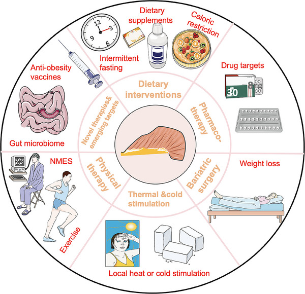
Exercise and physical therapy
Resistance training and its benefits on muscle mass and function
In WAT, consistent exercise results in a significant reduction in adipocyte size,ref. mco270030-bib-0725 an increase in mitochondrial biogenesis,ref. mco270030-bib-0726 control of adipokine release,ref. mco270030-bib-0727 resulting in an overall improvement in whole‐body metabolic health.ref. mco270030-bib-0728 Additionally, 12‐month treadmill exercise training induces adaptability in WAT. This is evidenced by increased FFA oxidation and a decrease in the inflammatory response, achieved through the regulation of proinflammatory and anti‐inflammatory gene expression, as well as a reduction in macrophage infiltrationref. mco270030-bib-0729 In elderly women, the number of p16INK4a‐positive cells in adipose tissue is negatively correlated with exercise capacity and cognitive function. However, combined treatment with physical exercise and calorie restriction can significantly reduce the number of p16INK4a‐positive cells in adipose tissue, promoting overall health.ref. mco270030-bib-0730
The majority of studies examine whether exercise primarily generates heat within the body while also reducing body weight. Consistent with these findings, preclinical data have shown that exercise directly influences BAT activity by boosting thermogenic gene expression and promoting WAT browning.ref. mco270030-bib-0731 Exercise is also known to diminish age‐related oxidative damage and curb chronic inflammation.ref. mco270030-bib-0732 Furthermore, myokines like IL‐6, induced by exercise, play a beneficial role in enhancing BAT activity, promoting browning of WAT, and reducing inflammation.ref. mco270030-bib-0733 In particular, mounting evidence has affirmed that irisin may serve as a positive regulator pertinent to this mechanism.ref. mco270030-bib-0734 Boström’s study revealed that plasma irisin levels increased after mice underwent 3 weeks of free wheel running. Similarly, circulating irisin showed a twofold increase with 10 weeks of endurance exercise training in healthy adults.ref. mco270030-bib-0527 Additionally, irisin has demonstrated the ability to induce WAT to adopt a BAT‐like phenotype, leading to heightened energy expenditure, reduced body weight, and enhanced glucose homeostasis, along with improved IR.ref. mco270030-bib-0527
Additionally, regular physical exercise also offers benefits for sarcopenia. Resistance training is particularly effective in combating muscle wasting and age‐related sarcopeniaref. mco270030-bib-0735 and improving muscle protein anabolism, as well as promoting specific metabolic and morphological muscular adaptations.ref. mco270030-bib-0736 Furthermore, multiple studies have affirmed that endurance exercise training leads to enhancements in maximal oxygen consumption, mitochondrial function, and insulin sensitivity, thereby mitigating sarcopenia progression.ref. mco270030-bib-0737, ref. mco270030-bib-0738, ref. mco270030-bib-0739, ref. mco270030-bib-0740 Indeed, myokines play a crucial role in exercise’s preventive effects against sarcopenia. For instance, irisin has been identified as increasing mitochondrial content and oxygen consumption while safeguarding skeletal muscle against metabolic stresses.ref. mco270030-bib-0537, ref. mco270030-bib-0538 According to clinical findings, reduced levels of irisin due to aging could serve as a predictor for sarcopenia,ref. mco270030-bib-0531, ref. mco270030-bib-0532 indicating that exercise‐enhanced serum irisin might prevent sarcopenia. Indeed, in support of this notion, studies have demonstrated that resistance training could serve as an effective intervention to increase irisin levels and prevent the age‐related decline in muscle function.ref. mco270030-bib-0530
Overall, it is clear that physical exercise is now commonly incorporated as a therapy for sarcopenia, possibly through regulating muscle, BAT and WAT functions as well as decreasing inflammation. How such changes might affect sarcopenia is still poorly understood, but we attempt to offer some insight here, suggesting that myokines are involved in this process. Further studies defining other mechanistic insight into the link among physical exercise, WAT regulation and sarcopenia are needed, especially in humans, and evidence of a direct causal relationship is thus also missing.
Neuromuscular stimulation and rehabilitation programs
Neuromuscular electrical stimulation (NMES) therapy is a physical therapy technique that employs muscle stimulation devices to deliver low‐frequency pulse currents through electrodes, directly targeting nerves or muscle tissue to induce skeletal muscle contraction. The primary aim of this therapy is to promote the recovery of neuromuscular function.ref. mco270030-bib-0741 Muscle plasticity in aged subjects undergoing NMES training is well documented, although many underlying mechanisms remain unexplored and warrant further investigation. Chronic application of NMES, when optimized for motor performance, can lead to increases in muscle mass,ref. mco270030-bib-0742 strength,ref. mco270030-bib-0743 power,ref. mco270030-bib-0744 activation,ref. mco270030-bib-0745 and endurance.ref. mco270030-bib-0746 For example, NMES has been identified as an effective alternative intervention for enhancing lower limb muscle mass and performance.ref. mco270030-bib-0747 Currently, NMES is utilized in sports training to boost muscle performance and in rehabilitation medicine to restore muscle properties following injury or surgery.ref. mco270030-bib-0748 This suggests that electrotherapy may hold promise for treating sarcopenia with minimal side effects, potentially paving the way for advancements in personalized bioelectronic medicine. For instance, Boutry‐Regard and his teamref. mco270030-bib-0749 proposed an innovative approach that combines NMES with nutritional interventions for sarcopenia. This method could help older adults overcome behavioral, physiological, and clinical challenges.
From a behavioral perspective, NMES can compensate for reduced physical activity and motivation to exercise. Clinically, it may help partially restore muscle strength and functional abilities, such as gait and balance, through chronic NMES applicationref. mco270030-bib-0748 thereby reducing fall risk.ref. mco270030-bib-0750 NMES training effectively addresses age‐related mobility issues, frailty, and fall risk while enhancing functional performance and ADL.ref. mco270030-bib-0751, ref. mco270030-bib-0752
Overall, physicians, therapists and physical conditioners/trainers could exploit this new knowledge in their professional practice to improve life conditions of frail and/or aged subjects.
Dietary interventions
Intermittent fasting
Fasting, whether it involves partial or total abstention from food or selective avoidance of certain foods, represents a potential nonpharmacological approach. It induces a metabolic shift from relying on glycogen for energy to utilizing fat stores, thereby effectively regulating body weight and reducing body fat levels.ref. mco270030-bib-0753, ref. mco270030-bib-0754 Moreover, fasting has been shown to enhance insulin sensitivity and glucose tolerance, diminish inflammatory responses, and mitigate oxidative stress. These effects contribute to the effective prevention and postponement of metabolic disorders like type II diabetes, myopathy, and cancer, ultimately promoting health and slowing down the aging process.ref. mco270030-bib-0753, ref. mco270030-bib-0754 Intermittent fasting (IF) refers to the regular maintenance of zero or very low‐calorie intake in a specific time, that is, fasting and free feeding altern IF involves regularly alternating between periods of zero or very low calorie intake and periods of unrestricted eating. Research suggests that IF can extend a healthy lifespan and may help in managing diseases associated with aging.ref. mco270030-bib-0755 Currently, research on the mechanisms underlying IF primarily focuses on factors such as IR, oxidative stress, and autophagy.ref. mco270030-bib-0756, ref. mco270030-bib-0757, ref. mco270030-bib-0758 Recent studies have demonstrated that fundamental autophagy processes are crucial for preserving muscle mass, as well as maintaining the integrity and adaptability of muscle fibers.ref. mco270030-bib-0759 An excessive activation of autophagy or its inhibition can result in muscle atrophy and potentially contribute to conditions like myofibrosis and muscle weakness.ref. mco270030-bib-0760 Furthermore, IF, as a dietary intervention, has been utilized in humans to address issues such as obesity and metabolic syndrome.ref. mco270030-bib-0761, ref. mco270030-bib-0762 Indeed, there is limited research on how IF influences myocyte renewal. Given its potential to systematically enhance autophagy and the significant role of autophagy in tissue and cell renewal, future investigations should delve into whether IF affects muscle cell renewal and whether it impacts sarcopenia through autophagy mechanisms.
Caloric restriction
Caloric restriction (CR) is indeed a dietary intervention known to delay aging and extend the healthy lifespan across various species.ref. mco270030-bib-0763 As aging progresses in mice, several changes occur including reduced potential for WAT browning, enlargement of adipocytes, decreased β‐oxidational function, and diminished thermogenic activity in BAT. Continuous CR through low calorie intake has been shown to decrease total fat mass and body weight, potentially through increased WAT browning. Additionally, small adipocytes following CR tend to secrete fewer proinflammatory cytokines like leptin and more adiponectin, resulting in reduced adiposity and improved metabolic outcomes.ref. mco270030-bib-0764 Indeed, during CR, there is an increase in M2 macrophage infiltration and eosinophils, along with enhanced type 2 cytokine signaling in fat tissues. These factors contribute to the development of functional beige fat induced by CR.ref. mco270030-bib-0765 Caloric excess, lack of exercise, and obesity can indeed disrupt long‐term energy metabolism balance, potentially resulting in increased production of ROS and reduced mitochondrial density in skeletal muscle.ref. mco270030-bib-0766
Initially, CR demonstrates a protective effect against muscular atrophy in rodents and primate mammals. After short‐term CR of 35 or 50% in 17‐month‐old rats, an increase in the number of satellite cells and the content of collagen VI was observed.ref. mco270030-bib-0767 Absolutely, muscle function depends not only on the structural integrity of the muscle tissue but also on the consumption of ATP to provide energy.ref. mco270030-bib-0768 Mitochondria play a central role in oxidative phosphorylation and ATP synthesis, serving as primary sites for energy production within cells. Furthermore, CR is employed to safeguard mitochondria and enhance proteolysis pathways, crucial for preserving the integrity of protein turnover mechanisms and maintaining skeletal muscle function.ref. mco270030-bib-0769 Previous studies have shown that CR reduces proton leakage and ROS generation in mitochondria within skeletal muscle, while also enhancing the expression of genes associated with ROS scavenging. Moreover, CR may alter the fatty acid composition of the mitochondrial membrane, thereby reducing lipid oxidation and proton leakage.ref. mco270030-bib-0770 Growing evidence suggests that apoptosis could play a fundamental role in both the onset and progression of sarcopenia.ref. mco270030-bib-0771 It is been proposed that CR may reduce the expression of various proapoptotic regulatory proteins, thereby diminishing the age‐related apoptotic potential.ref. mco270030-bib-0772 Specifically, in CR mice compared with control mice, there was a significant decrease observed in both the gene expression and cleavage of precaspase‐3 in the gastrocnemius muscle.ref. mco270030-bib-0773 CR has been demonstrated to be effective in preventing sarcopenic muscle loss in animals, which leads to a reduction in oxidative stress levels.ref. mco270030-bib-0774 CR has the potential to delay the onset of sarcopenia; however, it is essential to note its adverse effects in young mice (3 weeks old), including impaired skeletal growth, negative impacts on bone architecture and mass, and an increase in marrow adiposity.ref. mco270030-bib-0774 It is important to highlight that factor such as nutrition levels, dietary macronutrient composition, timing of interventions, and the use of different animal strains can significantly influence the observed effects of CR. These variations may contribute to the ongoing debate surrounding the efficacy of CR in delaying sarcopenia.
In the future, it will be essential to explore strategies for reconciling the apparent increase in brown adipocyte thermogenesis with the reduced energy expenditure (metabolic adaptation) that occurs during CR. Targeting WAT browning could emerge as a promising approach for promoting metabolic health during aging.
Dietary supplements
Dietary fats
Research indicates that dietary lipids play a dual role by not only providing energy but also regulating various cellular functions. Among dietary fatty acids, essential, polyunsaturated, saturated, and monounsaturated fats are key classifications. Moreover, recent findings suggest that consuming high levels of plant‐based essential fatty acids (ePUFAs) such as n‐6 linoleic acid (LA) and n‐3 α‐linolenic acid (ALA) may exert a more profound influence on body composition (including fat mass and lean mass) and glucose homeostasis compared with marine‐derived long‐chain n‐3 polyunsaturated fatty acids (PUFAs) like eicosapentaenoic acid (EPA) and docosahexaenoic acid (DHA).ref. mco270030-bib-0775 Furthermore, a high intake of both ePUFAs (LA and ALA) might exhibit anti‐inflammatory effects in humans. For example, a systemic review conducted by Bender et al.,ref. mco270030-bib-0776 which suggested that while supplementation of long‐chain n‐3 PUFA could modestly influence weight loss in the short term (<2 months), there was no evidence of associations with longer‐term intake. Further reinforcement for this argument can be found in a recent placebo‐controlled prospective RCT involving adults with overweight or obesity. The study indicated that 6 months of high‐dose marine n‐3 PUFA supplementation (3.9 g/day of EPA and DHA; 4.2 g total n‐3 per day) compared with placebo (4.2 g/day oleate) had no beneficial effects on adipose tissue lipolysis or inflammation.ref. mco270030-bib-0777 Indeed, a pooled analysis involving 20 prospective cohort studies conducted globally revealed that high levels of the biomarker for LA, measured in various forms such as adipose tissue, cholesterol ester, erythrocytes, or plasma phospholipids, and/or total plasma, were associated with dose‐dependent decreases in the incidence of T2DM.ref. mco270030-bib-0778 These findings are consistent with earlier cross‐sectional and intervention studies, which have shown that biomarkers of LA and ALA intake, rather than marine‐derived n‐3 PUFA, are linked to better glycemic control and/or insulin sensitivity, as well as a reduced risk of T2DM.ref. mco270030-bib-0779 Belury et al.ref. mco270030-bib-0780 found that erythrocyte LA (a biomarker of LA status) was positively associated with appendicular lean mass adjusted for BMI. Conversely, it was inversely related to trunk adipose mass, as well as with the homeostatic model assessment of IR and inflammation markers such as IL‐6.ref. mco270030-bib-0780 Research has demonstrated that compared with food‐restricted rats refed a high‐fat (lard) diet low in PUFA, those refed isoenergetically on diets enriched in LA or ALA, irrespective of the n‐6:n‐3 ratio, exhibit improved insulin sensitivity. Additionally, they tend to have lower fat mass and higher lean mass.ref. mco270030-bib-0781
Indeed, the benefits of ePUFA, particularly LA and ALA, as outlined, suggest that increasing ePUFA intake could have significant implications for public health and clinical medicine. These implications extend beyond the prevention of obesity to include the management of musculoskeletal and metabolic health throughout the lifespan. Given the observed positive associations with lean mass, improved insulin sensitivity, and reduced adiposity, promoting adequate ePUFA intake may be a valuable strategy for enhancing sarcopenia and overall health.
Resveratrol
Resveratrol, a prominent natural compound, is present in several plants, especially in the skin of red grapes and red wine. Its potential health benefits have gained significant attention, and it has traditionally been used in Chinese and Japanese medicine for its anti‐inflammatory and antiplatelet effects.ref. mco270030-bib-0782 Its anti‐inflammatory and antioxidant properties suggest that resveratrol may help prevent the development of diabetes and obesity.ref. mco270030-bib-0783 Aguirre et al.ref. mco270030-bib-0784 demonstrated that resveratrol can inhibit the early regulatory factor of adipogenesis, C/EBPβ, and effectively suppress adipogenesis in porcine, murine, and human preadipocytes at concentrations between 20 and 100 µM. Additionally, resveratrol exhibits lipolytic effects only at higher concentrations (50‐100 µM), but it can enhance the lipolytic effect of β‐adrenergic stimulation at any concentration.ref. mco270030-bib-0784 Research has shown that resveratrol supplementation can elevate levels of long‐chain and polyunsaturated fatty acids while reducing steroid levels. It may also decrease the size of abdominal subcutaneous adipocytes.ref. mco270030-bib-0785
Huang et al.ref. mco270030-bib-0786 discovered that resveratrol supplementation can prevent the buildup of intramuscular fat and increased body fat mass associated with a HFD, thereby helping to mitigate muscle loss. Resveratrol may enhance oxidative stress and mitochondrial function through the PKA/LKB1/AMPK pathway, which helps prevent HFD‐induced muscle atrophy in older rats. As a result, resveratrol holds potential for preventing sarcopenic obesity.ref. mco270030-bib-0786, ref. mco270030-bib-0787 Resveratrol may increase mitochondriogenesis and elevate UCP3 protein levels in skeletal muscle. Additionally, the upregulation of genes involved in mitochondrial oxidative phosphorylation suggests that resveratrol could improve the oxidative capacity of muscle fat.ref. mco270030-bib-0788 Reports indicate that resveratrol may boost exercise‐related nuclear and satellite cell numbers and enhance the fiber cross‐sectional area of exercise‐responsive muscle, potentially leading to improvements in muscle function.ref. mco270030-bib-0090 Human studies on resveratrol’s effects on sarcopenia, including RCTs, have been conducted. One trial with 30 healthy adults administered either a placebo or 500 mg/day of resveratrol for 12 weeks before and after exercise. Results showed that those receiving resveratrol with exercise experienced significant improvements compared with the placebo group, including better mitochondrial density, reduced muscle fatigue, increased mean muscle fiber area, and more total myonuclei. Additionally, the resveratrol group demonstrated enhanced knee extensor muscle peak energy, average peak energy, and muscle strength postexercise, while these parameters did not show significant improvement in the placebo group.ref. mco270030-bib-0090 A recent study by Toniolo et al.ref. mco270030-bib-0789 found that a 6‐month dietary supplementation of 0.04% resveratrol significantly improved muscle fatigue resistance. The study indicated that resveratrol enhanced capillarization in the skeletal muscle of aging mice, suggesting its potential to prevent capillary rarefaction in muscle tissue.ref. mco270030-bib-0789
While resveratrol appears promising in preventing sarcopenia by boosting muscle function and resistance to fatigue, some long‐term studies have found only modest or no significant improvements in muscle mass or inflammation.ref. mco270030-bib-0090, ref. mco270030-bib-0790 These findings highlight the complexity of studying dietary supplements like resveratrol and their effects on muscle health. They underscore the need for further research to better understand how resveratrol functions and its potential benefits, especially in relation to sarcopenia and muscle inflammation.
Pharmacotherapy
Hormone replacement therapy
As people age, circulating levels of various anabolic hormones decline, potentially leading to changes in muscle mass and function. Consequently, hormone modulation has been explored as a foundational strategy for treating sarcopenia, with testosterone supplementation being the most commonly studied option for enhancing muscle mass and promoting muscle‐protein anabolism.ref. mco270030-bib-0791, ref. mco270030-bib-0792 However, the findings from studies on testosterone replacement therapy in men vary based on factors such as age, pretreatment testosterone levels, and administration methods. These variables complicate the assessment of therapy’s impact on physical performance and disability. Additionally, potential side effects—such as peripheral edema, gynecomastia, polycythemia, and sleep apnea—must be weighed against the therapeutic benefits. A significant concern is that elevated testosterone levels may increase the risk of prostate cancer, necessitating careful planning and monitoring during treatment.ref. mco270030-bib-0792
Recently, selective androgen receptor modulators (SARMs) have emerged as a safer and more attractive alternative to long‐term testosterone therapy for combating muscle wasting. SARMs are nonsteroidal compounds that selectively bind to androgen receptors (AR), providing tissue‐selective anabolic effects similar to testosterone but with fewer side effects. These compounds also show promise for women due to their specific activity and improved pharmacokinetics.ref. mco270030-bib-0793, ref. mco270030-bib-0794 Other hormone modulation therapies also hold potential benefits. Dehydroepiandrosterone may enhance muscle mass and strength in both men and women, while tibolone can promote muscle anabolism by binding to ARs in muscle fibers and increasing levels of serum‐free testosterone, growth hormone, and IGF‐1. Estrogen may help suppress inflammatory cytokines and improve muscle strength in women, while growth hormone can stimulate the proliferation of muscle satellite cells and enhance muscle function. Ghrelin has also been shown to increase lean mass and physical performance.ref. mco270030-bib-0795 Despite the emerging evidence linking age‐related hormonal changes to sarcopenia, the clinical efficacy of hormone supplementation for treating this condition requires further investigation and validation in future studies.ref. mco270030-bib-0791
Metformin
Metformin’s widespread use as an oral antidiabetic drug is well established. Research indicates that metformin enhances proteins associated with mitochondrial biogenesis and VLDL clearance in BAT in mice, suggesting a direct effect on BAT. In human studies, metformin has been shown to reduce cellular respiration in brown adipocytes and effectively counteract obesity‐induced suppression of brown adipogenesis and thermogenesis. These findings suggest that BAT may be a target organ for metformin’s actions.ref. mco270030-bib-0796 Metformin’s influence on fatty acid metabolism offers a promising approach for lifespan extension. Additionally, its ability to reduce collagen gene expression suggests it might help mitigate ECM deposition related to aging, potentially improving adipose tissue regulation often disrupted by aging.ref. mco270030-bib-0797 Collagen buildup is linked to age‐related muscle loss, as excessive ECM deposition in aging skeletal muscles can stiffen myofibers, affecting myogenic progenitor cells and contributing to muscle aging. Metformin’s potential to reduce collagen gene expression and ECM deposition suggests it may help treat sarcopenia by addressing these age‐related changes in muscle tissue.ref. mco270030-bib-0798
Overall, metformin has been shown to guard against sarcopenia by influencing BAT and ECM dynamics, promoting the differentiation of skeletal muscle cells, and aiding in the maturation of myotubes, which enhances overall skeletal muscle function.ref. mco270030-bib-0799 Clinical trial data in elderly individuals without T2DM suggest that metformin may positively impact sarcopenia. While epidemiological studies indicate potential benefits, clinical trial results have shown varying consistency. In one study, metformin use led to a notable improvement in mean walking time by 0.39 s and a corresponding increase in gait speed of 0.13 m/s, highlighting its potential positive effects on physical performance in the context of sarcopenia.ref. mco270030-bib-0800 These findings indicate that metformin might enhance physical function and mobility in elderly individuals, even without T2DM. However, in a prospective open‐label observational study of overweight patients with both T2DM and chronic obstructive pulmonary disease, administration of metformin (850 mg twice daily) for 24 weeks resulted in a significant decrease in handgrip strength by 3.2%.ref. mco270030-bib-0801 Further clinical research is essential to determine the effectiveness of metformin in treating sarcopenia.
TRPM8 channel
Activating transient receptor potential (TRP) channels, like TRPM8, is an alternative method to indirectly stimulate BAT thermogenesis. TRPM8, a calcium‐gated channel responsive to heat and cold, functions as a key cold‐sensing molecular transducer in the peripheral nervous system of mice.ref. mco270030-bib-0802 Administering menthol activates TRPM8, leading to increased UCP‐1 expression and thermogenesis, which raises body temperature and provides protection against obesity in both mice and humans. Additionally, menthol intake enhances exercise endurance and energy metabolism by upregulating PGC‐1α in skeletal muscles through TRPM8 activation,ref. mco270030-bib-0803 improving muscle function. Therefore, targeting the TRPM8 channel could potentially be a viable intervention for improving both obesity management and skeletal muscle function.
βARs
In rodents, BAT thermogenesis is activated by sympathetic nervous system (SNS) input, with substantial evidence highlighting the critical role of β3‐AR in this process.ref. mco270030-bib-0804, ref. mco270030-bib-0805, ref. mco270030-bib-0806 Specifically, β3‐AR stimulation increased the gene expression of UCP1, linked to thermogenesis, as well as Cpt1b and Acox1, which are involved in β‐oxidation.ref. mco270030-bib-0807 Notably, β3‐AR mRNA levels in human BAT are lower than those in rodents, which makes the role of β3‐AR in humans less certain than in rodents.ref. mco270030-bib-0808 Blondin et al.ref. mco270030-bib-0809 discovered that β2‐AR mediates lipolysis and thermogenesis in BAT in response to sympathetic nervous stimulation in humans. As a result, the primary adrenergic receptor subtype responsible for regulating BAT thermogenesis in humans remains debated.
Currently, β2‐AR agonists show promise for stimulating skeletal muscle hypertrophy by boosting protein synthesis.ref. mco270030-bib-0810 β2‐AR agonists induce a significant shift in skeletal muscle fiber types from slow‐oxidative to fast‐glycolytic.ref. mco270030-bib-0811, ref. mco270030-bib-0812 This shift may partly explain their beneficial effects in sarcopenia,ref. mco270030-bib-0813 as age‐related muscle loss predominantly affects fast‐twitch fibers.ref. mco270030-bib-0814, ref. mco270030-bib-0815, ref. mco270030-bib-0816 A small clinical trial with patients who have brachial plexus injuries supports the effectiveness of β2‐AR agonists in reducing muscle mass loss due to denervation.ref. mco270030-bib-0817 Moreover, β2‐AR stimulation can accelerate skeletal muscle recovery following myotoxic injury, prompting the use of synthetic β2‐AR agonists to counteract or even reverse muscle weakness and wasting associated with aging and disease.ref. mco270030-bib-0814, ref. mco270030-bib-0818, ref. mco270030-bib-0819 Besides, activation of the β2‐AR–cAMP–PKA–mTORC2 pathway has been demonstrated to promote GLUT4 translocation to the plasma membrane and enhance glucose uptake independently of insulin.ref. mco270030-bib-0820 Additionally, β3‐AR agonists can also benefit muscle health. For example, Kern et al.ref. mco270030-bib-0821 treated obese individuals with IR using the β3‐AR agonist mirabegron (50 mg/day for 12 weeks), which resulted in an increase in type I muscle fibers and favorable changes in skeletal muscle gene expression, likely due to enhanced PGC‐1α expression. Previous research has demonstrated that PGC‐1α regulates genes related to mitochondrial function in various tissues, including the brain, heart, and skeletal muscle.ref. mco270030-bib-0822
Given that βARs influence adipose and muscle signaling differently across various pathological conditions, a deeper understanding of how βAR activation improves adipose and muscle function could help develop drugs that selectively target βAR signaling, potentially offering new treatment options for age‐related conditions like sarcopenia and metabolic diseases.
DPP4/GLP‐1R
It is worth noting that other treatments, such as glucagon‐like peptide 1 receptor (GLP‐1R) agonists and dipeptidyl peptidase‐4 inhibitors (DPP4i), have also been suggested as potentially beneficial for enhancing BAT activity.ref. mco270030-bib-0823, ref. mco270030-bib-0824 The introduction of GLP‐1R agonists and DPP4i has markedly changed the perspective on how T2DM impacts the regulation of musculoskeletal diseases.ref. mco270030-bib-0825, ref. mco270030-bib-0826, ref. mco270030-bib-0827 Exendin‐4, a GLP‐1R analog, may increase BAT activity, leading to faster plasma clearance of triacylglycerol and glucose, reduced food intake, and decreased body fat content, which can ultimately result in weight loss.ref. mco270030-bib-0828 Sitagliptin, a DPP4i, has been shown to increase energy expenditure by enhancing BAT activity involved in energy combustion.
Additionally, Nahon et al.ref. mco270030-bib-0829 demonstrated that sitagliptin can boost mitochondrial gene expression in skeletal muscle, enhancing muscle metabolism by upregulating PGC‐1β. Ogawa’s researchref. mco270030-bib-0830 found that elderly diabetic patients treated with a DPP4i had a significantly reduced risk of losing appendicular skeletal muscle mass, indicating a protective effect on muscle mass, particularly in the lower limbs. Epidemiological evidence suggests that elderly diabetic patients on DPP4i exhibit better sarcopenia parameters compared with those treated with other glucose‐lowering medications.ref. mco270030-bib-0831 Indeed, inconsistencies exist in the results. For example, a study found no significant changes in skeletal muscle mass and skeletal muscle index after 24 weeks of treatment with the DPP4i teneligliptin (20 mg daily) in overweight or obese patients with T2DM.
In contrast, treatment with the GLP‐1R agonist dulaglutide led to reductions in both markers of sarcopenia.ref. mco270030-bib-0832 Liraglutide treatment (GLP‐1R agonists, 3.0 mg daily) with over twenty‐four weeks is well tolerated and leads to fat mass reduction, particularly in android fat, while also improving the skeletal muscle index in patients with T2DM.ref. mco270030-bib-0833 Although the data are limited, they indicate that DPP4i, in addition to their established efficacy and safety for glucose control in elderly patients with T2DM, might be a favorable choice for managing sarcopenia risk due to their neutral effect on muscle mass.ref. mco270030-bib-0834 Based on these data, DPP‐4/GLP‐1R‐targeting medications may hold promise as potential treatments for sarcopenia. However, additional research is required to fully understand their efficacy and safety in addressing the condition.
Clearance of senescent cells
Senescent cells are more prevalent in older individuals than in younger ones, indicating that these cells accumulate in adipose tissues and muscle with age. This is supported by elevated levels of markers such as p16, p53, and SAβ‐gal found in the adipose tissue of elderly individuals with obesity.ref. mco270030-bib-0835, ref. mco270030-bib-0836 In people with obesity, senescence affects not only adipocytes but also extends to endothelial cells and preadipocytes within adipose tissue.ref. mco270030-bib-0837, ref. mco270030-bib-0838 In adipose tissue, cellular senescence leads to immune cell infiltration, which creates a proinflammatory environment for preadipocytes. Senescent preadipocytes produce a SASP, and activated macrophages secrete chemokines and cytokines, exacerbating the inflammation associated with aging in adipose tissue.ref. mco270030-bib-0130, ref. mco270030-bib-0839 The increased release of proinflammatory cytokines and chemokines by senescent cells in aging individuals is associated with age‐related metabolic dysfunctions.ref. mco270030-bib-0130, ref. mco270030-bib-0840, ref. mco270030-bib-0841
Studies have shown that senescent cells and the SASP can contribute to the progression of age‐related diseases such as osteoporosis, OA, and sarcopenia.ref. mco270030-bib-0050, ref. mco270030-bib-0826, ref. mco270030-bib-0842, ref. mco270030-bib-0843 Consequently, removing senescent cells has been shown to delay the aging phenotype in both human and mouse cells.ref. mco270030-bib-0842, ref. mco270030-bib-0844 Inducible depletion of p16INK4a cells in the BubR1 preaging mouse model led to delayed tissue dysfunction in adipose tissue, skeletal muscle, and the eye.ref. mco270030-bib-0844 In sarcopenia, there is a complex interplay between cellular senescence, SASP, and muscle dysfunction. Research by Jejurikar et al.,ref. mco270030-bib-0087 for example, shows that aged MuSCs are more prone to senescence or apoptosis than younger ones. Indeed, SASP factors have been linked to the upregulation of molecules like sarcolipin, which impairs myogenic differentiation and promotes skeletal muscle fibrosis. This contributes significantly to the development of sarcopenia.ref. mco270030-bib-0845 Transplanting a small number of senescent cells into young and aged mice caused decreased muscle strength and physical impairment. Existing evidence shows that eliminating senescent cells significantly mitigates sarcopenia and enhances muscle regeneration activity in aging and senescence mouse models.ref. mco270030-bib-0835, ref. mco270030-bib-0844 For example, treatment with Dasatinib and quercetin (D+Q) has been linked to lower levels of senescent cells and reduced production of proinflammatory cytokines, which may help alleviate physical dysfunction.ref. mco270030-bib-0143 Silencing p16INK4a cells may help preserve the regenerative capacity of satellite cells. Conversely, abnormal p16INK4a expression has been shown to obstruct the proliferation of satellite cells in injured young muscle within aged mice.ref. mco270030-bib-0846 In the future, clinical trials should investigate the potential of drugs that target and clear senescent cells as a means to alleviate sarcopenia.
Bariatric surgery
Bariatric surgery is currently recognized as the most effective approach for treating obesity. It not only leads to significant weight loss but also enhances insulin sensitivity and overall metabolic function in the body.ref. mco270030-bib-0847 Common surgical options include vertical sleeve gastrectomy (VSG), Roux‐en‐Y gastric bypass (RYGB), laparoscopic adjustable gastric banding, and biliopancreatic diversion with duodenal switch, which is less frequently performed.ref. mco270030-bib-0848 Following bariatric surgery, it is common to observe a significant reduction in adipose tissue mass, with up to half of the total adipose tissue being lost as the most notable effect.ref. mco270030-bib-0847 Furthermore, bariatric surgery has been shown to be effective in managing T2DM and hypertension, leading to reduced incidence rates and mortality associated with cardiovascular events.ref. mco270030-bib-0849 Obesity typically leads to decreased levels of adiponectin, which can be increased through metabolic surgery. Additionally, RYGB has been shown to decrease leptin levels, which is associated with both CR and weight loss.ref. mco270030-bib-0848 Research indicates that bariatric surgery can lead to significant weight loss in women due to continued mobilization of adipose tissue from intermuscular tissue and vWAT for up to 2 years postsurgery. However, between the 12th and 24th months after surgery, males tend to experience a rebound in weight gain across all adipose tissue depots.ref. mco270030-bib-0850
Studies have shown that bariatric surgery can result in a reduction of up to 32% of preoperative weight within two years after the procedure. This weight loss encompasses both fat mass and muscle mass, potentially leading to a decrease in basal metabolism, functional impairment, and a decline in overall QOL.ref. mco270030-bib-0851 Voican et al.ref. mco270030-bib-0852 reported that the prevalence of sarcopenia could increase significantly after bariatric surgery, rising from 8% at the time of surgery to one‐third of patients one year after undergoing VSG. Indeed, bariatric surgery can potentially elevate the risk of sarcopenia due to the loss of muscle mass. Optimizing exercise guidelines and ensuring adequate protein intake during the initial months following surgery could prove effective in mitigating muscle mass loss after undergoing bariatric surgery.ref. mco270030-bib-0851
Thermal and cold stimulation
BAT is primarily sensitive to thermal stimulation via the SNS.ref. mco270030-bib-0853 Acute heat stress can directly enhance the lipolytic response of bovine primary adipocytes by increasing protein kinase A phosphorylation of hormone‐sensitive lipase and perilipin. However, the reduced lipolytic response observed during heat stress in vivo might result from increased β‐adrenergic receptor downregulation or A1 receptor activation, attributable to the heightened sensitivity to β‐adrenergic receptor agonists.ref. mco270030-bib-0854 The application of localized heat can reduce FOXO3 mRNA levels, resulting in a decrease in proteolytic activity. Additionally, skeletal muscle heating may enhance the myogenic response to resistance exercise.ref. mco270030-bib-0855 Indeed, local muscle cooling combined with resistance training could induce proteolysis, thereby potentially resulting in a decrease in muscle growth.ref. mco270030-bib-0855
Cold stimulation can increase SNS activity, promoting BAT glucose uptake and lipolysis, consequently generating heat in BAT. Furthermore, SNS activation may decrease pancreatic insulin secretion while increasing lipolysis and glycolysis, thus elevating circulating fatty acids and glucose for uptake by brown adipocytes.ref. mco270030-bib-0853 Hence, cold exposure or activation of adrenergic signaling could serve as a potential complementary or adjunct therapeutic approach to prevent obesity development or the progression of T2DM.ref. mco270030-bib-0856 Zak et al.’sref. mco270030-bib-0857 report indicated that environmental temperature alone could not modify myogenetic or proteolytic expression. Indeed, Ivanova and Blondinref. mco270030-bib-0856 reported that cold exposure could enhance insulin sensitivity, glucose homeostasis, and the oxidation rate of fatty acids by stimulating thermogenic processes, particularly the recruitment of shivering skeletal muscles, indicating the beneficial effects of cold stimulation. Initiating the senescence pathway in young beige progenitors can trigger premature cellular senescence, hindering their capacity to produce cold‐induced beige adipocytes. Conversely, reversing cellular aging genetically or pharmacologically, notably through the p38/MAPK‐ p16INK4a pathway, in aged mouse or human beige progenitor cells rejuvenates cold‐induced beiging.ref. mco270030-bib-0858 Additionally, these treatments may have limited applications due to associated cardiovascular risks.ref. mco270030-bib-0859, ref. mco270030-bib-0860
It appears that temperature alone might not be adequate to induce changes in the gene expression of proteolysis and myogenesis. Therefore, combining local heat and cold stimulation with resistance exercise could be necessary to elicit significant gene expression changes.ref. mco270030-bib-0857 In the future, researchers should investigate how hot and cold stimulation impacts skeletal muscle function.
Combined therapy
As highlighted in the review, complex mechanisms are significant factors in sarcopenia’s development. To address these issues, a comprehensive approach is proposed, including dietary changes, bariatric surgery, pharmaceutical treatments, and physical exercise. Integrating these strategies could potentially slow the progression of sarcopenia by targeting the underlying mechanisms.ref. mco270030-bib-0861 This combined therapy approach appears promising for managing sarcopenia, as depicted in Figure 3. Moreover, while various drugs are being developed to target dysfunction muscle in aging individuals, additional research is necessary to discover new drug targets and therapies for addressing sarcopenia and related metabolic disorders.
THERAPEUTIC INTERVENTIONS FOR CACHEXIA
Anti‐inflammatory therapies
In recent years, as summarized above, clinical studies have implicated proinflammatory cytokines in the pathogenesis of cachexia, particularly cancer‐associated cachexia. Consequently, targeting these cytokines may prove effective in managing cachexia. For instance, in various cachectic mouse models, IL‐6/gp130‐dependent activation of STAT3 has been observed in atrophic skeletal muscle tissue. Pharmacological blockade of the JAK/STAT3 pathway has been shown to reduce muscle atrophy in C26‐driven cachexia, suggesting a significant role for this pathway in IL‐6/gp130‐mediated cachexia.ref. mco270030-bib-0332, ref. mco270030-bib-0333, ref. mco270030-bib-0862 Normalization of STAT3 activation levels through either homozygous or heterozygous genetic ablation of IL6 or STAT3 in mice has demonstrated protection against severe muscle wasting and weight loss, thereby improving survival.ref. mco270030-bib-0863, ref. mco270030-bib-0864 Additionally, a limited number of advanced lung cancer patients treated with neutralizing antibodies against IL‐6 or its receptor (IL‐6R) experienced amelioration of cachexia‐related symptoms.ref. mco270030-bib-0865, ref. mco270030-bib-0866 In cachectic models, pharmacological blockade of the JAK2/STAT3 pathway protected mice from weight loss, independent of tumor growth suppression.ref. mco270030-bib-0867Consistent with clinical findings, treatment using a humanized anti‐IL‐6 monoclonal antibody to globally block IL‐6 has shown protection against loss of LBM—a key measure of cachexia—without affecting tumor progression. Similarly, a case study involving the treatment of a cachectic advanced lung cancer patient with the anti‐IL‐6R monoclonal antibody tocilizumab resulted in significant weight gain, yet tumor growth continued unabated, leading to subsequent metastases.ref. mco270030-bib-0865The limited efficacy of global IL‐6 blockade in alleviating cancer cachexia symptoms without halting tumor progression is further supported by observations from the LLC cachexia model. Treatment with the monoclonal antibody MR16‐1, a rodent analogue of tocilizumab, preferentially protected against weight loss without affecting tumor growth.ref. mco270030-bib-0868 Moreover, a Phase II trial evaluating the safety, efficacy, and pharmacokinetics of the chimeric anti‐IL‐6 monoclonal antibody siltuximab in patients with advanced solid tumors, including non‐small cell lung cancer and Kras mutant colon cancer, revealed that global targeting of IL‐6 was well tolerated but did not impact tumor progression.ref. mco270030-bib-0869
TNF‐α is a crucial mediator of cachexia. In animal models, therapies aimed at neutralizing TNF‐α activity have shown significant anticachectic effects.ref. mco270030-bib-0870, ref. mco270030-bib-0871, ref. mco270030-bib-0872 For instance, gene knockout mice lacking the TNF‐α receptor type I exhibited less muscle wasting compared with wild‐type mice in a fast‐growing mouse tumor‐induced cachexia model. This underscores the role of TNF‐α in skeletal muscle depletion associated with cancer cachexia.ref. mco270030-bib-0873 Additionally, direct inhibition of NF‐κB has been shown to block cachexia in animal studies, suggesting a connection between NF‐κB and TNF‐α in this condition. However, evidence supporting the efficacy of anti‐TNF‐α therapy for treating cachexia in humans remains limited. Factors such as low patient accrual, small sample sizes, and the use of drugs that target only one of the multiple cytokines involved in cancer cachexia development may explain the lack of positive outcomes in these studies.
The strategy of using agents that target cytokines implicated in the pathogenesis of cancer cachexia has gained attention for managing cachexia. Unfortunately, most clinical trials assessing anticytokine therapies for cancer cachexia have not yielded favorable results. To enhance outcomes and address methodological challenges, future cancer cachexia clinical trials should incorporate standardized inclusion criteria, meaningful outcome measures, and low‐toxicity therapies that target multiple procachectic molecules and pathways, especially in the earlier phases of the cancer cachexia continuum. This approach may allow anticytokine treatments to be integrated into a multimodal strategy for managing the complex syndrome of cancer cachexia.
Nutritional interventions
Specialized nutritional formulations to counteract hypercatabolism
Multiple interventions can be considered for patients diagnosed with malnutrition, including dietary counseling, oral nutrition supplements, and enteral or parenteral feeding. Recommendations should be tailored to the patient’s specific disease and performance status. Early assessment of nutritional needs can have a lasting impact throughout the treatment duration. Immunonutrition—dietary supplementation with specific nutrients such as fat‐soluble vitamins, omega‐3 fatty acids, arginine, and glutamine—represents a novel strategy to modulate negative immune‐mediated inflammatory responses in advanced cancer and alleviate the physiological stress of surgery.ref. mco270030-bib-0874 Omega‐3 fatty acid intake may lead to significant increases in body weight and LBM, reductions in REE, and potentially prolonged survival.ref. mco270030-bib-0875 Nutritional formulations containing branched‐chain amino acids, arginine, and glutamine may help reduce cancer cachexia.ref. mco270030-bib-0876 However, no specific diet has been definitively proven beneficial in managing cancer cachexia or sarcopenia, partly due to the challenges associated with studying dietary interventions.ref. mco270030-bib-0877, ref. mco270030-bib-0878 A recent publication by the American Society of Clinical Oncology reported insufficient evidence to suggest that specific diets—such as low‐fat, high‐fiber, or ketogenic—impact oncologic outcomes, treatment toxicity, or QOL.ref. mco270030-bib-0879
Appetite stimulants
Many studies have also been conducted on the use of various appetite‐modulating drugs in cancer‐associated muscle cachexia. One appetite‐modulating approach is medroxyprogesterone acetate, a progesterone derivative similar to megestrol acetate (MA) that has been shown to improve anorexia, body weight, and QOL in phase II clinical trials.ref. mco270030-bib-0880 Endocannabinoids such as delta‐9‐tetrahydrocannabinol and nabilone have also been tested as appetite stimulants in phase II clinical trials, and results have generally shown increased appetite and QOL with no major side effects.ref. mco270030-bib-0881, ref. mco270030-bib-0882 Other appetite stimulants include corticosteroids such as dexamethasone and prednisolone, which are among the earliest drugs evaluated for cancer cachexia. In a randomized clinical trial, dexamethasone increased appetite but did not affect body weight due to its catabolic effects on muscle and bone.ref. mco270030-bib-0883 One preliminary study showed dexamethasone treatment significantly improved patient‐reported cluster scores for fatigue/anorexia–cachexia/depression symptoms after two weeks in advanced cancer patients.ref. mco270030-bib-0884 Another novel immunomodulatory, OHR118, affects the synthesis of chemokines and cytokines, including TNF‐α. In a phase 2 study, OHR118 significantly improved appetite, dyspepsia, and depression in cancer patients.ref. mco270030-bib-0885
Pharmacological treatments
Ghrelin agonists and anabolic steroids
Ghrelin is an endogenous hormone secreted by the stomach that stimulates appetite and plays a role in regulating energy expenditure by inhibiting sympathetic stimulation of BAT. It is believed to alleviate some symptoms of cancer cachexia by inhibiting MuRF‐1/MAFbx‐induced protein degradation in muscle tissue, promoting muscle protein synthesis via IGF‐1, and increasing fat accumulation.ref. mco270030-bib-0886 Due to its significant effects on appetite and metabolism, ghrelin has been the focus of several recent clinical trials. Anamorelin HCl, a ghrelin analogue with an extended half‐life, has undergone extensive testing in phase I, II, and III trials, demonstrating good tolerability and effectiveness in improving weight gain, lean muscle mass, and anorexic/cachectic symptoms in cachectic cancer patients.ref. mco270030-bib-0887, ref. mco270030-bib-0888, ref. mco270030-bib-0889, ref. mco270030-bib-0890 However, it did not show significant improvement in handgrip strength during phase III trials.ref. mco270030-bib-0889, ref. mco270030-bib-0890 Additionally, the potential side effects of anamorelin must be considered, as agents that elevate IGF‐1 or growth hormone levels can lead to diabetes or IR.ref. mco270030-bib-0891 Anabolic steroids have also been used effectively to treat muscle wasting,ref. mco270030-bib-0892, ref. mco270030-bib-0893 particularly in conditions like chronic heart failure, where nearly 20% of patients experience this issue.ref. mco270030-bib-0894 In heart failure patients, low levels of circulating anabolic hormones are linked to poor outcomes.ref. mco270030-bib-0895, ref. mco270030-bib-0896 However, the risks associated with anabolic steroid administration often outweigh their potential benefits.
Beta‐blockers to modulate catabolic signals
β‐Blockers can reduce body energy expenditure and enhance substrate utilization efficiency, with some exhibiting multiple pharmacological effects. Espindolol (MT‐102; PsiOxus Therapeutics) is a nonspecific β1/β2‐adrenergic receptor antagonist that also acts on β and central 5‐HT1α receptors, demonstrating proanabolic, anticatabolic, and appetite‐stimulating properties.ref. mco270030-bib-0021 The ACT‐ONE trial (NCT01238107) found that espindolol, administered at 10 mg twice daily, effectively reversed weight loss, improved fat‐free mass, maintained fat mass, and enhanced handgrip strength in cachectic patients with non‐small cell lung cancer or colorectal cancer.ref. mco270030-bib-0897, ref. mco270030-bib-0898
Multimodal interventions
Combination therapy
Due to the complexity of cancer cachexia syndrome, multidrug combination therapy targeting multiple underlying pathophysiological processes has been proposed. In a phase III RCT conducted in 2010, 332 patients with cachexia associated with cancer were randomized into five treatment arms: progesterone analogs, EPA, L‐carnitine, thalidomide, or a combination of all four interventions. The combination arm showed significant improvements in LBM, REE, fatigue, appetite, IL‐6 levels, Glasgow prognostic scores, and Eastern Cooperative Oncology Group performance scores.ref. mco270030-bib-0899
Conversely, a 2012 report indicated that a combination of l‐carnitine, celecoxib, and MA did not provide greater benefits than the two‐drug combination of l‐carnitine and celecoxib alone, as both groups demonstrated significant increases in LBM and physical performance compared with baseline.ref. mco270030-bib-0899 These findings suggest that additional treatments do not always enhance efficacy. Furthermore, the combination of olanzapine and MA was assessed in a 2010 RCT involving patients with advanced lung or gastrointestinal cancer, revealing significantly greater weight gain in the combination group compared with the MA‐only group.ref. mco270030-bib-0900
Multimodal intervention
As discussed, cancer cachexia is a complex metabolic disorder that is challenging to address with conventional nutritional therapy; thus, nutritional support tailored to its pathophysiology is crucial. The European Society for Clinical Nutrition and Metabolism and European Society for Medical Oncology guidelines recommend a multifaceted approach for the treatment of cancer cachexia, emphasizing the integration of nutrition, exercise, and pharmacotherapy.ref. mco270030-bib-0901, ref. mco270030-bib-0902, ref. mco270030-bib-0903 The evidence supporting nutritional counseling is also robust.ref. mco270030-bib-0901, ref. mco270030-bib-0904, ref. mco270030-bib-0905 In a randomized phase III trial involving 104 patients with advanced gynecologic cancer, a combination treatment of MA, l‐carnitine, celecoxib, and antioxidants was found to be more effective than MA alone in enhancing LBM, REE, fatigue, and QOL.ref. mco270030-bib-0906 Additionally, a multidisciplinary intervention that included omega‐3 polyunsaturated fatty acid nutraceuticals, exercise, and the anti‐inflammatory drug celecoxib was administered to 46 patients with lung and pancreatic cancer undergoing chemotherapy, effectively mitigating weight loss.ref. mco270030-bib-0907 Alongside anti‐inflammatory, metabolism‐enhancing, and appetite‐stimulating medications, high‐quality nutritional therapy and appropriate exercise tailored to each patient’s physical capabilities can improve physical function and increase skeletal muscle mass.ref. mco270030-bib-0908, ref. mco270030-bib-0909 Multimodal interventions are deemed effective and are currently under assessment in clinical studies, highlighting their increasing necessity in future treatments (Figure 4).
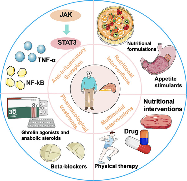
EMERGING THERAPIES AND FUTURE DIRECTIONS
Recent insights into the pathophysiology of sarcopenia and cachexia have revealed several promising drug targets and innovative treatments. These advancements offer potential solutions for addressing the global epidemic of sarcopenia and cachexia and their associated health challenges. In addition to pharmacological therapies, alternative approaches are being explored, including modulation of the gut microbiome, stem cell therapy, and gene therapy.
Gene therapy and molecular targets
Gene therapy represents a promising avenue for treating muscle wasting by specifically targeting and modifying the key molecular pathways involved in disease progression. This approach involves two methods: the first, known as the indirect method, enhances the secretion of therapeutic products derived from exogenous gene expression. The second method directly introduces target genes into cells using viral or nonviral vectors to express the desired functional proteins in vivo.
Viral vectors, such as adeno‐associated virus (AAV), are commonly used due to their low risk of genotoxicity and consistent long‐term expression.ref. mco270030-bib-0910, ref. mco270030-bib-0911 AAV has been employed to deliver therapeutic genes in both preclinical and clinical trials.ref. mco270030-bib-0912 Among various AAV types, AAV9 is particularly notable for its high delivery efficiency to skeletal muscle. Among multiple types of AAVs, AAV9 is one of the most widely used vectors for treating muscle diseases for its high delivery efficiency in muscle.ref. mco270030-bib-0913, ref. mco270030-bib-0914, ref. mco270030-bib-0915 Studies have demonstrated that AAV9‐mediated gene delivery, including miR‐23a/27a, significantly protects against muscle force loss and reverses dystrophic features in rodent models.ref. mco270030-bib-0916 Additionally, AAV9‐mediated gene therapies have shown promise in improving muscle histology in young adult Duchenne muscular dystrophy dogs.ref. mco270030-bib-0917
Furthermore, muscle‐specific AAV9‐mediated overexpression of NLS–PGC‐1α4 has alleviated aging‐associated sarcopenia and metabolic dysfunction in mouse models. However, these therapies have yet to be tested in humans, and the main drawback is the lack of extensive clinical trials. Current findings primarily focus on muscle atrophy impacts without thorough safety evaluations, highlighting the need for further research.
Stem cell therapy for muscle regeneration
Stem cell therapy, also known as cellular therapy or cytotherapy, utilizes stem cells to prevent and treat diseases. The efficacy of this approach has been explored across various conditions.ref. mco270030-bib-0918, ref. mco270030-bib-0919 In muscle tissue, satellite cells serve as the primary stem cells, typically located in the basal lamina or between muscle fibers, where they remain in a quiescent state. However, their self‐renewal ability is significantly diminished in patients with muscle atrophy.ref. mco270030-bib-0920 Therefore, increasing the number of satellite cells or enhancing their functionality could offer a promising theoretical solution for treating muscle atrophy.
In animal models, the transplantation of satellite cells into damaged muscle has demonstrated successful self‐renewal and regeneration.ref. mco270030-bib-0921, ref. mco270030-bib-0922 Early human studies have also indicated a potential role for mesenchymal stem cells in managing frailty.ref. mco270030-bib-0923 Nonetheless, challenges such as cost, regulatory constraints, and ethical considerations must be carefully addressed to facilitate the clinical application of stem cell therapy.
Targeting gut microbiota and metabolism
Modulation of microbiome could impact muscle metabolism and inflammation. For example, sarcopenia has been associated with alterations in gut microbiota composition, including higher levels of Lactobacillus, lower levels of Lachnospira, reduced microbial diversity, and decreased richness of microbial genes.ref. mco270030-bib-0924, ref. mco270030-bib-0925 Further, gut microbiota affects muscle properties that are fundamentally altered in cachexia by impacting muscle metabolism and contributing to inflammation. The gut microbiome is emerging as a new target for addressing sarcopenia and cachexia. Modulating the gut microbiota therapeutically could help prevent and treat metabolic disorders associated with aging.ref. mco270030-bib-0926, ref. mco270030-bib-0927, ref. mco270030-bib-0928 Selective modulation of the human gut microbiome offers a novel approach to treating sarcopenia. This can be done through dietary supplements like prebiotics and probiotics that impact bacterial growth, as well as through fecal microbiota transplantation.ref. mco270030-bib-0929, ref. mco270030-bib-0930, ref. mco270030-bib-0931, ref. mco270030-bib-0932
Moreover, in the context of cancer cachexia, a synbiotic containing inulin‐type fructans and Lactobacillus reuteri has been shown to counteract disturbances in gut microbiota composition in cachectic mice.ref. mco270030-bib-0933 This intervention also positively affected cancer progression, morbidity scores, and survival rates. Several features associated with cachexia, such as loss of muscle mass, skeletal muscle expression of Cathepsin L and LC3, as well as markers of intestinal permeability and immune function, showed slight improvements following synbiotic administration.
Current preclinical studies have primarily focused on the impact of gut microbiota on body weight, muscle properties, and proinflammatory cytokines. However, there is limited information on the relationships between gut microbiota and food intake, adipose tissue metabolism, and IR—key factors in cancer cachexia and sarcopenia. It is crucial to gather more data on these drivers of cachexia and sarcopenia in future research, as novel treatment approaches are urgently needed.
Precision medicine approaches
Customized drug combinations tailored to the specific genetic and molecular profile of a patient’s tumor or muscle can enhance efficacy while reducing toxicity. However, our current understanding of the genetic and epigenetic factors involved in sarcopenia and cachexia is limited. For instance, while muscle phenotypes exhibit strong heritability, the specific genetic factors underlying these traits remain unclear. Genetic factors alone do not solely determine the risk of muscle dysfunction; they interact with environmental influences, such as diet and physical activity, to shape individual risk.934
Understanding the potential connections between genes related to cachexia and sarcopenia could provide deeper insights into the pathogenesis of these two conditions. Ultimately, a patient‐centric approach is essential for managing sarcopenia and cachexia, highlighting the importance of personalized care strategies and patient education. Increasing awareness of these syndromes among patients, healthcare providers, and the general public is a crucial step toward developing more effective solutions to this growing clinical concern. Personalized therapeutic strategies based on genetic and molecular profiling are vital for improved outcomes.
CONCLUSION AND PERSPECTIVE
Cachexia is a complex systemic condition involving multiple metabolic pathways across various tissues and organs. It is characterized by systemic inflammation, progressive weight loss, and depletion of adipose tissue and skeletal muscle, which cannot be fully reversed through conventional nutritional support. Cachexia shares some similarities yet exhibits clear differences from other syndromes, such as age‐related muscle loss (sarcopenia), anorexia, malabsorption, hyperthyroidism, and starvation. Sarcopenia, a progressive and multifaceted disease, is associated with an increased burden of morbidity and mortality. While cachexia and sarcopenia can be defined as distinct clinical entities, they exhibit significant overlap, particularly in terms of inflammation and weakness. Since weakness/fatigue and inflammation are common features across various conditions, clinicians must look beyond these symptoms to identify unique characteristics of each condition, evaluating them within the context of a patient’s clinical history and personal profile (e.g., previous diseases, mental health status, age). Early and accurate diagnosis is critical for the effective management of both sarcopenia and cachexia. Future research should prioritize the discovery and clinical application of new biomarkers for early detection. Advanced imaging techniques and molecular diagnostics can be employed to monitor disease progression and assess treatment responses. Reliable biomarkers will facilitate personalized treatment strategies, allowing for timely and targeted interventions to slow or halt disease progression.
Understanding the pathophysiological foundations of cancer cachexia and sarcopenia is essential for distinguishing between the two conditions. This knowledge is crucial for establishing an early and accurate diagnosis and implementing timely therapeutic measures. Given the potential role of adipose tissue in the development of sarcopenia and cachexia, it is vital to investigate the interactions among different adipose depots (including intermuscular and intramuscular fat) and skeletal muscle, along with their associated molecular signaling pathways. This complex interplay involves various molecules, such as myokines, adipokines, lncRNAs, and miRs, that mediate the interactions between skeletal muscle and adipose tissue. The effects of these molecules on the homeostasis of both tissues may be closely linked to the molecular mechanisms underpinning sarcopenia and cachexia. Key questions remain regarding whether the regulation is direct or indirect—operating through an adipose–brain–muscle or an adipose–immune–muscle axis. Is there a master cytokine orchestrating these interactions? Are there “bystander” molecules that inadvertently function as “mediators”? Future studies should focus on defining the molecules produced by adipose and muscle cells and elucidating the intricate regulatory mechanisms involved. These considerations are vital for a comprehensive understanding of the therapeutic potential of these molecules in addressing sarcopenia and cachexia (Figure 5).
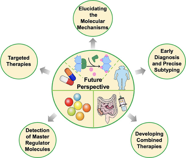
Due to the complex factors contributing to sarcopenia and cachexia, relying on a single treatment method is frequently insufficient. Consequently, a multifaceted treatment strategy that addresses various pathways and cellular interactions is essential. Combining medications with cell and gene therapies, as well as utilizing nanoparticles and gene‐editing technologies, can improve both the accuracy and effectiveness of treatments. Exploring the synergistic effects of different therapies could result in better outcomes and fewer side effects. Tailoring therapeutic approaches based on genetic and molecular profiles is crucial for achieving enhanced results.
AUTHOR CONTRIBUTIONS
Tiantian Wang was a major contributor to writing the manuscript, creating all figures and tables, and conducting the literature search. Tiantian Wang, Dong Zhou, and Zhen Hong made significant contributions to the manuscript’s design and critically revised it for important intellectual content. All authors have read and approved the final version of the manuscript.
CONFLICT OF INTEREST STATEMENT
The authors declare that they have no known competing financial interests or personal relationships that could have appeared to influence the work reported in this paper.
ETHICS STATEMENT
Not applicable.
References
- Summary comments.. Am J Clin Nutr., 1989
- Sarcopenia: revised European consensus on definition and diagnosis.. Age Ageing., 2019
- Sarcopenia: revised European consensus on definition and diagnosis.. Age Ageing., 2019. [PubMed]
- Sarcopenia.. Lancet., 2019. [PubMed]
- Sarcopenia ‐ Molecular mechanisms and open questions.. Ageing Res Rev., 2021. [PubMed]
- Cachexia: a new definition.. Clin Nutr., 2008. [PubMed]
- Definition and classification of cancer cachexia: an international consensus.. Lancet Oncol., 2011. [PubMed]
- Cachexia as a major public health problem: frequent, costly, and deadly.. J Cachexia Sarcopenia Muscle., 2013. [PubMed]
- Cachexia: A systemic consequence of progressive, unresolved disease.. Cell., 2023. [PubMed]
- Consensus definition of sarcopenia, cachexia and pre‐cachexia: joint document elaborated by Special Interest Groups (SIG) “cachexia‐anorexia in chronic wasting diseases” and “nutrition in geriatrics”.. Clin Nutr., 2010. [PubMed]
- Sarcopenia: European consensus on definition and diagnosis: report of the European Working Group on Sarcopenia in Older People.. Age Ageing., 2010. [PubMed]
- Cancer cachexia: molecular mechanisms and treatment strategies.. J Hematol Oncol., 2023. [PubMed]
- Loss of skeletal muscle mass in aging: examining the relationship of starvation, sarcopenia and cachexia.. Clin Nutr., 2007. [PubMed]
- Pancreatic cancer: let us focus on cachexia, not just sarcopenia!. Future Oncol., 2018. [PubMed]
- Pearls of optimizing nutrition and physical performance of older adults undergoing cancer therapy.. J Geriatr Oncol., 2017. [PubMed]
- Nutritional mechanisms of cancer cachexia.. Annu Rev Nutr., 2024. [PubMed]
- Understanding sarcopenia as a geriatric syndrome.. Curr Opin Clin Nutr Metab Care., 2010. [PubMed]
- Inflammation: roles in aging and sarcopenia.. JPEN J Parenter Enteral Nutr., 2008. [PubMed]
- Cancer cachexia: rationale for the MENAC (Multimodal‐Exercise, Nutrition and Anti‐inflammatory medication for Cachexia) trial.. BMJ Support Palliat Care., 2018
- Exercise for sarcopenia in older people: A systematic review and network meta‐analysis.. J Cachexia Sarcopenia Muscle., 2023. [PubMed]
- ACT‐ONE ‐ ACTION at last on cancer cachexia by adapting a novel action beta‐blocker.. J Cachexia Sarcopenia Muscle., 2016. [PubMed]
- Adipose tissue in older individuals: a contributing factor to sarcopenia.. Metabolism., 2024. [PubMed]
- Macrophage‐derived glutamine boosts satellite cells and muscle regeneration.. Nature., 2020. [PubMed]
- Stem cell aging in skeletal muscle regeneration and disease.. Inte J Mol Sci., 2020
- Regenerative decline of stem cells in sarcopenia.. Mol Aspects Med., 2016. [PubMed]
- Satellite cells are essential for skeletal muscle regeneration: the cell on the edge returns centre stage.. Development., 2012. [PubMed]
- Geriatric muscle stem cells switch reversible quiescence into senescence.. Nature., 2014. [PubMed]
- Inducible depletion of satellite cells in adult, sedentary mice impairs muscle regenerative capacity without affecting sarcopenia.. Nat Med., 2015. [PubMed]
- Skeletal muscle aging—stem cells in the spotlight.. Mech Ageing Dev., 2020. [PubMed]
- Stem cell aging in the skeletal muscle: the importance of communication.. Ageing Res Rev., 2022. [PubMed]
- Muscle stem cells and aging.. Curr Top Dev Biol., 2018. [PubMed]
- Age‐related differences in absolute numbers of skeletal muscle satellite cells.. Muscle Nerve., 1983. [PubMed]
- Satellite‐cell pool size does matter: defining the myogenic potency of aging skeletal muscle.. Dev Biol., 2006. [PubMed]
- Skeletal muscle satellite cells: changes in proliferation potential as a function of age.. Mech Ageing Dev., 1982. [PubMed]
- Activation of an adipogenic program in adult myoblasts with age.. Mech Ageing Dev., 2002. [PubMed]
- Increased Wnt signaling during aging alters muscle stem cell fate and increases fibrosis.. Science., 2007. [PubMed]
- Alterations in the in vitro and in vivo regulation of muscle regeneration in healthy ageing and the influence of sarcopenia.. J Cachexia Sarcopenia Muscle., 2018. [PubMed]
- Inflamm‐aging. An evolutionary perspective on immunosenescence.. Ann NY Acad Sci., 2000. [PubMed]
- Recent advances for measurement of protein synthesis rates, use of the ‘Virtual Biopsy’ approach, and measurement of muscle mass.. Curr Opin Clin Nutr Metab Care., 2017. [PubMed]
- Type I insulin‐like growth factor receptor signaling in skeletal muscle regeneration and hypertrophy.. J Musculoskelet Neuronal Interact., 2007. [PubMed]
- Association between sarcopenia and levels of growth hormone and insulin‐like growth factor‐1 in the elderly.. BMC Musculoskelet Disord., 2020. [PubMed]
- Do not neglect the role of circadian rhythm in muscle atrophy.. Ageing Res Rev., 2020. [PubMed]
- Restoration of epigenetic impairment in the skeletal muscle and chronic inflammation resolution as a therapeutic approach in sarcopenia.. Ageing Res Rev., 2024. [PubMed]
- Inflammaging: The ground for sarcopenia?. Exp Gerontol., 2022. [PubMed]
- Identification of ubiquitin ligases required for skeletal muscle atrophy.. Science., 2001. [PubMed]
- The integration of inflammaging in age‐related diseases.. Semin Immunol., 2018. [PubMed]
- TGFβ‐induced degradation of TRAF3 in mesenchymal progenitor cells causes age‐related osteoporosis.. Nat Commun., 2019. [PubMed]
- Inhibition of cellular communication network factor 1 (CCN1)‐driven senescence slows down cartilage inflammaging and osteoarthritis.. Bone., 2020. [PubMed]
- Searching for the link between inflammaging and sarcopenia.. Ageing Res Rev., 2022. [PubMed]
- Inflammaging: The ground for sarcopenia?. Exp Gerontol., 2022. [PubMed]
- Pro‐inflammatory cytokines: The link between obesity and osteoarthritis.. Cytokine Growth Factor Rev., 2018. [PubMed]
- TNF‐α and IL‐6: the link between immune and bone system.. Curr Drug Targets., 2020. [PubMed]
- Chronic inflammation in the etiology of disease across the life span.. Nat Med., 2019. [PubMed]
- Inflammaging and anti‐inflammaging: a systemic perspective on aging and longevity emerged from studies in humans.. Mech Ageing Dev., 2007. [PubMed]
- The continuum of aging and age‐related diseases: common mechanisms but different rates.. Front Med (Lausanne)., 2018. [PubMed]
- Frailty and sarcopenia: the potential role of an aged immune system.. Ageing Res Rev., 2017. [PubMed]
- Novel aspects of annexin 1 and glucocorticoid biology: intersection with nitric oxide and the lipoxin receptor.. Inflamm Allergy Drug Targets., 2006. [PubMed]
- Inflammation and sarcopenia: A focus on circulating inflammatory cytokines.. Exp Gerontol., 2021. [PubMed]
- Elevated hydrogen peroxide and decreased catalase and glutathione peroxidase protection are associated with aging sarcopenia.. BMC Geriatr., 2013. [PubMed]
- Sarcopenia: monitoring, molecular mechanisms, and physical intervention.. Physiol Res., 2014. [PubMed]
- Age‐related changes in skeletal muscle: changes to life‐style as a therapy.. Biogerontology., 2018. [PubMed]
- Mitochondrial function and apoptotic susceptibility in aging skeletal muscle.. Aging Cell., 2008. [PubMed]
- Skeletal muscle: molecular structure, myogenesis, biological functions, and diseases.. MedComm., 2024. [PubMed]
- Characteristics of secondary, primary, and compensated hypogonadism in aging men: evidence from the European Male Ageing Study.. J Clin Endocrinol Metab., 2010. [PubMed]
- Testosterone and leptin in older African‐American men: relationship to age, strength, function, and season.. Metabolism., 2000. [PubMed]
- Changes in muscle, fat and bone mass after 36 weeks of maximal androgen blockade for prostate cancer.. BJU Int., 2008. [PubMed]
- Effect of hormone therapy on muscle strength in postmenopausal women: a systematic review and meta‐analysis of randomized controlled trials.. Menopause., 2020. [PubMed]
- Association Between Hormone Therapy and Muscle Mass in Postmenopausal Women: A Systematic Review and Meta‐analysis.. JAMA Netw Open., 2019. [PubMed]
- Current and investigational medications for the treatment of sarcopenia.. Metabolism., 2023. [PubMed]
- Role of skeletal muscle in insulin resistance and glucose uptake.. Compr Physiol., 2020. [PubMed]
- Mechanisms of insulin action and insulin resistance.. Physiol Rev., 2018. [PubMed]
- Muscle insulin sensitivity and glucose metabolism are controlled by the intrinsic muscle clock.. Mol Metab., 2014. [PubMed]
- Muscle‐specific loss of Bmal1 leads to disrupted tissue glucose metabolism and systemic glucose homeostasis.. Skelet Muscle., 2016. [PubMed]
- The functional significance of the skeletal muscle clock: lessons from Bmal1 knockout models.. Skelet Muscle., 2016. [PubMed]
- Neuromuscular junction dysfunction in amyotrophic lateral sclerosis.. Mol Neurobiol., 2022. [PubMed]
- Multiple MuSK signaling pathways and the aging neuromuscular junction.. Neurosci Lett., 2020. [PubMed]
- Alterations of neuromuscular junctions in Duchenne muscular dystrophy.. Neurosci Lett., 2020. [PubMed]
- Sarcopenia–The search for emerging biomarkers.. Ageing Res Rev., 2015. [PubMed]
- Age changes in neuromuscular junction morphology and acetylcholine receptor distribution on rat skeletal muscle fibres.. J Physiol., 1981. [PubMed]
- Acetylcholine receptor binding properties at the rat neuromuscular junction during aging.. J Neurochem., 1987. [PubMed]
- Pathogenesis of sarcopenia and the relationship with fat mass: descriptive review.. J Cachexia Sarcopenia Muscle., 2022. [PubMed]
- Senescent cells and the incidence of age‐related diseases.. Aging Cell., 2021. [PubMed]
- Targeting normal and cancer senescent cells as a strategy of senotherapy.. Ageing Res Rev., 2019. [PubMed]
- Senescent cells: a therapeutic target for osteoporosis.. Cell Prolif., 2022. [PubMed]
- Cellular senescence links aging and diabetes in cardiovascular disease.. Am J Physiol Heart Circ Physiol., 2018. [PubMed]
- A senescent cell bystander effect: senescence‐induced senescence.. Aging Cell., 2012. [PubMed]
- Aging increases the susceptibility of skeletal muscle derived satellite cells to apoptosis.. Exp Gerontol., 2006. [PubMed]
- Characterization of cellular senescence in aging skeletal muscle.. Nat Aging., 2022. [PubMed]
- Sirt1 increases skeletal muscle precursor cell proliferation.. Eur J Cell Biol., 2009. [PubMed]
- Resveratrol enhances exercise‐induced cellular and functional adaptations of skeletal muscle in older men and women.. J Gerontol A Biol Sci Med Sci., 2017. [PubMed]
- Sirtuin 1 regulates skeletal myoblast survival and enhances differentiation in the presence of resveratrol.. Exp Physiol., 2012. [PubMed]
- Adipose tissue aging: mechanisms and therapeutic implications.. Cell Death Dis., 2022. [PubMed]
- Autophagy in Adipose Tissue Physiology and Pathophysiology.. Antioxid Redox Signal., 2019. [PubMed]
- Control of brown and beige fat development.. Nat Rev Mol Cell Biol., 2016. [PubMed]
- Attenuation of skeletal muscle and strength in the elderly: The Health ABC Study.. J Appl Physiol (1985)., 2001. [PubMed]
- Leg muscle mass and composition in relation to lower extremity performance in men and women aged 70 to 79: the health, aging and body composition study.. J Am Geriatr Soc., 2002. [PubMed]
- Aging human body: changes in bone, muscle and body fat with consequent changes in nutrient intake.. J Endocrinol., 2017. [PubMed]
- Sarcopenic obesity or obese sarcopenia: A cross talk between age‐associated adipose tissue and skeletal muscle inflammation as a main mechanism of the pathogenesis.. Ageing Res Rev., 2017. [PubMed]
- Intramuscular fat and inflammation differ in older adults: the impact of frailty and inactivity.. J Nutr Health Aging., 2014. [PubMed]
- Longitudinal study of muscle strength, quality, and adipose tissue infiltration.. Am J Clin Nutr., 2009. [PubMed]
- The crosstalk between BAT thermogenesis and skeletal muscle dysfunction.. Front Physiol., 2023. [PubMed]
- Factors involved in white‐to‐brown adipose tissue conversion and in thermogenesis: a review.. Obes Rev., 2017. [PubMed]
- Functional brown adipose tissue in healthy adults.. N Engl J Med., 2009. [PubMed]
- Brown adipose tissue: function and physiological significance.. Physiol Rev., 2004. [PubMed]
- Brown adipose tissue transplantation improves whole‐body energy metabolism.. Cell Res., 2013. [PubMed]
- Brown adipose tissue transplantation ameliorates polycystic ovary syndrome.. Proc Natl Acad Sci USA., 2016. [PubMed]
- Chronic mirabegron treatment increases human brown fat, HDL cholesterol, and insulin sensitivity.. J Clin Invest., 2020. [PubMed]
- Different metabolic responses of human brown adipose tissue to activation by cold and insulin.. Cell Metab., 2011. [PubMed]
- PTEN loss in the Myf5 lineage redistributes body fat and reveals subsets of white adipocytes that arise from Myf5 precursors.. Cell Metab., 2012. [PubMed]
- PRDM16 controls a brown fat/skeletal muscle switch.. Nature., 2008. [PubMed]
- Brown and beige adipose tissue and aging.. Front Endocrinol (Lausanne)., 2019. [PubMed]
- Outdoor temperature, age, sex, body mass index, and diabetic status determine the prevalence, mass, and glucose‐uptake activity of 18F‐FDG‐detected BAT in humans.. J Clin Endocrinol Metab., 2011. [PubMed]
- The serum levels of 17beta‐estradiol, progesterone and triiodothyronine correlate with brown adipose tissue thermogenic parameters during aging.. Cell Physiol Biochem., 2008. [PubMed]
- Influences of age and gender on human thermoregulatory responses to cold exposures.. J Appl Physiol (1985)., 1985. [PubMed]
- New insights into the secretory functions of brown adipose tissue.. J Endocrinol., 2019. [PubMed]
- An endocrine role for brown adipose tissue?. Am J Physiol Endocrinol Metab., 2013. [PubMed]
- Adipose tissue heterogeneity: implication of depot differences in adipose tissue for obesity complications.. Mol Aspects Med., 2013. [PubMed]
- Aging adipose: depot location dictates age‐associated expansion and dysfunction.. Ageing Res Rev., 2021. [PubMed]
- Biology of upper‐body and lower‐body adipose tissue–link to whole‐body phenotypes.. Nat Rev Endocrinol., 2015. [PubMed]
- Effects of weight loss on visceral and abdominal subcutaneous adipose tissue blood‐flow and insulin‐mediated glucose uptake in healthy obese subjects.. Ann Med., 2009. [PubMed]
- Changes in abdominal subcutaneous adipose tissue phenotype following menopause is associated with increased visceral fat mass.. Sci Rep., 2021. [PubMed]
- The adipose tissue microenvironment regulates depot‐specific adipogenesis in obesity.. Cell Metab., 2016. [PubMed]
- Inhibiting serotonin signaling through HTR2B in visceral adipose tissue improves obesity‐related insulin resistance.. J Clin Invest., 2021. [PubMed]
- Contribution of adipogenesis to healthy adipose tissue expansion in obesity.. J Clin Invest., 2019. [PubMed]
- Regional anatomic and age effects on cell function of human adipose‐derived stem cells.. Ann Plast Surg., 2008. [PubMed]
- Loss of white adipose hyperplastic potential is associated with enhanced susceptibility to insulin resistance.. Cell Metab., 2014. [PubMed]
- Aging results in paradoxical susceptibility of fat cell progenitors to lipotoxicity.. Am J Physiol Endocrinol Metab., 2007. [PubMed]
- Age, anatomic site, and the replication and differentiation of adipocyte precursors.. Am J Physiol., 1990. [PubMed]
- Altered expression of C/EBP family members results in decreased adipogenesis with aging.. Am J Physiol Regul Integr Comp Physiol., 2001. [PubMed]
- Fat tissue, aging, and cellular senescence.. Aging Cell., 2010. [PubMed]
- Growth hormone action predicts age‐related white adipose tissue dysfunction and senescent cell burden in mice.. Aging (Albany NY)., 2014. [PubMed]
- Targeting senescent cells enhances adipogenesis and metabolic function in old age.. Elife., 2015. [PubMed]
- Senolytics decrease senescent cells in humans: preliminary report from a clinical trial of Dasatinib plus Quercetin in individuals with diabetic kidney disease.. EBioMedicine., 2019. [PubMed]
- The immune landscape of visceral adipose tissue during obesity and aging.. Front Endocrinol (Lausanne)., 2020. [PubMed]
- Aging is associated with an increase in T cells and inflammatory macrophages in visceral adipose tissue.. J Immunol., 2011. [PubMed]
- Adipokines and aging.. J Atheroscler Thromb., 2011. [PubMed]
- Bidirectional roles of skeletal muscle fibro‐adipogenic progenitors in homeostasis and disease.. Ageing research reviews., 2022. [PubMed]
- Myosteatosis: diagnosis, pathophysiology and consequences in metabolic dysfunction‐associated steatotic liver disease.. JHEP Rep., 2023. [PubMed]
- Mesenchymal Bmp3b expression maintains skeletal muscle integrity and decreases in age‐related sarcopenia.. J Clin Invest., 2021. [PubMed]
- Aging disrupts muscle stem cell function by impairing matricellular WISP1 secretion from fibro‐adipogenic progenitors.. Cell Stem Cell., 2019. [PubMed]
- Interleukin‐15 facilitates muscle regeneration through modulation of fibro/adipogenic progenitors.. Cell Commun Signal., 2018. [PubMed]
- Fibro‐adipogenic progenitors in skeletal muscle homeostasis, regeneration and diseases.. Open Biol., 2021. [PubMed]
- Senolytics improve physical function and increase lifespan in old age.. Nat Med., 2018. [PubMed]
- Muscle injury activates resident fibro/adipogenic progenitors that facilitate myogenesis.. Nat Cell Biol., 2010. [PubMed]
- Nilotinib reduces muscle fibrosis in chronic muscle injury by promoting TNF‐mediated apoptosis of fibro/adipogenic progenitors.. Nat Med., 2015. [PubMed]
- Poor repair of skeletal muscle in aging mice reflects a defect in local, interleukin‐33‐dependent accumulation of regulatory T cells.. Immunity., 2016. [PubMed]
- Control of muscle fibro‐adipogenic progenitors by myogenic lineage is altered in aging and duchenne muscular dystrophy.. Cell Physiol Biochem., 2019. [PubMed]
- Satellite cells, connective tissue fibroblasts and their interactions are crucial for muscle regeneration.. Development., 2011. [PubMed]
- Fibroadipogenic progenitors are responsible for muscle loss in limb girdle muscular dystrophy 2B.. Nat Commun., 2019. [PubMed]
- Fibro‐adipogenic progenitors of dystrophic mice are insensitive to NOTCH regulation of adipogenesis.. Life Sci Alliance., 2019. [PubMed]
- Wnt10b deficiency promotes coexpression of myogenic and adipogenic programs in myoblasts.. Mol Biol Cell., 2005. [PubMed]
- Nitric oxide controls fat deposition in dystrophic skeletal muscle by regulating fibro‐adipogenic precursor differentiation.. Stem Cells., 2014. [PubMed]
- Age‐related loss of nitric oxide synthase in skeletal muscle causes reductions in calpain S‐nitrosylation that increase myofibril degradation and sarcopenia.. Aging Cell., 2012. [PubMed]
- Fibro‐adipogenic remodeling of the diaphragm in obesity‐associated respiratory dysfunction.. Diabetes., 2019. [PubMed]
- Adipogenic progenitors from obese human skeletal muscle give rise to functional white adipocytes that contribute to insulin resistance.. Int J Obes (Lond)., 2016. [PubMed]
- Characterization of adipocytes derived from fibro/adipogenic progenitors resident in human skeletal muscle.. Cell Death Dis., 2015. [PubMed]
- Thrombospondin‐1 regulates adiposity and metabolic dysfunction in diet‐induced obesity enhancing adipose inflammation and stimulating adipocyte proliferation.. Am J Physiol Endocrinol Metab., 2013. [PubMed]
- Diabetic mice exhibited a peculiar alteration in body composition with exaggerated ectopic fat deposition after muscle injury due to anomalous cell differentiation.. J Cachexia Sarcopenia Muscle., 2016. [PubMed]
- Adipose tissue infiltration in skeletal muscle: age patterns and association with diabetes among men of African ancestry.. Am J Clin Nutr., 2008. [PubMed]
- Muscle mass, muscle strength, and muscle fat infiltration as predictors of incident mobility limitations in well‐functioning older persons.. J Gerontol A Biol Sci Med Sci., 2005. [PubMed]
- Intramuscular adipose tissue in the quadriceps is more strongly related to recovery of activities of daily living than muscle mass in older inpatients.. J Cachexia Sarcopenia Muscle., 2021. [PubMed]
- Excessive adipose tissue infiltration in skeletal muscle in individuals with obesity, diabetes mellitus, and peripheral neuropathy: association with performance and function.. Phys Ther., 2008. [PubMed]
- Sarcopenia and increased adipose tissue infiltration of muscle in elderly African American women.. Am J Clin Nutr., 2004. [PubMed]
- Intermuscular adipose tissue (IMAT): association with other adipose tissue compartments and insulin sensitivity.. J Magn Reson Imaging., 2009. [PubMed]
- Intermuscular adipose tissue rivals visceral adipose tissue in independent associations with cardiovascular risk.. Int J Obes (2005)., 2007
- Inflammation and race and gender differences in computerized tomography‐measured adipose depots.. Obesity (Silver Spring)., 2009. [PubMed]
- Reduced physical activity increases intermuscular adipose tissue in healthy young adults.. Am J Clin Nutr., 2007. [PubMed]
- Short‐term disuse promotes fatty acid infiltration into skeletal muscle.. J Cachexia Sarcopenia Muscle., 2018. [PubMed]
- The impact of ageing, physical activity, and pre‐frailty on skeletal muscle phenotype, mitochondrial content, and intramyocellular lipids in men.. J Cachexia Sarcopenia Muscle., 2017. [PubMed]
- Atrophy and intramuscular fat in specific muscles of the thigh: associated weakness and hyperinsulinemia in stroke survivors.. Neurorehabil Neural Repair., 2011. [PubMed]
- Lower physical activity is associated with higher intermuscular adipose tissue in people with type 2 diabetes and peripheral neuropathy.. Phys Ther., 2011. [PubMed]
- Intermuscular fat: a review of the consequences and causes.. Int J Endocrinol., 2014. [PubMed]
- Diet and exercise reduce low‐grade inflammation and macrophage infiltration in adipose tissue but not in skeletal muscle in severely obese subjects.. Am J Physiol Endocrinol Metab., 2006. [PubMed]
- Exercise but not diet‐induced weight loss decreases skeletal muscle inflammatory gene expression in frail obese elderly persons.. J Appl Physiol (1985)., 2008. [PubMed]
- Skeletal muscle fat infiltration: impact of age, inactivity, and exercise.. J Nutr Health Aging., 2010. [PubMed]
- A periodized training attenuates thigh intermuscular fat and improves muscle quality in patients with knee osteoarthritis: results from a randomized controlled trial.. Clin Rheumatol., 2020. [PubMed]
- Chronic exercise preserves lean muscle mass in masters athletes.. Phys Sportsmed., 2011. [PubMed]
- Intramyocellular lipid and impaired myofiber contraction in normal weight and obese older adults.. J Gerontol A Biol Sci Med Sci., 2016. [PubMed]
- Skeletal muscle lipid content and insulin resistance: evidence for a paradox in endurance‐trained athletes.. J Clin Endocrinol Metab., 2001. [PubMed]
- Insulin resistance and sarcopenia: mechanistic links between common co‐morbidities.. J Endocrinol., 2016. [PubMed]
- Mitochondrial and cellular mechanisms for managing lipid excess.. Front Physiol., 2014. [PubMed]
- Skeletal muscle ceramides and daily fat oxidation in obesity and diabetes.. Metabolism., 2018. [PubMed]
- Skeletal muscle insulin resistance: role of mitochondria and other ROS sources.. J Endocrinol., 2017. [PubMed]
- Skeletal muscle inflammation and insulin resistance in obesity.. J Clin Invest., 2017. [PubMed]
- Diminished anabolic signaling response to insulin induced by intramuscular lipid accumulation is associated with inflammation in aging but not obesity.. Am J Physiol Regul Integr Comp Physiol., 2016. [PubMed]
- Regulation of metabolism by the innate immune system.. Nat Rev Endocrinol., 2016. [PubMed]
- Proapoptotic factor Bax is increased in satellite cells in the tibialis anterior muscles of old rats.. Muscle Nerve., 2006. [PubMed]
- Aging alters macrophage properties in human skeletal muscle both at rest and in response to acute resistance exercise.. Exp Gerontol., 2006. [PubMed]
- Aging of the immune system causes reductions in muscle stem cell populations, promotes their shift to a fibrogenic phenotype, and modulates sarcopenia.. FASEB J., 2019. [PubMed]
- Lymphocyte immunophenotyping in inflammatory myositis: a review.. Curr Opin Rheumatol., 2021. [PubMed]
- CD8 T cells are involved in skeletal muscle regeneration through facilitating MCP‐1 secretion and Gr1(high) macrophage infiltration.. J Immunol., 2014. [PubMed]
- Relationship of Peripheral Lymphocyte Subsets and Skeletal Muscle Mass Index in Sarcopenia: A Cross‐Sectional Study.. J Nutr Health Aging., 2020. [PubMed]
- CD28‐mediated co‐stimulation: a quantitative support for TCR signalling.. Nat Rev Immunol., 2003. [PubMed]
- Functional properties of CD4+ CD28‐ T cells in the aging immune system.. Mech Ageing Dev., 1998. [PubMed]
- The lymphocyte secretome from young adults enhances skeletal muscle proliferation and migration, but effects are attenuated in the secretome of older adults.. Physiol Rep., 2015. [PubMed]
- Age‐related impairment of T cell‐induced skeletal muscle precursor cell function.. Am J Physiol Cell Physiol., 2011. [PubMed]
- Regulatory T cells: mechanisms of differentiation and function.. Annu Rev Immunol., 2012. [PubMed]
- Regulatory T cells suppress muscle inflammation and injury in muscular dystrophy.. Sci Transl Med., 2014
- Differentially activated macrophages orchestrate myogenic precursor cell fate during human skeletal muscle regeneration.. Stem Cells., 2013. [PubMed]
- The immune system and the repair of skeletal muscle.. Pharmacol Res., 2008. [PubMed]
- IFN‐γ promotes muscle damage in the mdx mouse model of Duchenne muscular dystrophy by suppressing M2 macrophage activation and inhibiting muscle cell proliferation.. J Immunol., 2011. [PubMed]
- p38/MKP‐1‐regulated AKT coordinates macrophage transitions and resolution of inflammation during tissue repair.. J Cell Biol., 2011. [PubMed]
- Increases of M2a macrophages and fibrosis in aging muscle are influenced by bone marrow aging and negatively regulated by muscle‐derived nitric oxide.. Aging Cell., 2015. [PubMed]
- Aging is associated with chronic innate immune activation and dysregulation of monocyte phenotype and function.. Aging Cell., 2012. [PubMed]
- Impaired functions of peripheral blood monocyte subpopulations in aged humans.. J Clin Immunol., 2010. [PubMed]
- Age‐associated elevation in TLR5 leads to increased inflammatory responses in the elderly.. Aging Cell., 2012. [PubMed]
- Sarcopenic obesity: Time to meet the challenge.. Clin Nutr., 2018. [PubMed]
- Human adipocytes induce inflammation and atrophy in muscle cells during obesity.. Diabetes., 2015. [PubMed]
- Inflammatory mechanisms linking obesity and metabolic disease.. J Clin Investig., 2017. [PubMed]
- From neutrophils to macrophages: differences in regional adipose tissue depots.. Obes Rev., 2016. [PubMed]
- The role of adipose tissue immune cells in obesity and low‐grade inflammation.. J Endocrinol., 2014. [PubMed]
- Inflammation and macrophage modulation in adipose tissues.. Cell Microbiol., 2014. [PubMed]
- Adipocyte death defines macrophage localization and function in adipose tissue of obese mice and humans.. J Lipid Res., 2005. [PubMed]
- Dead adipocytes, detected as crown‐like structures, are prevalent in visceral fat depots of genetically obese mice.. J Lipid Res., 2008. [PubMed]
- Adipose macrophage infiltration is associated with insulin resistance and vascular endothelial dysfunction in obese subjects.. Arterioscler Thromb Vasc Biol., 2008. [PubMed]
- Monocyte chemoattractant protein 1 in obesity and insulin resistance.. Proc Natl Acad Sci USA., 2003. [PubMed]
- Chemokines and atherosclerosis.. Ann Med., 2004
- Hematopoietic cell‐specific deletion of toll‐like receptor 4 ameliorates hepatic and adipose tissue insulin resistance in high‐fat‐fed mice.. Cell Metab., 2009. [PubMed]
- Obesity‐related upregulation of monocyte chemotactic factors in adipocytes: involvement of nuclear factor‐kappaB and c‐Jun NH2‐terminal kinase pathways.. Diabetes., 2009. [PubMed]
- Normalization of obesity‐associated insulin resistance through immunotherapy.. Nat Med., 2009. [PubMed]
- Lean, but not obese, fat is enriched for a unique population of regulatory T cells that affect metabolic parameters.. Nat Med., 2009. [PubMed]
- CD8+ effector T cells contribute to macrophage recruitment and adipose tissue inflammation in obesity.. Nat Med., 2009. [PubMed]
- B cells promote insulin resistance through modulation of T cells and production of pathogenic IgG antibodies.. Nat Med., 2011. [PubMed]
- The expression of TNF alpha by human muscle. Relationship to insulin resistance.. J Clin Invest., 1996. [PubMed]
- IL‐1β‐ and IL‐4‐polarized macrophages have opposite effects on adipogenesis of intramuscular fibro‐adipogenic progenitors in humans.. Sci Rep., 2018. [PubMed]
- Short‐term high‐fat diet increases macrophage markers in skeletal muscle accompanied by impaired insulin signalling in healthy male subjects.. Clin Sci (Lond)., 2015. [PubMed]
- Weight gain reveals dramatic increases in skeletal muscle extracellular matrix remodeling.. J Clin Endocrinol Metab., 2014. [PubMed]
- Intermuscular and perimuscular fat expansion in obesity correlates with skeletal muscle T cell and macrophage infiltration and insulin resistance.. Int J Obes (Lond)., 2015. [PubMed]
- Pro‐inflammatory macrophages increase in skeletal muscle of high fat‐fed mice and correlate with metabolic risk markers in humans.. Obesity (Silver Spring)., 2014. [PubMed]
- A paracrine loop between adipocytes and macrophages aggravates inflammatory changes: role of free fatty acids and tumor necrosis factor alpha.. Arterioscler Thromb Vasc Biol., 2005. [PubMed]
- T‐cell accumulation and regulated on activation, normal T cell expressed and secreted upregulation in adipose tissue in obesity.. Circulation., 2007. [PubMed]
- Insulin resistance is associated with MCP1‐mediated macrophage accumulation in skeletal muscle in mice and humans.. PLoS One., 2014. [PubMed]
- Systemic inflammation correlates with increased expression of skeletal muscle ubiquitin but not uncoupling proteins in cancer cachexia.. Oncol Rep., 2005. [PubMed]
- The systemic inflammatory response, weight loss, performance status and survival in patients with inoperable non‐small cell lung cancer.. Br J Cancer., 2002. [PubMed]
- Targeting cancer cachexia: Molecular mechanisms and clinical study.. MedComm., 2022. [PubMed]
- A macrophage factor inhibits adipocyte gene expression: an in vitro model of cachexia.. Science., 1985. [PubMed]
- Weight loss associated with an endotoxin‐induced mediator from peritoneal macrophages: the role of cachectin (tumor necrosis factor).. Immunol Lett., 1985. [PubMed]
- Cancer cachexia and its pathophysiology: links with sarcopenia, anorexia and asthenia.. J Cachexia Sarcopenia Muscle., 2020. [PubMed]
- Inflammation based regulation of cancer cachexia.. Biomed Res Int., 2014. [PubMed]
- Serum levels of leptin and proinflammatory cytokines in patients with advanced‐stage cancer at different sites.. J Mol Med (Berl)., 2000. [PubMed]
- Cytokines in the pathogenesis of cancer cachexia.. Curr Opin Clin Nutr Metab Care., 2003. [PubMed]
- Understanding cachexia as a cancer metabolism syndrome.. Oncogenesis., 2016. [PubMed]
- Cachexia and anorexia in malignancy.. Hematol Oncol Clin North Am., 1996. [PubMed]
- Cancer cachexia is regulated by selective targeting of skeletal muscle gene products.. J Clin Invest., 2004. [PubMed]
- A switch from white to brown fat increases energy expenditure in cancer‐associated cachexia.. Cell Metab., 2014. [PubMed]
- The ubiquitin‐proteasome system in regulation of the skeletal muscle homeostasis and atrophy: from basic science to disorders.. J Physiol Sci., 2020. [PubMed]
- Muscle protein breakdown and the critical role of the ubiquitin‐proteasome pathway in normal and disease states.. J Nutr., 1999. [PubMed]
- Molecular pathways leading to cancer cachexia.. Physiology (Bethesda)., 2005. [PubMed]
- Cancer cachexia: understanding the molecular basis.. Nat Rev Cancer., 2014. [PubMed]
- Molecular and cellular mechanisms of skeletal muscle atrophy: an update.. J Cachexia Sarcopenia Muscle., 2012. [PubMed]
- Skeletal muscle wasting in cachexia and sarcopenia: molecular pathophysiology and impact of exercise training.. J Cachexia Sarcopenia Muscle., 2010. [PubMed]
- The expression of genes in the ubiquitin‐proteasome proteolytic pathway is increased in skeletal muscle from patients with cancer.. Surgery., 1999. [PubMed]
- Increased muscle proteasome activity correlates with disease severity in gastric cancer patients.. Ann Surg., 2003. [PubMed]
- Activation of caspase‐3 is an initial step triggering accelerated muscle proteolysis in catabolic conditions.. J Clin Invest., 2004. [PubMed]
- Molecular mechanisms of cancer cachexia‑induced muscle atrophy (Review).. Mol Med Rep., 2020. [PubMed]
- Skeletal muscle wasting in cachexia and sarcopenia: molecular pathophysiology and impact of exercise training.. J Cachexia Sarcopenia Muscle., 2015. [PubMed]
- The role of GABARAPL1/GEC1 in autophagic flux and mitochondrial quality control in MDA‐MB‐436 breast cancer cells.. Autophagy., 2014. [PubMed]
- Autophagic‐lysosomal pathway is the main proteolytic system modified in the skeletal muscle of esophageal cancer patients.. Am J Clin Nutr., 2013. [PubMed]
- Human cachexia induces changes in mitochondria, autophagy and apoptosis in the skeletal muscle.. Cancers (Basel)., 2019. [PubMed]
- Nuclear transcription factor κ B activation and protein turnover adaptations in skeletal muscle of patients with progressive stages of lung cancer cachexia.. Am J Clin Nutr., 2013. [PubMed]
- Autophagy is induced in the skeletal muscle of cachectic cancer patients.. Sci Rep., 2016. [PubMed]
- NF‐kappaB‐induced loss of MyoD messenger RNA: possible role in muscle decay and cachexia.. Science., 2000. [PubMed]
- STAT3 promotes IFNγ/TNFα‐induced muscle wasting in an NF‐κB‐dependent and IL‐6‐independent manner.. EMBO Mol Med., 2017. [PubMed]
- Molecular pathways leading to loss of skeletal muscle mass in cancer cachexia–can findings from animal models be translated to humans?. BMC Cancer., 2016. [PubMed]
- Molecular mechanisms of cancer cachexia‐related loss of skeletal muscle mass: data analysis from preclinical and clinical studies.. J Cachexia Sarcopenia Muscle., 2023. [PubMed]
- Cachexia: a problem of energetic inefficiency.. J Cachexia Sarcopenia Muscle., 2014. [PubMed]
- Hypermetabolism is an independent prognostic factor of survival in metastatic non‐small cell lung cancer patients.. Clin Nutr., 2020. [PubMed]
- Relation between hypermetabolism, cachexia, and survival in cancer patients: a prospective study in 390 cancer patients before initiation of anticancer therapy.. Am J Clin Nutr., 2017. [PubMed]
- Assessing metabolic dysregulation in muscle during cachexia.. Methods Mol Biol., 2019. [PubMed]
- Nuclear magnetic resonance in conjunction with functional genomics suggests mitochondrial dysfunction in a murine model of cancer cachexia.. Int J Mol Med., 2011. [PubMed]
- Mitochondrial and sarcoplasmic reticulum abnormalities in cancer cachexia: altered energetic efficiency?. Biochim Biophys Acta., 2013. [PubMed]
- Altered mitochondrial quality control signaling in muscle of old gastric cancer patients with cachexia.. Exp Gerontol., 2017. [PubMed]
- Body composition and time course changes in regional distribution of fat and lean tissue in unselected cancer patients on palliative care–correlations with food intake, metabolism, exercise capacity, and hormones.. Cancer., 2005. [PubMed]
- Adipose tissue pathways involved in weight loss of cancer cachexia.. Br J Cancer., 2010. [PubMed]
- Enhanced ZAG production by subcutaneous adipose tissue is linked to weight loss in gastrointestinal cancer patients.. Br J Cancer., 2011. [PubMed]
- Adipose tissue fibrosis in human cancer cachexia: the role of TGFβ pathway.. BMC Cancer., 2017. [PubMed]
- Cachexia‐associated adipose tissue morphological rearrangement in gastrointestinal cancer patients.. J Cachexia Sarcopenia Muscle., 2016. [PubMed]
- Tumour necrosis factor‐alpha in human adipose tissue – from signalling mechanisms to clinical implications.. J Intern Med., 2007. [PubMed]
- Lipolysis and lipid oxidation in weight‐losing cancer patients and healthy subjects.. Metabolism., 2000. [PubMed]
- Mechanism of increased lipolysis in cancer cachexia.. Cancer Res., 2007. [PubMed]
- Adipose triglyceride lipase contributes to cancer‐associated cachexia.. Science., 2011. [PubMed]
- An AMP‐activated protein kinase‐stabilizing peptide ameliorates adipose tissue wasting in cancer cachexia in mice.. Nat Med., 2016. [PubMed]
- Intramyocellular lipid droplets increase with progression of cachexia in cancer patients.. J Cachexia Sarcopenia Muscle., 2011. [PubMed]
- Adipose tissue: between the extremes.. EMBO J., 2017. [PubMed]
- Cancer cachexia: more than skeletal muscle wasting.. Trends Cancer., 2018. [PubMed]
- Activation of thermogenesis in brown adipose tissue and dysregulated lipid metabolism associated with cancer cachexia in mice.. Cancer Res., 2012. [PubMed]
- Hypothalamic actions of tumor necrosis factor alpha provide the thermogenic core for the wastage syndrome in cachexia.. Endocrinology., 2010. [PubMed]
- Induction of uncoupling protein 1 by central interleukin‐6 gene delivery is dependent on sympathetic innervation of brown adipose tissue and underlies one mechanism of body weight reduction in rats.. Neuroscience., 2002. [PubMed]
- Tumour‐derived PTH‐related protein triggers adipose tissue browning and cancer cachexia.. Nature., 2014. [PubMed]
- Parathyroid hormone‐related protein, human adipose‐derived stem cells adipogenic capacity and healthy obesity.. J Clin Endocrinol Metab., 2015. [PubMed]
- Molecular basis of parathyroid hormone receptor signaling and trafficking: a family B GPCR paradigm.. Cell Mol Life Sci., 2011. [PubMed]
- Lipolytic and thermogenic depletion of adipose tissue in cancer cachexia.. Semin Cell Dev Biol., 2016. [PubMed]
- Anorexia‐cachexia syndrome in hepatoma tumour‐bearing rats requires the area postrema but not vagal afferents and is paralleled by increased MIC‐1/GDF15.. J Cachexia Sarcopenia Muscle., 2017. [PubMed]
- Tumor‐derived cytokines impair myogenesis and alter the skeletal muscle immune microenvironment.. Cytokine., 2018. [PubMed]
- A CXCL1 paracrine network links cancer chemoresistance and metastasis.. Cell., 2012. [PubMed]
- Pleiotropic effects of CXC chemokines in gastric carcinoma: differences in CXCL8 and CXCL1 expression between diffuse and intestinal types of gastric carcinoma.. Clin Exp Immunol., 2003. [PubMed]
- A key role for leukemia inhibitory factor in C26 cancer cachexia.. J Biol Chem., 2015. [PubMed]
- Tumour‐derived leukaemia inhibitory factor is a major driver of cancer cachexia and morbidity in C26 tumour‐bearing mice.. J Cachexia Sarcopenia Muscle., 2018. [PubMed]
- Mediators of cachexia in cancer patients.. Nutrition., 2019. [PubMed]
- Cancer secretome and inflammation: the bright and the dark sides of NF‐κB.. Semin Cell Dev Biol., 2018. [PubMed]
- Targeting of Fn14 prevents cancer‐induced cachexia and prolongs survival.. Cell., 2015. [PubMed]
- Cancer‐associated muscle wasting‐candidate mechanisms and molecular pathways.. Int J Mol Sci., 2020. [PubMed]
- Reversal of cancer cachexia and muscle wasting by ActRIIB antagonism leads to prolonged survival.. Cell., 2010. [PubMed]
- Autocrine activin A signalling in ovarian cancer cells regulates secretion of interleukin 6, autophagy, and cachexia.. J Cachexia Sarcopenia Muscle., 2020. [PubMed]
- Role of Activin A and myostatin in human cancer cachexia.. J Clin Endocrinol Metab., 2015. [PubMed]
- Identification of prognostic inflammatory factors in colorectal liver metastases.. BMC Cancer., 2014. [PubMed]
- Is IL‐6 the best pro‐inflammatory biomarker of clinical outcomes of cancer cachexia?. Clin Nutr., 2012. [PubMed]
- Growth of ovarian cancer xenografts causes loss of muscle and bone mass: a new model for the study of cancer cachexia.. J Cachexia Sarcopenia Muscle., 2018. [PubMed]
- Elevated expression of activins promotes muscle wasting and cachexia.. FASEB J., 2014. [PubMed]
- Skeletal muscle hypertrophy and atrophy signaling pathways.. Int J Biochem Cell Biol., 2005. [PubMed]
- Regulation of skeletal muscle mass in mice by a new TGF‐beta superfamily member.. Nature., 1997. [PubMed]
- Acute inhibition of myostatin‐family proteins preserves skeletal muscle in mouse models of cancer cachexia.. Biochem Biophys Res Commun., 2010. [PubMed]
- Regulation of myostatin activity and muscle growth.. Proc Natl Acad Sci USA., 2001. [PubMed]
- Myostatin gene inactivation prevents skeletal muscle wasting in cancer.. Cancer Res., 2014. [PubMed]
- Smad3 induces atrogin‐1, inhibits mTOR and protein synthesis, and promotes muscle atrophy in vivo.. Mol Endocrinol., 2013. [PubMed]
- Perturbed BMP signaling and denervation promote muscle wasting in cancer cachexia.. Sci Transl Med., 2021. [PubMed]
- Elevated circulating activin A levels in patients with malignant pleural mesothelioma are related to cancer cachexia and reduced response to platinum‐based chemotherapy.. Clin Lung Cancer., 2020. [PubMed]
- Activin‐βC modulates cachexia by repressing the ubiquitin‐proteasome and autophagic degradation pathways.. J Cachexia Sarcopenia Muscle., 2015. [PubMed]
- Twist1 activation in muscle progenitor cells causes muscle loss akin to cancer cachexia.. Dev Cell., 2018. [PubMed]
- Treating cachexia using soluble ACVR2B improves survival, alters mTOR localization, and attenuates liver and spleen responses.. J Cachexia Sarcopenia Muscle., 2018. [PubMed]
- Differential effects of IL6 and activin A in the development of cancer‐associated cachexia.. Cancer Res., 2016. [PubMed]
- Skeletal muscle fibrosis in pancreatic cancer patients with respect to survival.. JNCI Cancer Spectr., 2018. [PubMed]
- New genetic signatures associated with cancer cachexia as defined by low skeletal muscle index and weight loss.. J Cachexia Sarcopenia Muscle., 2017. [PubMed]
- Age‐associated NF‐κB signaling in myofibers alters the satellite cell niche and re‐strains muscle stem cell function.. Aging (Albany NY)., 2016. [PubMed]
- A selective novel low‐molecular‐weight inhibitor of IkappaB kinase‐beta (IKK‐beta) prevents pulmonary inflammation and shows broad anti‐inflammatory activity.. Br J Pharmacol., 2005. [PubMed]
- Sustained NFκB inhibition improves insulin sensitivity but is detrimental to muscle health.. Aging Cell., 2017. [PubMed]
- NF‐kappa B‐mediated MyoD decay during muscle wasting requires nitric oxide synthase mRNA stabilization, HuR protein, and nitric oxide release.. Mol Cell Biol., 2005. [PubMed]
- Consequences of late‐stage non‐small‐cell lung cancer cachexia on muscle metabolic processes.. Clin Lung Cancer., 2017. [PubMed]
- The Stat3‐Fam3a axis promotes muscle stem cell myogenic lineage progression by inducing mitochondrial respiration.. Nat Commun., 2019. [PubMed]
- Inflammatory signalling regulates eccentric contraction‐induced protein synthesis in cachectic skeletal muscle.. J Cachexia Sarcopenia Muscle., 2018. [PubMed]
- Interleukin‐6 and cachexia in ApcMin/+ mice.. Am J Physiol Regul Integr Comp Physiol., 2008. [PubMed]
- STAT3 activation in skeletal muscle links muscle wasting and the acute phase response in cancer cachexia.. PLoS One., 2011. [PubMed]
- JAK/STAT3 pathway inhibition blocks skeletal muscle wasting downstream of IL‐6 and in experimental cancer cachexia.. Am J Physiol Endocrinol Metab., 2012. [PubMed]
- Sepsis induces interleukin 6, gp130/JAK2/STAT3, and muscle wasting.. J Cachexia Sarcopenia Muscle., 2022. [PubMed]
- Insulin‐like growth factor‐1 deficiency and metabolic syndrome.. J Transl Med., 2016. [PubMed]
- Mechanisms of IGF‐1‐mediated regulation of skeletal muscle hypertrophy and atrophy.. Cells., 2020. [PubMed]
- Association of IGF‐I levels with muscle strength and mobility in older women.. J Clin Endocrinol Metab., 2001. [PubMed]
- Predictors of skeletal muscle mass in elderly men and women.. Mech Ageing Dev., 1999. [PubMed]
- Longitudinal effects of aging on serum total and free testosterone levels in healthy men. Baltimore Longitudinal Study of Aging.. J Clin Endocrinol Metab., 2001. [PubMed]
- Testosterone and growth hormone improve body composition and muscle performance in older men.. J Clin Endocrinol Metab., 2009. [PubMed]
- mTOR signaling in growth control and disease.. Cell., 2012. [PubMed]
- An emerging role of mTOR in lipid biosynthesis.. Curr Biol., 2009. [PubMed]
- mTOR at the nexus of nutrition, growth, ageing and disease.. Nat Rev Mol Cell Biol., 2020. [PubMed]
- Sustained activation of mTORC1 in skeletal muscle inhibits constitutive and starvation‐induced autophagy and causes a severe, late‐onset myopathy.. Cell Metab., 2013. [PubMed]
- mTOR signaling in growth, metabolism, and disease.. Cell., 2017. [PubMed]
- regulation of peroxisome proliferator‐activated receptor‐gamma activity by mammalian target of rapamycin and amino acids in adipogenesis.. Diabetes., 2004. [PubMed]
- Adipose‐specific knockout of raptor results in lean mice with enhanced mitochondrial respiration.. Cell Metab., 2008. [PubMed]
- mTOR inhibition and BMP signaling act synergistically to reduce muscle fibrosis and improve myofiber regeneration.. JCI Insight., 2016. [PubMed]
- Lost in translation: regulation of skeletal muscle protein synthesis.. Curr Opin Pharmacol., 2012. [PubMed]
- Activity of the Akt‐dependent anabolic and catabolic pathways in muscle and liver samples in cancer‐related cachexia.. J Mol Med (Berl)., 2007. [PubMed]
- The regulation of skeletal muscle protein turnover during the progression of cancer cachexia in the Apc(Min/+) mouse.. PLoS One., 2011. [PubMed]
- Effect of tumor burden and subsequent surgical resection on skeletal muscle mass and protein turnover in colorectal cancer patients.. Am J Clin Nutr., 2012. [PubMed]
- Maintenance of skeletal muscle mitochondria in health, exercise, and aging.. Annu Rev Physiol., 2019. [PubMed]
- Targeting mitochondrial quality control for treating sarcopenia: lessons from physical exercise.. Expert Opin Ther Targets., 2019. [PubMed]
- Mitochondrial dynamics and mitophagy in skeletal muscle health and aging.. Int J Mol Sci., 2021. [PubMed]
- Mitochondrial dysfunction and sarcopenia of aging: from signaling pathways to clinical trials.. Int J Biochem Cell Biol., 2013. [PubMed]
- SIRT1 and aging related signaling pathways.. Mech Ageing Dev., 2020. [PubMed]
- Regulation of energy metabolism by long‐chain fatty acids.. Prog Lipid Res., 2014. [PubMed]
- Mitochondrial energetics in the kidney.. Nat Rev Nephrol., 2017. [PubMed]
- Exercise training attenuates aging‐associated mitochondrial dysfunction in rat skeletal muscle: role of PGC‐1α.. Exp Gerontol., 2013. [PubMed]
- Markers of human skeletal muscle mitochondrial biogenesis and quality control: effects of age and aerobic exercise training.. J Gerontol A Biol Sci Med Sci., 2014. [PubMed]
- Muscle wasting in cancer: the role of mitochondria.. Curr Opin Clin Nutr Metab Care., 2015. [PubMed]
- Cancer and chemotherapy contribute to muscle loss by activating common signaling pathways.. Front Physiol., 2016. [PubMed]
- Mitochondrial dynamics in cancer‐induced cachexia.. Biochim Biophys Acta Rev Cancer., 2018. [PubMed]
- Autophagy exacerbates muscle wasting in cancer cachexia and impairs mitochondrial function.. J Mol Biol., 2019. [PubMed]
- The mitochondrial metabolic reprogramming agent trimetazidine as an ‘exercise mimetic’ in cachectic C26‐bearing mice.. J Cachexia Sarcopenia Muscle., 2017. [PubMed]
- Cancer‐associated cachexia ‐ understanding the tumour macroenvironment and microenvironment to improve management.. Nat Rev Clin Oncol., 2023. [PubMed]
- Muscles, exercise and obesity: skeletal muscle as a secretory organ.. Nat Rev Endocrinol., 2012. [PubMed]
- Physical activity and muscle‐brain crosstalk.. Nat Rev Endocrinol., 2019. [PubMed]
- Adipose tissue, adipokines, and inflammation.. J Allergy Clin Immunol., 2005. [PubMed]
- Biomarkers of physical frailty and sarcopenia: coming up to the place?. Int J Mol Sci., 2020. [PubMed]
- The mechanisms and treatments for sarcopenia: could exosomes be a perspective research strategy in the future?. J Cachexia Sarcopenia Muscle., 2020. [PubMed]
- Adipose extracellular vesicles in intercellular and inter‐organ crosstalk in metabolic health and diseases.. Front Immunol., 2021. [PubMed]
- Extracellular vesicles from adipose stem cells prevent muscle damage and inflammation in a mouse model of hind limb ischemia: role of neuregulin‐1.. Arterioscler Thromb Vasc Biol., 2020. [PubMed]
- Tissue‐specific effects of leptin on glucose and lipid metabolism.. Endocr Rev., 2021. [PubMed]
- Adipocyte‐selective reduction of the leptin receptors induced by antisense RNA leads to increased adiposity, dyslipidemia, and insulin resistance.. J Biol Chem., 2003. [PubMed]
- Human obese gene expression. Adipocyte‐specific expression and regional differences in the adipose tissue.. Diabetes., 1995. [PubMed]
- Leptin impairs insulin signaling in rat adipocytes.. Diabetes., 2004. [PubMed]
- Leptin impairs metabolic actions of insulin in isolated rat adipocytes.. J Biol Chem., 1997. [PubMed]
- Leptin raises defended body temperature without activating thermogenesis.. Cell Rep., 2016. [PubMed]
- A leptin‐BDNF pathway regulating sympathetic innervation of adipose tissue.. Nature., 2020. [PubMed]
- Leptin administration favors muscle mass accretion by decreasing FoxO3a and increasing PGC‐1alpha in ob/ob mice.. PLoS One., 2009. [PubMed]
- Leptin administration activates irisin‐induced myogenesis via nitric oxide‐dependent mechanisms, but reduces its effect on subcutaneous fat browning in mice.. Int J Obes (Lond)., 2015. [PubMed]
- Leptin impairs myogenesis in C2C12 cells through JAK/STAT and MEK signaling pathways.. Cytokine., 2013. [PubMed]
- Muscle satellite cells are impaired in type 2 diabetic mice by elevated extracellular adenosine.. Cell Rep., 2022. [PubMed]
- Direct evidence for leptin‐induced lipid oxidation independent of long‐form leptin receptor.. Biochim Biophys Acta., 2010. [PubMed]
- SUMO‐specific protease 2 mediates leptin‐induced fatty acid oxidation in skeletal muscle.. Metabolism., 2019. [PubMed]
- Leptin directly stimulates thermogenesis in skeletal muscle.. FEBS Lett., 2002. [PubMed]
- Acute and chronic effects of leptin on glucose utilization in lean mice.. Biochem Biophys Res Commun., 1998. [PubMed]
- Comparing effects of leptin and insulin on glucose metabolism in skeletal muscle: evidence for an effect of leptin on glucose uptake and decarboxylation.. Int J Obes Relat Metab Disord., 1999. [PubMed]
- Leptin stimulates glucose transport and glycogen synthesis in C2C12 myotubes: evidence for a P13‐kinase mediated effect.. Diabetologia., 1997. [PubMed]
- Leptin in sarcopenic visceral obesity: possible link between adipocytes and myocytes.. PLoS One., 2011. [PubMed]
- Circulating factors associated with sarcopenia during ageing and after intensive lifestyle intervention.. J Cachexia Sarcopenia Muscle., 2019. [PubMed]
- Serum leptin concentration is associated with incident frailty in older adults.. Aging Dis., 2017. [PubMed]
- Influence of age on leptin induced skeletal muscle signalling.. Acta Physiol (Oxf)., 2014. [PubMed]
- Elevated serum leptin levels are associated with low vitamin D, sarcopenic obesity, poor muscle strength, and physical performance in knee osteoarthritis().. Biomarkers., 2017. [PubMed]
- Regulatory role of leptin in glucose and lipid metabolism in skeletal muscle.. Indian J Endocrinol Metab., 2012. [PubMed]
- Leptin resistance and diet‐induced obesity: central and peripheral actions of leptin.. Metabolism., 2015. [PubMed]
- Intramuscular injection of exogenous leptin induces adiposity, glucose intolerance and fatty liver by repressing the JAK2‐STAT3/PI3K pathway in a rat model.. Gen Compar Endocrinol., 2017
- Sarcopenic obesity: molecular clues to a better understanding of its pathogenesis?. Biogerontology., 2015. [PubMed]
- Targeting fatty acid metabolism to improve glucose metabolism.. Obes Rev., 2015. [PubMed]
- Fatty infiltration of skeletal muscle: mechanisms and comparisons with bone marrow adiposity.. Front Endocrinol (Lausanne)., 2016. [PubMed]
- The role of adipokines as regulators of skeletal muscle fatty acid metabolism and insulin sensitivity.. Acta Physiol (Oxf)., 2006. [PubMed]
- Leptin resistance exacerbates diet‐induced obesity and is associated with diminished maximal leptin signalling capacity in rats.. Diabetologia., 2005. [PubMed]
- Recent advances in the relationship between obesity, inflammation, and insulin resistance.. Eur Cytokine Netw., 2006. [PubMed]
- Adiponectin, leptin, and fatty acids in the maintenance of metabolic homeostasis through adipose tissue crosstalk.. Cell Metab., 2016. [PubMed]
- Obesity and its metabolic complications: the role of adipokines and the relationship between obesity, inflammation, insulin resistance, dyslipidemia and nonalcoholic fatty liver disease.. Int J Mol Sci., 2014. [PubMed]
- Novel insights into the genetically obese (ob/ob) and diabetic (db/db) mice: two sides of the same coin.. Microbiome., 2021. [PubMed]
- Attenuation of hypercholesterolemia and hyperglycemia in ob/ob mice by NPY Y2 receptor ablation.. Peptides., 2002. [PubMed]
- Brown fat expresses adiponectin in humans.. Int J Endocrinol., 2013. [PubMed]
- PPARγ‐induced upregulation of subcutaneous fat adiponectin secretion, glyceroneogenesis and BCAA oxidation requires mTORC1 activity.. Biochim Biophys Acta Mol Cell Biol Lipids., 2021. [PubMed]
- Exercise restores muscle stem cell mobilization, regenerative capacity and muscle metabolic alterations via adiponectin/AdipoR1 activation in SAMP10 mice.. J Cachexia Sarcopenia Muscle., 2017. [PubMed]
- Small molecule adiponectin receptor agonist GTDF protects against skeletal muscle atrophy.. Mol Cell Endocrinol., 2017. [PubMed]
- Low‐ and high‐thermogenic brown adipocyte subpopulations coexist in murine adipose tissue.. J Clin Invest., 2020. [PubMed]
- Adiponectin increases fatty acid oxidation in skeletal muscle cells by sequential activation of AMP‐activated protein kinase, p38 mitogen‐activated protein kinase, and peroxisome proliferator‐activated receptor alpha.. Diabetes., 2006. [PubMed]
- Adiponectin and adiponectin receptors.. Endocr Rev., 2005. [PubMed]
- Adiponectin resistance in skeletal muscle: pathophysiological implications in chronic heart failure.. J Cachexia Sarcopenia Muscle., 2016. [PubMed]
- Globular adiponectin increases GLUT4 translocation and glucose uptake but reduces glycogen synthesis in rat skeletal muscle cells.. Diabetologia., 2005. [PubMed]
- Activation of autophagy by globular adiponectin is required for muscle differentiation.. Biochim Biophys Acta., 2016. [PubMed]
- The adiponectin receptors AdipoR1 and AdipoR2 activate ERK1/2 through a Src/Ras‐dependent pathway and stimulate cell growth.. Biochemistry., 2008. [PubMed]
- Adiponectin and AdipoR1 regulate PGC‐1alpha and mitochondria by Ca(2+) and AMPK/SIRT1.. Nature., 2010. [PubMed]
- Serum markers of inflammation and oxidative stress in sarcopenia.. Aging Clin Exp Res., 2017. [PubMed]
- The adiponectin paradox in the elderly: associations with body composition, physical functioning, and mortality.. J Gerontol A Biol Sci Med Sci., 2019. [PubMed]
- Associations of abdominal muscle area and radiodensity with adiponectin and leptin: the multiethnic study of atherosclerosis.. Obesity (Silver Spring)., 2018. [PubMed]
- Influence of skeletal muscle mass and fat mass on the metabolic and inflammatory profile in sarcopenic and non‐sarcopenic overfat elderly.. Aging Clin Exp Res., 2019. [PubMed]
- Myokines and adipokines in sarcopenia: understanding cross‐talk between skeletal muscle and adipose tissue and the role of exercise.. Curr Opin Pharmacol., 2020. [PubMed]
- High circulating adiponectin levels predict decreased muscle strength among older adults aged 70 years and over: A prospective cohort study.. Nutr Metab Cardiovasc Dis., 2015. [PubMed]
- Adiponectin enhances cold‐induced browning of subcutaneous adipose tissue via promoting M2 macrophage proliferation.. Cell Metab., 2015. [PubMed]
- The fat‐derived hormone adiponectin reverses insulin resistance associated with both lipoatrophy and obesity.. Nat Med., 2001. [PubMed]
- Targeted disruption of AdipoR1 and AdipoR2 causes abrogation of adiponectin binding and metabolic actions.. Nat Med., 2007. [PubMed]
- The effect of cumulative endurance exercise on leptin and adiponectin and their role as markers to monitor training load.. Biol Sport., 2016. [PubMed]
- Benefits of regular exercise on inflammatory and cardiovascular risk markers in normal weight, overweight and obese adults.. PLoS One., 2015. [PubMed]
- Heterogeneous response of adipose tissue to cancer cachexia.. Braz J Med Biol Res., 2001. [PubMed]
- Adipose tissue in Walker 256 tumour‐induced cachexia: possible association between decreased leptin concentration and mononuclear cell infiltration.. Cell Tissue Res., 2004. [PubMed]
- The hormone resistin links obesity to diabetes.. Nature., 2001. [PubMed]
- A cysteine‐rich adipose tissue‐specific secretory factor inhibits adipocyte differentiation.. J Biol Chem., 2001. [PubMed]
- Regulation of fatty acid uptake and metabolism in L6 skeletal muscle cells by resistin.. FEBS Lett., 2005. [PubMed]
- Regulation of insulin signalling, glucose uptake and metabolism in rat skeletal muscle cells upon prolonged exposure to resistin.. Diabetologia., 2006. [PubMed]
- Obese subcutaneous adipose tissue impairs human myogenesis, particularly in old skeletal muscle, via resistin‐mediated activation of NFκB.. Sci Rep., 2018. [PubMed]
- Human resistin inhibits myogenic differentiation and induces insulin resistance in myocytes.. Biomed Res Int., 2013. [PubMed]
- Association of plasma levels of resistin with subcutaneous fat mass and markers of inflammation but not with metabolic determinants or insulin resistance.. J Korean Med Sci., 2009. [PubMed]
- Circulating levels of adipokines and IGF‐1 are associated with skeletal muscle strength of young and old healthy subjects.. Biogerontology., 2013. [PubMed]
- Adipose tissue adipokines and lipokines: functions and regulatory mechanism in skeletal muscle development and homeostasis.. Metabolism., 2023. [PubMed]
- Resistin, an adipokine with potent proinflammatory properties.. J Immunol., 2005. [PubMed]
- Sarcopenia, obesity, and natural killer cell immune senescence in aging: altered cytokine levels as a common mechanism.. Aging (Albany NY)., 2012. [PubMed]
- Circulating resistin levels and obesity‐related cancer risk: a meta‐analysis.. Oncotarget., 2016. [PubMed]
- Serum leptin, adiponectin, and resistin concentration in colorectal adenoma and carcinoma (CC) patients.. Int J Colorectal Dis., 2009. [PubMed]
- Effects of serum leptin and resistin levels on cancer cachexia in patients with advanced‐stage non‐small cell lung cancer.. Clin Med Insights Oncol., 2017. [PubMed]
- Adipocytokine levels in gastric cancer patients: resistin and visfatin as biomarkers of gastric cancer.. J Gastroenterol., 2009. [PubMed]
- IL‐6 in inflammation, immunity, and disease.. Cold Spring Harb Perspect Biol., 2014. [PubMed]
- Mesenchymal progenitors distinct from satellite cells contribute to ectopic fat cell formation in skeletal muscle.. Nat Cell Biol., 2010. [PubMed]
- Interleukin‐6 myokine signaling in skeletal muscle: a double‐edged sword?. FEBS J., 2013. [PubMed]
- Interleukin‐6 is an essential regulator of satellite cell‐mediated skeletal muscle hypertrophy.. Cell Metab., 2008. [PubMed]
- STAT3 signaling controls satellite cell expansion and skeletal muscle repair.. Nat Med., 2014. [PubMed]
- Transcriptional activation of the IL‐6 gene in human contracting skeletal muscle: influence of muscle glycogen content.. FASEB J., 2001. [PubMed]
- Interleukin‐6 production in contracting human skeletal muscle is influenced by pre‐exercise muscle glycogen content.. J Physiol., 2001. [PubMed]
- Glucose ingestion attenuates interleukin‐6 release from contracting skeletal muscle in humans.. J Physiol., 2003. [PubMed]
- Interleukin‐6 increases insulin‐stimulated glucose disposal in humans and glucose uptake and fatty acid oxidation in vitro via AMP‐activated protein kinase.. Diabetes., 2006. [PubMed]
- Interleukin‐6 directly increases glucose metabolism in resting human skeletal muscle.. Diabetes., 2007. [PubMed]
- Cytokine regulation of skeletal muscle fatty acid metabolism: effect of interleukin‐6 and tumor necrosis factor‐alpha.. Am J Physiol Endocrinol Metab., 2004. [PubMed]
- Skeletal muscle IL‐6 regulates muscle substrate utilization and adipose tissue metabolism during recovery from an acute bout of exercise.. PLoS One., 2017. [PubMed]
- AMPK activity is diminished in tissues of IL‐6 knockout mice: the effect of exercise.. Biochem Biophys Res Commun., 2004. [PubMed]
- Myostatin knockout drives browning of white adipose tissue through activating the AMPK‐PGC1α‐Fndc5 pathway in muscle.. FASEB J., 2013. [PubMed]
- Role of IL‐6 in exercise training‐ and cold‐induced UCP1 expression in subcutaneous white adipose tissue.. PLoS One., 2014. [PubMed]
- Skeletal muscle‐secreted myokine interleukin‐6 induces white adipose tissue conversion into beige adipose tissue in myostatin gene knockout pigs.. Domest Anim Endocrinol., 2022. [PubMed]
- Adipocyte‐specific deletion of IL‐6 does not attenuate obesity‐induced weight gain or glucose intolerance in mice.. Am J Physiol Endocrinol Metab., 2019. [PubMed]
- Interleukins 6 and 15 levels are higher in subcutaneous adipose tissue, but obesity is associated with their increased content in visceral fat depots.. Int J Mol Sci., 2015. [PubMed]
- The influence of body fat distribution on the incidence of diabetes mellitus. 13.5 years of follow‐up of the participants in the study of men born in 1913.. Diabetes., 1985. [PubMed]
- Association between intra‐abdominal fat and hypertension in android obese diabetics.. Ann Med Interne (Paris)., 1996. [PubMed]
- Skeletal muscle as potential central link between sarcopenia and immune senescence.. EBioMedicine., 2019. [PubMed]
- Biomarkers shared by frailty and sarcopenia in older adults: a systematic review and meta‐analysis.. Ageing Res Rev., 2022. [PubMed]
- Study on relationship between elderly sarcopenia and inflammatory cytokine IL‐6, anti‐inflammatory cytokine IL‐10.. BMC Geriatr., 2018. [PubMed]
- Inflammatory markers and loss of muscle mass (sarcopenia) and strength.. Am J Med., 2006
- IL‐6 regulation on skeletal muscle mitochondrial remodeling during cancer cachexia in the ApcMin/+ mouse.. Skelet Muscle., 2012. [PubMed]
- Interleukin‐6 induces proteolysis by activating intracellular proteases (cathepsins B and L, proteasome) in C2C12 myotubes.. Clin Sci (Lond)., 1995. [PubMed]
- Denervation‐activated STAT3‐IL‐6 signalling in fibro‐adipogenic progenitors promotes myofibres atrophy and fibrosis.. Nat Cell Biol., 2018. [PubMed]
- IL‐6 impairs myogenic differentiation by downmodulation of p90RSK/eEF2 and mTOR/p70S6K axes, without affecting AKT activity.. Biomed Res Int., 2014. [PubMed]
- Regulation of skeletal muscle DRP‐1 and FIS‐1 protein expression by IL‐6 signaling.. Oxid Med Cell Longev., 2019. [PubMed]
- The emerging role of skeletal muscle oxidative metabolism as a biological target and cellular regulator of cancer‐induced muscle wasting.. Semin Cell Dev Biol., 2016. [PubMed]
- Molecular mechanisms for mitochondrial adaptation to exercise training in skeletal muscle.. FASEB J., 2016. [PubMed]
- Mitochondrial fission and remodelling contributes to muscle atrophy.. EMBO J., 2010. [PubMed]
- Metabolic regulation of mitochondrial dynamics.. J Cell Biol., 2016. [PubMed]
- Mitochondrial fission contributes to mitochondrial dysfunction and insulin resistance in skeletal muscle.. Mol Cell Biol., 2012. [PubMed]
- Mitochondrial dynamics and metabolic regulation.. Trends Endocrinol Metab., 2016. [PubMed]
- Response of mitochondrial fusion and fission protein gene expression to exercise in rat skeletal muscle.. Biochim Biophys Acta., 2010. [PubMed]
- Sepsis in mice stimulates muscle proteolysis in the absence of IL‐6.. Am J Physiol., 1998. [PubMed]
- Identification of possible genetic polymorphisms involved in cancer cachexia: a systematic review.. J Genet., 2011. [PubMed]
- An overview of the role of interleukin‐8 in colorectal cancer.. Cytokine., 2020. [PubMed]
- Chemokines are elevated in plasma after strenuous exercise in humans.. Eur J Appl Physiol., 2001. [PubMed]
- Cytokine changes after a marathon race.. J Appl Physiol (1985)., 2001. [PubMed]
- Carbohydrate ingestion influences skeletal muscle cytokine mRNA and plasma cytokine levels after a 3‐h run.. J Appl Physiol (1985)., 2003. [PubMed]
- Attenuated strength gains during prolonged resistance exercise training in older adults with high inflammatory status.. Exp Gerontol., 2018. [PubMed]
- Relationships between markers of inflammation and muscle mass, strength and function: findings from the hertfordshire cohort study.. Calcif Tissue Int., 2018. [PubMed]
- Altered levels of circulating mitochondrial DNA in elderly people with sarcopenia: association with mitochondrial impairment.. Exp Gerontol., 2022. [PubMed]
- Excessive secretion of IL‐8 by skeletal muscle in type 2 diabetes impairs tube growth: potential role of PI3K and the Tie2 receptor.. Am J Physiol Endocrinol Metab., 2015. [PubMed]
- Sarcopenic obesity: Myokines as potential diagnostic biomarkers and therapeutic targets?. Exp Gerontol., 2020. [PubMed]
- Higher production of IL‐8 in visceral vs. subcutaneous adipose tissue. Implication of nonadipose cells in adipose tissue.. Am J Physiol Endocrinol Metab., 2004. [PubMed]
- Plasma interleukin‐8 concentrations are increased in obese subjects and related to fat mass and tumor necrosis factor‐alpha system.. J Clin Endocrinol Metab., 2002. [PubMed]
- Estrogen reduces pro‐inflammatory cytokines in rodent adipose tissue: studies in vivo and in vitro.. Horm Metab Res., 2003. [PubMed]
- Adipose tissue IL‐8 is increased in normal weight women after menopause and reduced after gastric bypass surgery in obese women.. Clin Endocrinol (Oxf)., 2012. [PubMed]
- The anti‐diabetic AMPK activator AICAR reduces IL‐6 and IL‐8 in human adipose tissue and skeletal muscle cells.. Mol Cell Endocrinol., 2008. [PubMed]
- Inhibitory effect of IL‐8 on insulin action in human adipocytes via MAP kinase pathway.. J Inflamm (Lond)., 2009. [PubMed]
- Subcutaneous and visceral adipose tissue gene expression of serum adipokines that predict type 2 diabetes.. Obesity (Silver Spring)., 2010. [PubMed]
- Circulating levels of MCP‐1 and IL‐8 are elevated in human obese subjects and associated with obesity‐related parameters.. Int J Obes., 2006
- Chemokines control fat accumulation and leptin secretion by cultured human adipocytes.. Mol Cell Endocrinol., 2001. [PubMed]
- Elevated serum interleukin‐8 level correlates with cancer‐related cachexia and sarcopenia: an indicator for pancreatic cancer outcomes.. J Clin Med., 2018. [PubMed]
- Association of interleukin‐8 with cachexia from patients with low‐third gastric cancer.. Comp Funct Genomics., 2009. [PubMed]
- Association of interleukin‐8 gene polymorphism with cachexia from patients with gastric cancer.. J Interferon Cytokine Res., 2010. [PubMed]
- Upregulation of circulating IL‐15 by treadmill running in healthy individuals: is IL‐15 an endocrine mediator of the beneficial effects of endurance exercise?. Endocr J., 2011. [PubMed]
- IL‐15 promotes human myogenesis and mitigates the detrimental effects of TNFα on myotube development.. Sci Rep., 2017. [PubMed]
- Overexpression of interleukin‐15 induces skeletal muscle hypertrophy in vitro: implications for treatment of muscle wasting disorders.. Exp Cell Res., 2002. [PubMed]
- Interleukin‐15 antagonizes muscle protein waste in tumour‐bearing rats.. Br J Cancer., 2000. [PubMed]
- Interleukin‐15 decreases proteolysis in skeletal muscle: a direct effect.. Int J Mol Med., 2005. [PubMed]
- The effect of exercise induced cytokines on insulin stimulated glucose transport in C2C12 cells.. Cytokine., 2011. [PubMed]
- IL‐15 activates the Jak3/STAT3 signaling pathway to mediate glucose uptake in skeletal muscle cells.. Front Physiol., 2016. [PubMed]
- Exercise‐stimulated interleukin‐15 is controlled by AMPK and regulates skin metabolism and aging.. Aging Cell., 2015. [PubMed]
- Overexpression of interleukin‐15 in mice promotes resistance to diet‐induced obesity, increased insulin sensitivity, and markers of oxidative skeletal muscle metabolism.. Int J Interferon Cytokine Mediat Res., 2011. [PubMed]
- Interleukin‐15 modulates adipose tissue by altering mitochondrial mass and activity.. PLoS One., 2014. [PubMed]
- Interleukin‐15 stimulates adiponectin secretion by 3T3‐L1 adipocytes: evidence for a skeletal muscle‐to‐fat signaling pathway.. Cell Biol Int., 2005. [PubMed]
- Ciliary hedgehog signaling restricts injury‐induced adipogenesis.. Cell., 2017. [PubMed]
- The relationship between plasma interleukin‐15 levels and sarcopenia in outpatient older people.. Aging Clin Exp Res., 2018. [PubMed]
- Serum and muscle interleukin‐15 levels decrease in aging mice: correlation with declines in soluble interleukin‐15 receptor alpha expression.. Exp Gerontol., 2010. [PubMed]
- Interleukin‐15: a muscle‐derived cytokine regulating fat‐to‐lean body composition.. J Anim Sci., 2008. [PubMed]
- Association between interleukin‐15 and obesity: interleukin‐15 as a potential regulator of fat mass.. J Clin Endocrinol Metab., 2008. [PubMed]
- Serum interleukin‐15 levels in cancer patients with cachexia.. Oncol Rep., 2012. [PubMed]
- Exercise‐linked FNDC5/irisin rescues synaptic plasticity and memory defects in Alzheimer’s models.. Nat Med., 2019. [PubMed]
- A PGC1‐α‐dependent myokine that drives brown‐fat‐like development of white fat and thermogenesis.. Nature., 2012. [PubMed]
- FNDC5/irisin is not only a myokine but also an adipokine.. PLoS One., 2013. [PubMed]
- FNDC5 and irisin in humans: I. Predictors of circulating concentrations in serum and plasma and II. mRNA expression and circulating concentrations in response to weight loss and exercise.. Metabolism., 2012. [PubMed]
- Resistance exercise training increases the expression of irisin concomitant with improvement of muscle function in aging mice and humans.. Exp Gerontol., 2015. [PubMed]
- Circulating irisin levels as a predictive biomarker for sarcopenia: a cross‐sectional community‐based study.. Geriatr Gerontol Int., 2017. [PubMed]
- The novel myokine irisin: clinical implications and potential role as a biomarker for sarcopenia in postmenopausal women.. Endocrine., 2019. [PubMed]
- Irisin stimulates muscle growth‐related genes and regulates adipocyte differentiation and metabolism in humans.. Int J Obes (Lond)., 2014. [PubMed]
- Irisin prevents dexamethasone‐induced atrophy in C2C12 myotubes.. Pflugers Arch., 2020. [PubMed]
- Exercise‐induced irisin secretion is independent of age or fitness level and increased irisin may directly modulate muscle metabolism through AMPK activation.. J Clin Endocrinol Metab., 2014. [PubMed]
- Irisin improves fatty acid oxidation and glucose utilization in type 2 diabetes by regulating the AMPK signaling pathway.. Int J Obes (Lond)., 2016. [PubMed]
- Irisin, a unique non‐inflammatory myokine in stimulating skeletal muscle metabolism.. J Physiol Biochem., 2015. [PubMed]
- Characterization of the metabolic effects of irisin on skeletal muscle in vitro.. Diabetes Obes Metab., 2014. [PubMed]
- Irisin is a pro‐myogenic factor that induces skeletal muscle hypertrophy and rescues denervation‐induced atrophy.. Nat Commun., 2017. [PubMed]
- FNDC5 overexpression and irisin ameliorate glucose/lipid metabolic derangements and enhance lipolysis in obesity.. Biochim Biophys Acta., 2015. [PubMed]
- Metformin promotes irisin release from murine skeletal muscle independently of AMP‐activated protein kinase activation.. Acta Physiol (Oxf)., 2015. [PubMed]
- Circulating levels of irisin in patients with anorexia nervosa and different stages of obesity–correlation with body mass index.. Peptides., 2013. [PubMed]
- Circulating irisin in relation to insulin resistance and the metabolic syndrome.. J Clin Endocrinol Metab., 2013. [PubMed]
- Irisin is expressed and produced by human muscle and adipose tissue in association with obesity and insulin resistance.. J Clin Endocrinol Metab., 2013. [PubMed]
- Irisin ameliorates age‐associated sarcopenia and metabolic dysfunction.. J Cachexia Sarcopenia Muscle., 2023. [PubMed]
- Irisin treatment improves healing of dystrophic skeletal muscle.. Oncotarget., 2017. [PubMed]
- Myostatin negatively regulates satellite cell activation and self‐renewal.. J Cell Biol., 2003. [PubMed]
- Treatment of sarcopenia with bimagrumab: results from a phase II, randomized, controlled, proof‐of‐concept study.. J Am Geriatr Soc., 2017. [PubMed]
- Bimagrumab vs optimized standard of care for treatment of sarcopenia in community‐dwelling older adults: a randomized clinical trial.. JAMA Netw Open., 2020. [PubMed]
- Changes in muscle myostatin expression in obese subjects after weight loss.. J Clin Endocrinol Metab., 2004. [PubMed]
- Myostatin knockout in mice increases myogenesis and decreases adipogenesis.. Biochem Biophys Res Commun., 2002. [PubMed]
- Suppression of body fat accumulation in myostatin‐deficient mice.. J Clin Invest., 2002. [PubMed]
- Inhibition of myostatin protects against diet‐induced obesity by enhancing fatty acid oxidation and promoting a brown adipose phenotype in mice.. Diabetologia., 2012. [PubMed]
- Myostatin inhibits myogenesis and promotes adipogenesis in C3H 10T(1/2) mesenchymal multipotent cells.. Endocrinology., 2005. [PubMed]
- Myostatin signals through a transforming growth factor beta‐like signaling pathway to block adipogenesis.. Mol Cell Biol., 2003. [PubMed]
- Myostatin regulates preadipocyte differentiation and lipid metabolism of adipocyte via ERK1/2.. Cell Biol Int., 2011. [PubMed]
- Inhibition of myostatin reverses muscle fibrosis through apoptosis.. J Cell Sci., 2012. [PubMed]
- Myostatin directly regulates skeletal muscle fibrosis.. J Biol Chem., 2008. [PubMed]
- The pathway to muscle fibrosis depends on myostatin stimulating the differentiation of fibro/adipogenic progenitor cells in chronic kidney disease.. Kidney Int., 2017. [PubMed]
- Inhibition of in vitro and in vivo brown fat differentiation program by myostatin.. Obesity., 2013. [PubMed]
- Myostatin knockout drives browning of white adipose tissue through activating the AMPK‐PGC1α‐Fndc5 pathway in muscle.. FASEB J., 2013. [PubMed]
- Myostatin inhibits brown adipocyte differentiation via regulation of Smad3‐mediated β‐catenin stabilization.. Int J Biochem Cell Biol., 2012. [PubMed]
- Blockade of the activin receptor IIb activates functional brown adipogenesis and thermogenesis by inducing mitochondrial oxidative metabolism.. Mol Cell Biol., 2012. [PubMed]
- Inhibition of in vitro and in vivo brown fat differentiation program by myostatin.. Obesity (Silver Spring)., 2013. [PubMed]
- Brown adipose tissue controls skeletal muscle function via the secretion of myostatin.. Cell Metab., 2018. [PubMed]
- Increased secretion and expression of myostatin in skeletal muscle from extremely obese women.. Diabetes., 2009. [PubMed]
- Myostatin inhibition in muscle, but not adipose tissue, decreases fat mass and improves insulin sensitivity.. PLoS One., 2009. [PubMed]
- Skeletal muscle mass adjusted by height correlated better with muscular functions than that adjusted by body weight in defining sarcopenia.. Sci Rep., 2016. [PubMed]
- Human sarcopenia reveals an increase in SOCS‐3 and myostatin and a reduced efficiency of Akt phosphorylation.. Rejuvenation Res., 2008. [PubMed]
- Myogenic gene expression at rest and after a bout of resistance exercise in young (18‐30 yr) and old (80‐89 yr) women.. J Appl Physiol (1985)., 2006. [PubMed]
- Serum concentrations of myostatin and myostatin‐interacting proteins do not differ between young and sarcopenic elderly men.. J Gerontol A Biol Sci Med Sci., 2011. [PubMed]
- Insulin‐like growth factor‐1 and myostatin mRNA expression in muscle: comparison between 62–77 and 21–31 yr old men.. Exp Gerontol., 2002. [PubMed]
- Targeting the myostatin signaling pathway to treat muscle loss and metabolic dysfunction.. J Clin Invest., 2021. [PubMed]
- Meteorin‐like is a hormone that regulates immune‐adipose interactions to increase beige fat thermogenesis.. Cell., 2014. [PubMed]
- The novel cytokine Metrnl/IL‐41 is elevated in Psoriatic Arthritis synovium and inducible from both entheseal and synovial fibroblasts.. Clin Immunol., 2019. [PubMed]
- Meteorin‐like/meteorin‐β is a novel immunoregulatory cytokine associated with inflammation.. J Immunol., 2018. [PubMed]
- A PGC‐1α isoform induced by resistance training regulates skeletal muscle hypertrophy.. Cell., 2012. [PubMed]
- Meteorin‐like facilitates skeletal muscle repair through a Stat3/IGF‐1 mechanism.. Nat Metab., 2020. [PubMed]
- Meteorin‐like is an injectable peptide that can enhance regeneration in aged muscle through immune‐driven fibro/adipogenic progenitor signaling.. Nat Commun., 2022. [PubMed]
- Meteorin‐like is an injectable peptide that can enhance regeneration in aged muscle through immune‐driven fibro/adipogenic progenitor signaling.. Nat Commun., 2022. [PubMed]
- METRNL attenuates lipid‐induced inflammation and insulin resistance via AMPK or PPARδ‐dependent pathways in skeletal muscle of mice.. Exp Mol Med., 2018
- The myokine meteorin‐like (metrnl) improves glucose tolerance in both skeletal muscle cells and mice by targeting AMPKα2.. FEBS J., 2020. [PubMed]
- Adipocyte Metrnl antagonizes insulin resistance through PPARγ signaling.. Diabetes., 2015. [PubMed]
- Serum Meteorin‐like is associated with weight loss in the elderly patients with chronic heart failure.. J Cachexia Sarcopenia Muscle., 2022. [PubMed]
- β‐Aminoisobutyric acid induces browning of white fat and hepatic β‐oxidation and is inversely correlated with cardiometabolic risk factors.. Cell Metab., 2014. [PubMed]
- Beta‐aminoisobutyric acid prevents diet‐induced obesity in mice with partial leptin deficiency.. Obesity (Silver Spring)., 2008. [PubMed]
- β‐aminoisobutyric acid attenuates LPS‐induced inflammation and insulin resistance in adipocytes through AMPK‐mediated pathway.. J Biomed Sci., 2018. [PubMed]
- BAIBA attenuates insulin resistance and inflammation induced by palmitate or a high fat diet via an AMPK‐PPARδ‐dependent pathway in mice.. Diabetologia., 2015. [PubMed]
- Identification of a lipokine, a lipid hormone linking adipose tissue to systemic metabolism.. Cell., 2008. [PubMed]
- A diurnal serum lipid integrates hepatic lipogenesis and peripheral fatty acid use.. Nature., 2013. [PubMed]
- 12,13‐diHOME: an exercise‐induced lipokine that increases skeletal muscle fatty acid uptake.. Cell Metab., 2018. [PubMed]
- The cold‐induced lipokine 12,13‐diHOME promotes fatty acid transport into brown adipose tissue.. Nat Med., 2017. [PubMed]
- Dietary annatto‐extracted tocotrienol reduces inflammation and oxidative stress, and improves macronutrient metabolism in obese mice: a metabolic profiling study.. Nutrients., 2021. [PubMed]
- Pro‐inflammatory cytokines: new potential therapeutic targets for obesity‐related bone disorders.. Current drug targets., 2017. [PubMed]
- A study on relationship between elderly sarcopenia and inflammatory factors IL‐6 and TNF‐α.. Eur J Med Res., 2017. [PubMed]
- Myeloid cell‐derived tumor necrosis factor‐alpha promotes sarcopenia and regulates muscle cell fusion with aging muscle fibers.. Aging Cell., 2018. [PubMed]
- siRNA‐mediated reduction of inhibitor of nuclear factor‐kappaB kinase prevents tumor necrosis factor‐alpha‐induced insulin resistance in human skeletal muscle.. Diabetes., 2008. [PubMed]
- Muscle inflammatory response and insulin resistance: synergistic interaction between macrophages and fatty acids leads to impaired insulin action.. Am J Physiol Endocrinol Metab., 2009. [PubMed]
- Altered myokine secretion is an intrinsic property of skeletal muscle in type 2 diabetes.. PLoS One., 2016. [PubMed]
- Skeletal muscle regeneration is modulated by inflammation.. J Orthop Translat., 2018. [PubMed]
- Role of TNF‐{alpha} signaling in regeneration of cardiotoxin‐injured muscle.. Am J Physiol Cell Physiol., 2005. [PubMed]
- Altered pathological progression of diaphragm and quadriceps muscle in TNF‐deficient, dystrophin‐deficient mice.. Neuromuscul Disord., 2000. [PubMed]
- Physiological role of tumor necrosis factor alpha in traumatic muscle injury.. FASEB J., 2002. [PubMed]
- Pharmacological blockage of fibro/adipogenic progenitor expansion and suppression of regenerative fibrogenesis is associated with impaired skeletal muscle regeneration.. Stem Cell Res., 2016. [PubMed]
- Muscle resting and TGF‐β inhibitor treatment prevent fatty infiltration following skeletal muscle injury.. Cell Physiol Biochem., 2019. [PubMed]
- Adipose tissue‐derived tumor necrosis factor activity correlates with fat cell size but not insulin action in aging rats.. Endocrinology., 1998. [PubMed]
- The expression of tumor necrosis factor in human adipose tissue. Regulation by obesity, weight loss, and relationship to lipoprotein lipase.. J Clin Invest., 1995. [PubMed]
- The role of TNF‐alpha in human adipose tissue: prevention of weight gain at the expense of insulin resistance?. Horm Metab Res., 1999. [PubMed]
- High fat diets elevate adipose tissue‐derived tumor necrosis factor‐alpha activity.. Endocrinology., 1997. [PubMed]
- Subcutaneous adipose tissue releases interleukin‐6, but not tumor necrosis factor‐alpha, in vivo.. J Clin Endocrinol Metab., 1997. [PubMed]
- TNF‐α and cancer cachexia: Molecular insights and clinical implications.. Life Sci., 2017. [PubMed]
- In the rat, tumor necrosis factor alpha administration results in an increase in both UCP2 and UCP3 mRNAs in skeletal muscle: a possible mechanism for cytokine‐induced thermogenesis?. FEBS Lett., 1998. [PubMed]
- Understanding tumor anabolism and patient catabolism in cancer‐associated cachexia.. Am J Cancer Res., 2017. [PubMed]
- Tumor necrosis factor‐alpha promotes the accumulation of neutrophils and macrophages in skeletal muscle.. J Appl Physiol (1985)., 2006. [PubMed]
- The importance of tissue environment surrounding the tumor on the development of cancer cachexia.. Int J Oncol., 2014. [PubMed]
- The therapeutic potential of FGF21 in metabolic diseases: from bench to clinic.. Nat Rev Endocrinol., 2020. [PubMed]
- Fibroblast growth factor 21 controls mitophagy and muscle mass.. J Cachexia Sarcopenia Muscle., 2019. [PubMed]
- Human aging and longevity are characterized by high levels of mitokines.. J Gerontol A Biol Sci Med Sci., 2019. [PubMed]
- Association of FGF‐19 and FGF‐21 levels with primary sarcopenia.. Geriatr Gerontol Int., 2021. [PubMed]
- Association of plasma FGF21 levels with muscle mass and muscle strength in a national multicentre cohort study: Korean Frailty and Aging Cohort Study.. Age Ageing., 2021. [PubMed]
- Fibroblast growth factor‐21 potentiates glucose transport in skeletal muscle fibers.. J Mol Endocrinol., 2020
- Fibroblast growth factor 21 regulates lipid accumulation and adipogenesis in goat intramuscular adipocyte.. Anim Biotechnol., 2021. [PubMed]
- Autophagy deficiency leads to protection from obesity and insulin resistance by inducing Fgf21 as a mitokine.. Nat Med., 2013. [PubMed]
- OPA1 deficiency promotes secretion of FGF21 from muscle that prevents obesity and insulin resistance.. EMBO J., 2017. [PubMed]
- FGF21 regulates PGC‐1α and browning of white adipose tissues in adaptive thermogenesis.. Genes Dev., 2012. [PubMed]
- FGF21 attenuates high‐fat diet‐induced cognitive impairment via metabolic regulation and anti‐inflammation of obese mice.. Mol Neurobiol., 2018. [PubMed]
- The metabolic state of diabetic monkeys is regulated by fibroblast growth factor‐21.. Endocrinology., 2007. [PubMed]
- A long‐acting FGF21 molecule, PF‐05231023, decreases body weight and improves lipid profile in non‐human primates and type 2 diabetic subjects.. Cell Metab., 2016. [PubMed]
- A novel Fc‐FGF21 with improved resistance to proteolysis, increased affinity toward β‐klotho, and enhanced efficacy in mice and cynomolgus monkeys.. Endocrinology., 2017. [PubMed]
- Serum FGF21 levels are increased in obesity and are independently associated with the metabolic syndrome in humans.. Diabetes., 2008. [PubMed]
- High plasma level of fibroblast growth factor 21 is an Independent predictor of type 2 diabetes: a 5.4‐year population‐based prospective study in Chinese subjects.. Diabetes Care., 2011. [PubMed]
- Distinct changes in serum fibroblast growth factor 21 levels in different subtypes of diabetes.. J Clin Endocrinol Metab., 2012. [PubMed]
- Adiponectin mediates the metabolic effects of FGF21 on glucose homeostasis and insulin sensitivity in mice.. Cell Metab., 2013. [PubMed]
- Circulating miR‐130b mediates metabolic crosstalk between fat and muscle in overweight/obesity.. Diabetologia., 2013. [PubMed]
- Expression analysis of porcine miR‐33a/b in liver, adipose tissue and muscle and its potential role in fatty acid metabolism.. PLoS One., 2021. [PubMed]
- Genetic ablation of miR‐33 increases food intake, enhances adipose tissue expansion, and promotes obesity and insulin resistance.. Cell Rep., 2018. [PubMed]
- MicroRNA‐33a negatively regulates myoblast proliferation by targeting IGF1, follistatin and cyclin D1.. Biosci Rep., 2020. [PubMed]
- The role of microRNA‐1 and microRNA‐133 in skeletal muscle proliferation and differentiation.. Nat Genet., 2006. [PubMed]
- microRNA‐133a regulates cardiomyocyte proliferation and suppresses smooth muscle gene expression in the heart.. Genes Dev., 2008. [PubMed]
- Role of miR‐1 and miR‐133a in myocardial ischemic postconditioning.. J Biomed Sci., 2011. [PubMed]
- Myomir dysregulation and reactive oxygen species in aged human satellite cells.. Biochem Biophys Res Commun., 2016. [PubMed]
- Acceleration of muscle regeneration by local injection of muscle‐specific microRNAs in rat skeletal muscle injury model.. J Cell Mol Med., 2010. [PubMed]
- Expression patterns of muscle‐specific miR‐133b and miR‐206 correlate with nutritional status and sarcopenia.. Nutrients., 2020. [PubMed]
- Aging and microRNA expression in human skeletal muscle: a microarray and bioinformatics analysis.. Physiol Genomics., 2011. [PubMed]
- Differential expression of microRNAs in adipose tissue after long‐term high‐fat diet‐induced obesity in mice.. PLoS One., 2012. [PubMed]
- MicroRNA‐133 controls brown adipose determination in skeletal muscle satellite cells by targeting Prdm16.. Cell Metab., 2013. [PubMed]
- Notch3 and Mef2c proteins are mutually antagonistic via Mkp1 protein and miR‐1/206 microRNAs in differentiating myoblasts.. J Biol Chem., 2012. [PubMed]
- MIR‐206 regulates connexin43 expression during skeletal muscle development.. Nucleic Acids Res., 2006. [PubMed]
- A mutation creating a potential illegitimate microRNA target site in the myostatin gene affects muscularity in sheep.. Nat Genet., 2006. [PubMed]
- microRNA‐1 and microRNA‐206 regulate skeletal muscle satellite cell proliferation and differentiation by repressing Pax7.. J Cell Biol., 2010. [PubMed]
- MicroRNA‐206 regulates cell proliferation by targeting G6PD in skeletal muscle.. FASEB J., 2019. [PubMed]
- Involvement of plasma miRNAs, muscle miRNAs and mitochondrial miRNAs in the pathophysiology of frailty.. Exp Gerontol., 2019. [PubMed]
- Genome‐wide profiling of the microRNA‐mRNA regulatory network in skeletal muscle with aging.. Aging (Albany NY)., 2014. [PubMed]
- The adipokine leptin increases skeletal muscle mass and significantly alters skeletal muscle miRNA expression profile in aged mice.. Biochem Biophys Res Commun., 2010. [PubMed]
- Aging differentially affects human skeletal muscle microRNA expression at rest and after an anabolic stimulus of resistance exercise and essential amino acids.. Am J Physiol Endocrinol Metab., 2008. [PubMed]
- Circulating microRNAs are deregulated in overweight/obese children: preliminary results of the I.Family study.. Genes Nutr., 2016. [PubMed]
- Distinct expression of muscle‐specific microRNAs (myomirs) in brown adipocytes.. J Cell Physiol., 2009. [PubMed]
- Differential regulatory roles of MicroRNAs in porcine intramuscular and subcutaneous adipocytes.. J Agric Food Chem., 2020. [PubMed]
- miR‐322‐5p targets IGF‐1 and is suppressed in the heart of rats with pulmonary hypertension.. FEBS Open Bio., 2018
- The GH/IGF1 axis and signaling pathways in the muscle and bone: mechanisms underlying age‐related skeletal muscle wasting and osteoporosis.. J Endocrinol., 2010. [PubMed]
- miR‐424‐5p reduces ribosomal RNA and protein synthesis in muscle wasting.. J Cachexia Sarcopenia Muscle., 2018. [PubMed]
- H19X‐encoded microRNAs induced by IL‐4 in adipocyte precursors regulate proliferation to facilitate differentiation.. Biol Direct., 2023. [PubMed]
- Differential expression of miRNAs related to angiogenesis and adipogenesis in subcutaneous fat of obese and nonobese women.. Mol Biol Rep., 2019. [PubMed]
- The H19 long noncoding RNA gives rise to microRNAs miR‐675‐3p and miR‐675‐5p to promote skeletal muscle differentiation and regeneration.. Genes Dev., 2014. [PubMed]
- The imprinted H19 lncRNA antagonizes let‐7 microRNAs.. Mol Cell., 2013. [PubMed]
- Recent advances on the role of long non‐coding RNA H19 in regulating mammalian muscle growth and development.. Yi Chuan., 2017. [PubMed]
- LncRNA H19 ameliorates myocardial ischemia‐reperfusion injury by targeting miR‐22‐3P.. Int J Cardiol., 2019. [PubMed]
- Functional significance of gain‐of‐function H19 lncRNA in skeletal muscle differentiation and anti‐obesity effects.. Genome Med., 2021. [PubMed]
- Long non‐coding RNA H19 promotes glucose metabolism and cell growth in malignant melanoma via miR‐106a‐5p/E2F3 axis.. J Cancer Res Clin Oncol., 2018. [PubMed]
- The H19/let‐7 double‐negative feedback loop contributes to glucose metabolism in muscle cells.. Nucleic Acids Res., 2014. [PubMed]
- LncRNAH19 improves insulin resistance in skeletal muscle by regulating heterogeneous nuclear ribonucleoprotein A1.. Cell Commun Signal., 2020. [PubMed]
- Myostatin‐induced inhibition of the long noncoding RNA Malat1 is associated with decreased myogenesis.. Am J Physiol Cell Physiol., 2013. [PubMed]
- Long non‐coding RNA MALAT1 is depleted with age in skeletal muscle in vivo and MALAT1 silencing increases expression of TGF‐β1 in vitro.. Front Physiol., 2021. [PubMed]
- Malat1 regulates serum response factor through miR‐133 as a competing endogenous RNA in myogenesis.. FASEB J., 2015. [PubMed]
- Malat1 regulates myogenic differentiation and muscle regeneration through modulating MyoD transcriptional activity.. Cell Discov., 2017. [PubMed]
- The long noncoding RNA MALAT1 modulates adipose loss in cancer‐associated cachexia by suppressing adipogenesis through PPAR‐γ.. Nutr Metab., 2021
- Adipocyte‐derived extracellular vesicles modulate appetite and weight through mTOR signalling in the hypothalamus.. Acta Physiolog (Oxford, England)., 2020
- Fibronectin type III domain containing 5 expression in skeletal muscle in chronic heart failure‐relevance of inflammatory cytokines.. J Cachexia Sarcopenia Muscle., 2015. [PubMed]
- Tumor necrosis factor‐alpha contributes to obesity‐related hyperleptinemia by regulating leptin release from adipocytes.. J Clin Investig., 1997. [PubMed]
- Tumour necrosis factor‐alpha exerts dual effects on human adipose leptin synthesis and release.. Mol Cell Endocrinol., 2000. [PubMed]
- Autocrine inhibition of leptin production by tumor necrosis factor‐alpha (TNF‐alpha) through TNF‐alpha type‐I receptor in vitro.. Biochem Biophys Res Commun., 1998. [PubMed]
- Leptin administration activates irisin‐induced myogenesis via nitric oxide‐dependent mechanisms, but reduces its effect on subcutaneous fat browning in mice.. Int J Obes., 2015
- PRDM16: the interconvertible adipo‐myocyte switch.. Trends Cell Biol., 2009. [PubMed]
- Role of PRDM16 in the activation of brown fat programming. Relevance to the development of obesity.. Histol Histopathol., 2013. [PubMed]
- Ablation of PRDM16 and beige adipose causes metabolic dysfunction and a subcutaneous to visceral fat switch.. Cell., 2014. [PubMed]
- Increased energy expenditure and leptin sensitivity account for low fat mass in myostatin‐deficient mice.. Am J Physiol Endocrinol Metab., 2011. [PubMed]
- Micro(RNA)‐managing muscle wasting.. J Appl Physiol (1985)., 2019. [PubMed]
- Interorgan communication by exosomes, adipose tissue, and adiponectin in metabolic syndrome.. J Clin Invest., 2019. [PubMed]
- Mechanical overload‐induced muscle‐derived extracellular vesicles promote adipose tissue lipolysis.. FASEB J., 2021. [PubMed]
- MicroRNAs in obesity, sarcopenia, and commonalities for sarcopenic obesity: a systematic review.. J Cachexia Sarcopenia Muscle., 2022. [PubMed]
- HDAC‐regulated myomiRs control BAF60 variant exchange and direct the functional phenotype of fibro‐adipogenic progenitors in dystrophic muscles.. Genes Dev., 2014. [PubMed]
- The reality of pervasive transcription.. PLoS Biol., 2011. [PubMed]
- Involvements of long noncoding RNAs in obesity‐associated inflammatory diseases.. Obes Rev., 2021. [PubMed]
- Recent advances and future avenues in understanding the role of adipose tissue cross talk in mediating skeletal muscle mass and function with ageing.. Geroscience., 2021. [PubMed]
- Prevalence and overlap of sarcopenia, frailty, cachexia and malnutrition in older medical inpatients.. BMC Geriatr., 2019. [PubMed]
- Assessing cachexia in older patients: Different definitions ‐ But which one is the most practical for clinical routine?. Arch Gerontol Geriatr., 2020. [PubMed]
- Sarcopenia as prognostic factor in lung cancer patients: a systematic review and meta‐analysis.. Anticancer Res., 2019. [PubMed]
- Differentiating sarcopenia and cachexia among patients with cancer.. Nutr Clin Pract., 2017. [PubMed]
- The malnutrition overlap syndromes of cachexia and sarcopenia: a malnutrition conundrum.. Am J Clin Nutr., 2018. [PubMed]
- Relationship between markers of malnutrition and clinical outcomes in older adults with cancer: systematic review, narrative synthesis and meta‐analysis.. Eur J Clin Nutr., 2020. [PubMed]
- Validated screening tools for the assessment of cachexia, sarcopenia, and malnutrition: a systematic review.. Am J Clin Nutr., 2018. [PubMed]
- Cachexia and sarcopenia in older adults with cancer: a comprehensive review.. Cancers (Basel)., 2019. [PubMed]
- Alternative definitions of sarcopenia, lower extremity performance, and functional impairment with aging in older men and women.. J Am Geriatr Soc., 2007. [PubMed]
- The loss of skeletal muscle strength, mass, and quality in older adults: the health, aging and body composition study.. J Gerontol A Biol Sci Med Sci., 2006. [PubMed]
- Diagnosis and outcomes of cachexia in Asia: Working Consensus Report from the Asian Working Group for Cachexia.. J Cachexia Sarcopenia Muscle., 2023. [PubMed]
- Consensus on cachexia definitions.. J Am Med Dir Assoc., 2010. [PubMed]
- Assessing skeletal muscle mass: historical overview and state of the art.. J Cachexia Sarcopenia Muscle., 2014. [PubMed]
- Prevalence of sarcopenia estimated using a bioelectrical impedance analysis prediction equation in community‐dwelling elderly people in Taiwan.. J Am Geriatr Soc., 2008. [PubMed]
- Finding good biomarkers for sarcopenia.. J Cachexia Sarcopenia Muscle., 2012. [PubMed]
- Biomarkers of sarcopenia in clinical trials‐recommendations from the International Working Group on Sarcopenia.. J Cachexia Sarcopenia Muscle., 2012. [PubMed]
- Bioelectrical impedance analysis in body composition measurement: National Institutes of Health Technology Assessment Conference Statement.. Am J Clin Nutr., 1996. [PubMed]
- Estimation of skeletal muscle mass by bioelectrical impedance analysis.. J Appl Physiol (1985)., 2000. [PubMed]
- Application of bioelectrical impedance analysis to elderly populations.. J Gerontol A Biol Sci Med Sci., 1997. [PubMed]
- Single prediction equation for bioelectrical impedance analysis in adults aged 20–94 years.. Nutrition., 2001. [PubMed]
- Functional outcomes for clinical trials in frail older persons: time to be moving.. J Gerontol A Biol Sci Med Sci., 2008. [PubMed]
- Active ghrelin levels and active to total ghrelin ratio in cancer‐induced cachexia.. J Clin Endocrinol Metab., 2005. [PubMed]
- Effects of intra‐VMN mianserin and IL‐1ra on meal number in anorectic tumor‐bearing rats.. J Investig Med., 2000
- Ghrelin increases energy intake in cancer patients with impaired appetite: acute, randomized, placebo‐controlled trial.. J Clin Endocrinol Metab., 2004. [PubMed]
- Obestatin: an interesting but controversial gut hormone.. Ann Nutr Metab., 2011. [PubMed]
- Emerging markers of cachexia predict survival in cancer patients.. BMC Cancer., 2014. [PubMed]
- Loss of muscle mass: current developments in cachexia and sarcopenia focused on biomarkers and treatment.. Int J Cardiol., 2016. [PubMed]
- Loss of muscle mass: current developments in cachexia and sarcopenia focused on biomarkers and treatment.. J Cachexia Sarcopenia Muscle., 2015. [PubMed]
- Metabolomics evaluation of serum markers for cachexia and their intra‐day variation in patients with advanced pancreatic cancer.. PLoS One., 2014. [PubMed]
- Alterations in inflammatory biomarkers and energy intake in cancer cachexia: a prospective study in patients with inoperable pancreatic cancer.. Med Oncol., 2016. [PubMed]
- A novel role for subcutaneous adipose tissue in exercise‐induced improvements in glucose homeostasis.. Diabetes., 2015. [PubMed]
- Exercise training induces mitochondrial biogenesis and glucose uptake in subcutaneous adipose tissue through eNOS‐dependent mechanisms.. Diabetes., 2014. [PubMed]
- Voluntary wheel running decreases adipose tissue mass and expression of leptin mRNA in Osborne‐Mendel rats.. Diabetes., 1997. [PubMed]
- Effects of exercise on brown and beige adipocytes.. Biochim Biophys Acta Mol Cell Biol Lipids., 2019. [PubMed]
- Differential remodeling of subcutaneous white and interscapular brown adipose tissue by long‐term exercise training in aged obese female mice.. J Physiol Biochem., 2023. [PubMed]
- Cellular senescence biomarker p16INK4a+ cell burden in thigh adipose is associated with poor physical function in older women.. J Gerontol A Biol Sci Med Sci., 2018. [PubMed]
- Strength training and aerobic exercise alter mitochondrial parameters in brown adipose tissue and equally reduce body adiposity in aged rats.. J Physiol Biochem., 2019. [PubMed]
- Physical activity and exercise: strategies to manage frailty.. Redox Biol., 2020. [PubMed]
- Exercise‐induced ‘browning’ of adipose tissues.. Metabolism., 2018. [PubMed]
- Aerobic exercise alleviates oxidative stress‐induced apoptosis in kidneys of myocardial infarction mice by inhibiting ALCAT1 and activating FNDC5/Irisin signaling pathway.. Free Radic Biol Med., 2020. [PubMed]
- Effects of resistance training on functional strength and muscle mass in 70‐year‐old individuals with pre‐sarcopenia: a randomized controlled trial.. J Am Med Dir Assoc., 2019. [PubMed]
- Intramuscular anabolic signaling and endocrine response following resistance exercise: implications for muscle hypertrophy.. Sports Med., 2016. [PubMed]
- Exercise training increases anabolic and attenuates catabolic and apoptotic processes in aged skeletal muscle of male rats.. Exp Gerontol., 2015. [PubMed]
- The role of continuous versus fractionated physical training on muscle oxidative stress parameters and calcium‐handling proteins in aged rats.. Aging Clin Exp Res., 2016. [PubMed]
- Muscle tissue changes with aging.. Curr Opin Clin Nutr Metab Care., 2004. [PubMed]
- Sarcopenia, frailty and their prevention by exercise.. Free Radic Biol Med., 2019. [PubMed]
- Neuromuscular electrical stimulation. An overview and its application in the treatment of sports injuries.. Sports Med., 1992. [PubMed]
- Effects of electrical stimulation of different frequencies on the myonuclei and fiber size in human muscle.. Int J Sports Med., 1987. [PubMed]
- Dose‐response relationship between neuromuscular electrical stimulation and muscle function in people with rheumatoid arthritis.. Phys Ther., 2019. [PubMed]
- Effects of two types of neuromuscular electrical stimulation training on vertical jump performance.. J Strength Cond Res., 2008. [PubMed]
- Electromyostimulation training effects on neural drive and muscle architecture.. Med Sci Sports Exerc., 2005. [PubMed]
- Training effects of electrically induced dynamic contractions in human quadriceps muscle.. Aviat Space Environ Med., 1995. [PubMed]
- The contribution of neuromuscular stimulation in elucidating muscle plasticity revisited.. Eur J Transl Myol., 2017. [PubMed]
- Neuromuscular electrical stimulation and voluntary exercise.. Sports Med., 1992. [PubMed]
- Supplementation with whey protein, omega‐3 fatty acids and polyphenols combined with electrical muscle stimulation increases muscle strength in elderly adults with limited mobility: a randomized controlled trial.. Nutrients., 2020. [PubMed]
- Relationship between muscle function, muscle typology and postural performance according to different postural conditions in young and older adults.. Front Physiol., 2017. [PubMed]
- Electrical stimulation counteracts muscle decline in seniors.. Front Aging Neurosci., 2014. [PubMed]
- Muscle plasticity of aged subjects in response to electrical stimulation training and inversion and/or limitation of the sarcopenic process.. Ageing Res Rev., 2018. [PubMed]
- Impact of intermittent fasting on health and disease processes.. Ageing Res Rev., 2017. [PubMed]
- Effects of intermittent fasting on health, aging, and disease.. N Engl J Med., 2019. [PubMed]
- Health benefits of fasting and caloric restriction.. Curr Diab Rep., 2017. [PubMed]
- Intermittent fasting promotes white adipose browning and decreases obesity by shaping the gut microbiota.. Cell Metab., 2017. [PubMed]
- Early time‐restricted feeding improves insulin sensitivity, blood pressure, and oxidative stress even without weight loss in men with prediabetes.. Cell Metab., 2018. [PubMed]
- Early time‐restricted feeding improves 24‐hour glucose levels and affects markers of the circadian clock, aging, and autophagy in humans.. Nutrients., 2019. [PubMed]
- Autophagy impairment in muscle induces neuromuscular junction degeneration and precocious aging.. Cell Rep., 2014. [PubMed]
- Role of autophagy in muscle disease.. Mol Aspects Med., 2021. [PubMed]
- Alternate day calorie restriction improves clinical findings and reduces markers of oxidative stress and inflammation in overweight adults with moderate asthma.. Free Radic Biol Med., 2007. [PubMed]
- The effect of intermittent energy and carbohydrate restriction v. daily energy restriction on weight loss and metabolic disease risk markers in overweight women.. Br J Nutr., 2013. [PubMed]
- Aging and caloric restriction impact adipose tissue, adiponectin, and circulating lipids.. Aging Cell., 2017. [PubMed]
- Differential responses of white adipose tissue and brown adipose tissue to calorie restriction during aging.. J Gerontol A Biol Sci Med Sci., 2021. [PubMed]
- Caloric restriction leads to browning of white adipose tissue through type 2 immune signaling.. Cell Metab., 2016. [PubMed]
- Effects of physical activity and weight loss on skeletal muscle mitochondria and relationship with glucose control in type 2 diabetes.. Diabetes., 2007. [PubMed]
- The effect of calorie restriction on mouse skeletal muscle is sex, strain and time‐dependent.. Sci Rep., 2017. [PubMed]
- Size, history‐dependent, activation and three‐dimensional effects on the work and power produced during cyclic muscle contractions.. Integr Comp Biol., 2018. [PubMed]
- Molecular and functional networks linked to sarcopenia prevention by caloric restriction in rhesus monkeys.. Cell Syst., 2020. [PubMed]
- Effects of short‐ and medium‐term calorie restriction on muscle mitochondrial proton leak and reactive oxygen species production.. Am J Physiol Endocrinol Metab., 2004. [PubMed]
- Modulation of age‐induced apoptotic signaling and cellular remodeling by exercise and calorie restriction in skeletal muscle.. Free Radic Biol Med., 2008. [PubMed]
- Sarcopenia: the role of apoptosis and modulation by caloric restriction.. Exerc Sport Sci Rev., 2008. [PubMed]
- Aging and lifelong calorie restriction result in adaptations of skeletal muscle apoptosis repressor, apoptosis‐inducing factor, X‐linked inhibitor of apoptosis, caspase‐3, and caspase‐12.. Free Radic Biol Med., 2004. [PubMed]
- Caloric restriction.. Mol Aspects Med., 2011. [PubMed]
- Polyunsaturated fatty acids as modulators of fat mass and lean mass in human body composition regulation and cardiometabolic health.. Obes Rev., 2021. [PubMed]
- Fish or n3‐PUFA intake and body composition: a systematic review and meta‐analysis.. Obes Rev., 2014. [PubMed]
- Very‐long‐chain ω‐3 fatty acid supplements and adipose tissue functions: a randomized controlled trial.. Am J Clin Nutr., 2017. [PubMed]
- Omega‐6 fatty acid biomarkers and incident type 2 diabetes: pooled analysis of individual‐level data for 39 740 adults from 20 prospective cohort studies.. Lancet Diabetes Endocrinol., 2017. [PubMed]
- Association of plasma phospholipid n‐3 and n‐6 polyunsaturated fatty acids with type 2 diabetes: The EPIC‐InterAct Case‐Cohort Study.. PLoS Med., 2016. [PubMed]
- Erythrocyte linoleic acid, but not oleic acid, is associated with improvements in body composition in men and women.. Mol Nutr Food Res., 2016. [PubMed]
- Dietary modulation of body composition and insulin sensitivity during catch‐up growth in rats: effects of oils rich in n‐6 or n‐3 PUFA.. Br J Nutr., 2011. [PubMed]
- Wine, alcohol, platelets, and the French paradox for coronary heart disease.. Lancet., 1992. [PubMed]
- The beneficial effects of quercetin, curcumin, and resveratrol in obesity.. Oxid Med Cell Longev., 2017. [PubMed]
- Resveratrol: anti‐obesity mechanisms of action.. Molecules., 2014. [PubMed]
- Resveratrol and its human metabolites‐effects on metabolic health and obesity.. Nutrients., 2019. [PubMed]
- Resveratrol prevents sarcopenic obesity by reversing mitochondrial dysfunction and oxidative stress via the PKA/LKB1/AMPK pathway.. Aging (Albany NY)., 2019. [PubMed]
- Antiobesity effects of resveratrol: which tissues are involved?. Ann N Y Acad Sci., 2017. [PubMed]
- Long‐term resveratrol treatment improves the capillarization in the skeletal muscles of ageing C57BL/6J mice.. Int J Food Sci Nutr., 2021. [PubMed]
- Impact of β‐adrenergic signaling in PGC‐1α‐mediated adaptations in mouse skeletal muscle.. Am J Physiol Endocrinol Metab., 2018. [PubMed]
- Pharmacological interventions for treatment of sarcopenia: current status of drug development for sarcopenia.. Ann Geriatr Med Res., 2019. [PubMed]
- The current landscape of pharmacotherapies for sarcopenia.. Drugs Aging., 2024. [PubMed]
- Current pharmacotherapies for sarcopenia.. Expert Opin Pharmacother., 2019. [PubMed]
- Current nutritional and pharmacological approaches for attenuating sarcopenia.. Cells., 2023. [PubMed]
- Emerging therapeutic strategies in sarcopenia: an updated review on pathogenesis and treatment advances.. Int J Mol Sci., 2024. [PubMed]
- Metformin targets brown adipose tissue in vivo and reduces oxygen consumption in vitro.. Diabetes Obes Metab., 2018. [PubMed]
- Metformin regulates metabolic and nonmetabolic pathways in skeletal muscle and subcutaneous adipose tissues of older adults.. Aging Cell., 2018. [PubMed]
- Effect of resistance training on extracellular matrix adaptations in skeletal muscle of older rats.. Front Physiol., 2018. [PubMed]
- The function of metformin in aging‐related musculoskeletal disorders.. Front Pharmacol., 2022. [PubMed]
- Effect of metformin on handgrip strength, gait speed, myostatin serum level, and health‐related quality of life: a double blind randomized controlled trial among non‐diabetic pre‐frail elderly patients.. Acta Med Indones., 2017. [PubMed]
- Respiratory effects of insulin sensitisation with metformin: a prospective observational study.. Copd., 2014. [PubMed]
- The menthol receptor TRPM8 is the principal detector of environmental cold.. Nature., 2007. [PubMed]
- TRPM8 activation improves energy expenditure in skeletal muscle and exercise endurance in mice.. Gene., 2018. [PubMed]
- Norepinephrine increases glucose transport in brown adipocytes via beta3‐adrenoceptors through a cAMP, PKA, and PI3‐kinase‐dependent pathway stimulating conventional and novel PKCs.. Endocrinology., 2004. [PubMed]
- Coexisting beta‐adrenoceptor subtypes: significance for thermogenic process in brown fat cells.. Am J Physiol., 1994. [PubMed]
- Activation of β3‐adrenoceptors increases in vivo free fatty acid uptake and utilization in brown but not white fat depots in high‐fat‐fed rats.. Am J Physiol Endocrinol Metab., 2016. [PubMed]
- Human adipose beiging in response to cold and mirabegron.. JCI Insight., 2018
- Expression of beta3‐adrenoceptors with low lipolytic action in human subcutaneous white adipocytes.. J Lipid Res., 1996. [PubMed]
- Human brown adipocyte thermogenesis is driven by β2‐AR stimulation.. Cell Metab., 2020. [PubMed]
- β2‐Adrenergic agonists and the treatment of skeletal muscle wasting disorders.. Int J Biochem Cell Biol., 2013. [PubMed]
- Slow to fast alterations in skeletal muscle fibers caused by clenbuterol, a beta 2‐receptor agonist.. Am J Physiol., 1988. [PubMed]
- Skeletal muscle myosin heavy chain isoforms and energy metabolism after clenbuterol treatment in the rat.. Am J Physiol Regul Integr Comp Physiol., 2000. [PubMed]
- The β‐arrestin‐biased β‐adrenergic receptor blocker carvedilol enhances skeletal muscle contractility.. Proc Natl Acad Sci U S A., 2020. [PubMed]
- Beta2‐agonist administration increases sarcoplasmic reticulum Ca2+‐ATPase activity in aged rat skeletal muscle.. Am J Physiol Endocrinol Metab., 2005. [PubMed]
- Novel sarcopenia‐related alterations in sarcomeric protein post‐translational modifications (PTMs) in skeletal muscles identified by top‐down proteomics.. Mol Cell Proteomics., 2018. [PubMed]
- Aging‐related changes in skeletal muscle. Mechanisms and interventions.. Drugs Aging., 2000. [PubMed]
- Randomized, double‐blind, and placebo‐controlled trial of clenbuterol in denervated muscle atrophy.. ISRN Pharm., 2011. [PubMed]
- Intramuscular beta2‐agonist administration enhances early regeneration and functional repair in rat skeletal muscle after myotoxic injury.. J Appl Physiol (1985)., 2008. [PubMed]
- Beta2‐adrenoceptor agonist fenoterol enhances functional repair of regenerating rat skeletal muscle after injury.. J Appl Physiol (1985)., 2004. [PubMed]
- Improving type 2 diabetes through a distinct adrenergic signaling pathway involving mTORC2 that mediates glucose uptake in skeletal muscle.. Diabetes., 2014. [PubMed]
- The β3‐adrenergic receptor agonist mirabegron improves glucose homeostasis in obese humans.. J Clin Invest., 2020. [PubMed]
- Natural products, PGC‐1 α, and Duchenne muscular dystrophy.. Acta Pharm Sin B., 2020. [PubMed]
- The dipeptidyl peptidase‐4 (DPP‐4) inhibitor teneligliptin enhances brown adipose tissue function, thereby preventing obesity in mice.. FEBS Open Bio., 2018
- GLP‐1 agonism stimulates brown adipose tissue thermogenesis and browning through hypothalamic AMPK.. Diabetes., 2014. [PubMed]
- A new blood glucose management algorithm for type 2 diabetes: a position statement of the Australian Diabetes Society.. Med J Aust., 2014. [PubMed]
- Targeting cellular senescence prevents glucocorticoid‐induced bone loss through modulation of the DPP4‐GLP‐1 axis.. Signal Transduct Target Ther., 2021. [PubMed]
- Anagliptin increases insulin‐induced skeletal muscle glucose uptake via an NO‐dependent mechanism in mice.. Diabetologia., 2016. [PubMed]
- Central GLP‐1 receptor signalling accelerates plasma clearance of triacylglycerol and glucose by activating brown adipose tissue in mice.. Diabetologia., 2015. [PubMed]
- Effect of sitagliptin on energy metabolism and brown adipose tissue in overweight individuals with prediabetes: a randomised placebo‐controlled trial.. Diabetologia., 2018. [PubMed]
- Dipeptidyl peptidase 4 inhibitors attenuates the decline of skeletal muscle mass in patients with type 2 diabetes.. Diabetes Metab Res Rev., 2018
- Sarcopenia in elderly diabetic patients: role of dipeptidyl peptidase 4 inhibitors.. J Am Med Dir Assoc., 2016. [PubMed]
- The effect of dulaglutide on body composition in type 2 diabetes mellitus patients on hemodialysis.. J Diabetes Complications., 2018. [PubMed]
- Liraglutide and obesity in elderly: efficacy in fat loss and safety in order to prevent sarcopenia. A perspective case series study.. Aging Clin Exp Res., 2016. [PubMed]
- Comparative effectiveness of glucose‐lowering drugs for type 2 diabetes: a systematic review and network meta‐analysis.. Ann Intern Med., 2020. [PubMed]
- Opposing roles for p16Ink4a and p19Arf in senescence and ageing caused by BubR1 insufficiency.. Nat Cell Biol., 2008. [PubMed]
- Apigenin exhibits protective effects in a mouse model of d‐galactose‐induced aging via activating the Nrf2 pathway.. Food Funct., 2017. [PubMed]
- Adipose tissue endothelial cells from obese human subjects: differences among depots in angiogenic, metabolic, and inflammatory gene expression and cellular senescence.. Diabetes., 2010. [PubMed]
- Senescent cells in human adipose tissue: a cross‐sectional study.. Obesity (Silver Spring)., 2021. [PubMed]
- Aging‐related inflammation in osteoarthritis.. Osteoarthritis Cartilage., 2015. [PubMed]
- Aged skeletal stem cells generate an inflammatory degenerative niche.. Nature., 2021. [PubMed]
- Targeting senescent cells alleviates obesity‐induced metabolic dysfunction.. Aging Cell., 2019. [PubMed]
- Senescent cells: A therapeutic target for osteoporosis.. Cell Prolif., 2022. [PubMed]
- Pulsed electromagnetic fields attenuate glucocorticoid‐induced bone loss by targeting senescent LepR(+) bone marrow mesenchymal stromal cells.. Biomater Adv., 2022. [PubMed]
- Clearance of p16Ink4a‐positive senescent cells delays ageing‐associated disorders.. Nature., 2011. [PubMed]
- Effect of sarcolipin‐mediated cell transdifferentiation in sarcopenia‐associated skeletal muscle fibrosis.. Exp Cell Res., 2020. [PubMed]
- Geriatric muscle stem cells switch reversible quiescence into senescence.. Nature., 2014. [PubMed]
- Does bariatric surgery improve adipose tissue function?. Obes Rev., 2016. [PubMed]
- Metabolic effects of bariatric surgery.. Clin Chem., 2018. [PubMed]
- Improvement of glucose metabolism following rapid weight loss after bariatric surgery and its impact on reduction of visceral abdominal fat versus free fat muscle.. Surg Obes Relat Dis., 2021. [PubMed]
- Continued loss in visceral and intermuscular adipose tissue in weight‐stable women following bariatric surgery.. Obesity (Silver Spring)., 2015. [PubMed]
- The magnitude and progress of lean body mass, fat‐free mass, and skeletal muscle mass loss following bariatric surgery: a systematic review and meta‐analysis.. Obes Rev., 2022. [PubMed]
- Predictive score of sarcopenia occurrence one year after bariatric surgery in severely obese patients.. PLoS One., 2018. [PubMed]
- Reduced brown adipose tissue‐associated skin temperature following cold stimulation in children and adolescents with type 1 diabetes.. Pediatr Diabetes., 2021. [PubMed]
- Effects of acute heat stress on lipid metabolism of bovine primary adipocytes.. J Dairy Sci., 2015. [PubMed]
- Resistance exercise performed with a heated versus cooled muscle alters proteolytic gene response.. Med Sci Sports Exercise., 2015
- Examining the benefits of cold exposure as a therapeutic strategy for obesity and type 2 diabetes.. J Appl Physiol (1985)., 2021. [PubMed]
- Impact of hot and cold exposure on human skeletal muscle gene expression.. Appl Physiol Nutr Metab., 2017. [PubMed]
- Cellular aging contributes to failure of cold‐induced beige adipocyte formation in old mice and humans.. Cell Metab., 2017. [PubMed]
- β(3) receptors: role in cardiometabolic disorders.. Ther Adv Endocrinol Metab., 2011. [PubMed]
- The impact of winter cold weather on acute myocardial infarctions in Portugal.. Environ Pollut., 2013. [PubMed]
- Diet for the prevention and management of sarcopenia.. Metabolism., 2023. [PubMed]
- Skeletal muscle glycoprotein 130’s role in Lewis lung carcinoma‐induced cachexia.. FASEB J., 2014. [PubMed]
- Interleukin‐6 promotes pulmonary emphysema associated with apoptosis in mice.. Am J Respir Cell Mol Biol., 2011. [PubMed]
- Deregulated Stat3 signaling dissociates pulmonary inflammation from emphysema in gp130 mutant mice.. Am J Physiol Lung Cell Mol Physiol., 2012. [PubMed]
- Possible role for tocilizumab, an anti‐interleukin‐6 receptor antibody, in treating cancer cachexia.. J Clin Oncol., 2013. [PubMed]
- A humanized anti‐IL‐6 antibody (ALD518) in non‐small cell lung cancer.. Expert Opin Biol Ther., 2011. [PubMed]
- Blockade of the IL‐6 trans‐signalling/STAT3 axis suppresses cachexia in Kras‐induced lung adenocarcinoma.. Oncogene., 2017. [PubMed]
- Tocilizumab, a proposed therapy for the cachexia of Interleukin6‐expressing lung cancer.. PLoS One., 2014. [PubMed]
- A phase I/II, multiple‐dose, dose‐escalation study of siltuximab, an anti‐interleukin‐6 monoclonal antibody, in patients with advanced solid tumors.. Clin Cancer Res., 2014. [PubMed]
- Anti‐TNF treatment reverts increased muscle ubiquitin gene expression in tumour‐bearing rats.. Biochem Biophys Res Commun., 1996. [PubMed]
- The cachexia associated with Trypanosoma cruzi acute infection in mice is attenuated by anti‐TNF‐alpha, but not by anti‐IL‐6 or anti‐IFN‐gamma antibodies.. Parasite Immunol., 1995. [PubMed]
- Anti‐TNF‐alpha antibody allows healing of joint damage in polyarthritic transgenic mice.. Arthritis Res., 2002. [PubMed]
- Role of TNF receptor 1 in protein turnover during cancer cachexia using gene knockout mice.. Mol Cell Endocrinol., 1998. [PubMed]
- Impact of enteral immunonutrition on infectious complications and immune and inflammatory markers in cancer patients undergoing chemotherapy: a systematic review of randomised controlled trials.. Clin Nutr., 2022. [PubMed]
- The consumption of omega‐3 polyunsaturated fatty acids improves clinical outcomes and prognosis in pancreatic cancer patients: a systematic evaluation.. Nutr Cancer., 2015. [PubMed]
- Protein and amino acid metabolism in cancer cachexia: investigative techniques and therapeutic interventions.. Crit Rev Clin Lab Sci., 1993. [PubMed]
- Association of National Expansion of Insurance Coverage of Medically Tailored Meals With Estimated Hospitalizations and Health Care Expenditures in the US.. JAMA Netw Open., 2022. [PubMed]
- Association between receipt of a medically tailored meal program and health care use.. JAMA Intern Med., 2019. [PubMed]
- Exercise, diet, and weight management during cancer treatment: ASCO Guideline.. J Clin Oncol., 2022. [PubMed]
- Medroxyprogesterone acetate in the management of cancer cachexia.. Expert Opin Pharmacother., 2009. [PubMed]
- Delta‐9‐tetrahydrocannabinol may palliate altered chemosensory perception in cancer patients: results of a randomized, double‐blind, placebo‐controlled pilot trial.. Ann Oncol., 2011. [PubMed]
- The effect of nabilone on appetite, nutritional status, and quality of life in lung cancer patients: a randomized, double‐blind clinical trial.. Support Care Cancer., 2018. [PubMed]
- Randomized comparison of megestrol acetate versus dexamethasone versus fluoxymesterone for the treatment of cancer anorexia/cachexia.. J Clin Oncol., 1999. [PubMed]
- Effects of dexamethasone and placebo on symptom clusters in advanced cancer patients: a preliminary report.. Oncologist., 2016. [PubMed]
- Phase II study of the novel peptide‐nucleic acid OHR118 in the management of cancer‐related anorexia/cachexia.. J Am Med Dir Assoc., 2011. [PubMed]
- Prokinetics and ghrelin for the management of cancer cachexia syndrome.. Ann Palliat Med., 2019. [PubMed]
- Effect on body weight and safety of RC‐1291, a novel, orally available ghrelin mimetic and growth hormone secretagogue: results of a phase I, randomized, placebo‐controlled, multiple‐dose study in healthy volunteers.. Oncologist., 2007. [PubMed]
- Anamorelin (ONO‐7643) in Japanese patients with non‐small cell lung cancer and cachexia: results of a randomized phase 2 trial.. Support Care Cancer., 2016. [PubMed]
- Anamorelin in patients with non‐small‐cell lung cancer and cachexia (ROMANA 1 and ROMANA 2): results from two randomised, double‐blind, phase 3 trials.. Lancet Oncol., 2016. [PubMed]
- ROMANA 3: a phase 3 safety extension study of anamorelin in advanced non‐small‐cell lung cancer (NSCLC) patients with cachexia.. Ann Oncol., 2017. [PubMed]
- Exenatide treatment causes suppression of serum ghrelin levels following mixed meal test in obese diabetic women.. J Diabetes Res., 2016. [PubMed]
- Effect of long‐acting testosterone treatment on functional exercise capacity, skeletal muscle performance, insulin resistance, and baroreflex sensitivity in elderly patients with chronic heart failure a double‐blind, placebo‐controlled, randomized study.. J Am Coll Cardiol., 2009. [PubMed]
- Testosterone therapy in women with chronic heart failure: a pilot double‐blind, randomized, placebo‐controlled study.. J Am Coll Cardiol., 2010. [PubMed]
- Muscle wasting in patients with chronic heart failure: results from the studies investigating co‐morbidities aggravating heart failure (SICA‐HF).. Eur Heart J., 2013. [PubMed]
- Hormonal changes and catabolic/anabolic imbalance in chronic heart failure and their importance for cardiac cachexia.. Circulation., 1997. [PubMed]
- Circulating estradiol and mortality in men with systolic chronic heart failure.. Jama., 2009. [PubMed]
- Espindolol for the treatment and prevention of cachexia in patients with stage III/IV non‐small cell lung cancer or colorectal cancer: a randomized, double‐blind, placebo‐controlled, international multicentre phase II study (the ACT‐ONE trial).. J Cachexia Sarcopenia Muscle., 2016. [PubMed]
- Novel targeted therapies for cancer cachexia.. Biochem J., 2017. [PubMed]
- Randomized phase III clinical trial of a combined treatment with carnitine + celecoxib ± megestrol acetate for patients with cancer‐related anorexia/cachexia syndrome.. Clin Nutr., 2012. [PubMed]
- Treatment of cancer‐related anorexia with olanzapine and megestrol acetate: a randomized trial.. Support Care Cancer., 2010. [PubMed]
- Cancer cachexia in adult patients: ESMO Clinical Practice Guidelines(☆).. ESMO Open., 2021. [PubMed]
- ESPEN expert group recommendations for action against cancer‐related malnutrition.. Clin Nutr., 2017. [PubMed]
- ESPEN practical guideline: clinical nutrition in cancer.. Clin Nutr., 2021. [PubMed]
- Role of personalized nutrition in chronic‐degenerative diseases.. Nutrients., 2019. [PubMed]
- Nutrition intervention approaches to reduce malnutrition in oncology patients: a systematic review.. Support Care Cancer., 2016. [PubMed]
- A randomized phase III clinical trial of a combined treatment for cachexia in patients with gynecological cancers: evaluating the impact on metabolic and inflammatory profiles and quality of life.. Gynecol Oncol., 2012. [PubMed]
- A randomized phase II feasibility trial of a multimodal intervention for the management of cachexia in lung and pancreatic cancer.. J Cachexia Sarcopenia Muscle., 2017. [PubMed]
- A multimodal approach to cancer‐related cachexia: from theory to practice.. Expert Rev Anticancer Ther., 2021. [PubMed]
- Cancer cachexia: its mechanism and clinical significance.. Int J Mol Sci., 2021. [PubMed]
- Engineering adeno‐associated virus vectors for gene therapy.. Nat Rev Genet., 2020. [PubMed]
- Adeno‐associated viral vector mediated expression of broadly‐ neutralizing antibodies against HIV‐hitting a fast‐moving target.. Curr HIV Res., 2020. [PubMed]
- AAV gene delivery to the spinal cord: serotypes, methods, candidate diseases, and clinical trials.. Expert Opin Biol Ther., 2018. [PubMed]
- MicroRNA‐206 downregulation improves therapeutic gene expression and motor function in mdx mice.. Mol Ther Nucleic Acids., 2018. [PubMed]
- Treatment of a mouse model of ALS by in vivo base editing.. Mol Ther., 2020. [PubMed]
- AAV9 edits muscle stem cells in normal and dystrophic adult mice.. Mol Ther., 2019. [PubMed]
- miRNA‐23a/27a attenuates muscle atrophy and renal fibrosis through muscle‐kidney crosstalk.. J Cachexia Sarcopenia Muscle., 2018. [PubMed]
- Safe and bodywide muscle transduction in young adult Duchenne muscular dystrophy dogs with adeno‐associated virus.. Hum Mol Genet., 2015. [PubMed]
- Pluripotent stem cell‐based cell therapy‐promise and challenges.. Cell Stem Cell., 2020. [PubMed]
- Induced pluripotent stem cells (iPSC)‐derived retinal cells in disease modeling and regenerative medicine.. J Chem Neuroanat., 2019. [PubMed]
- Molecular circuitry of stem cell fate in skeletal muscle regeneration, ageing and disease.. Nat Rev Mol Cell Biol., 2016. [PubMed]
- Stem cell function, self‐renewal, and behavioral heterogeneity of cells from the adult muscle satellite cell niche.. Cell., 2005. [PubMed]
- Self‐renewal and expansion of single transplanted muscle stem cells.. Nature., 2008. [PubMed]
- Allogeneic mesenchymal stem cells ameliorate aging frailty: a phase ii randomized, double‐blind, placebo‐controlled clinical trial.. J Gerontol A Biol Sci Med Sci., 2017. [PubMed]
- (Dis)Trust your gut: the gut microbiome in age‐related inflammation, health, and disease.. Microbiome., 2017. [PubMed]
- Alterations in intestinal microbiota diversity, composition, and function in patients with sarcopenia.. Sci Rep., 2021. [PubMed]
- Lactobacillus plantarum TWK10 supplementation improves exercise performance and increases muscle mass in mice.. Nutrients., 2016. [PubMed]
- Effect of Lactobacillus Plantarum TWK10 on improving endurance performance in humans.. Chin J Physiol., 2018. [PubMed]
- Heat‐killed Bifidobacterium breve B‐3 enhances muscle functions: possible involvement of increases in muscle mass and mitochondrial biogenesis.. Nutrients., 2020. [PubMed]
- Beneficial bacteria inhibit cachexia.. Oncotarget., 2016. [PubMed]
- Nutritional influence on bone: role of gut microbiota.. Aging Clin Exp Res., 2019. [PubMed]
- Minerals and sarcopenia; the role of calcium, iron, magnesium, phosphorus, potassium, selenium, sodium, and zinc on muscle mass, muscle strength, and physical performance in older adults: a systematic review.. J Am Med Dir Assoc., 2018. [PubMed]
- Lactobacillus paracasei PS23 decelerated age‐related muscle loss by ensuring mitochondrial function in SAMP8 mice.. Aging (Albany NY)., 2019. [PubMed]
- Synbiotic approach restores intestinal homeostasis and prolongs survival in leukaemic mice with cachexia.. ISME J., 2016. [PubMed]
- Gene‐environment interactions in obesity: implication for future applications in preventive medicine.. J Hum Genet., 2016. [PubMed]