
Cannabidiol (CBD) Inhibits Streptococcus oralis Growth and Biofilm Formation, While Maintaining Human Gingival Epithelial Cell Viability: An In Vitro Study
Abstract
Background:
The oral ecosystem harbors multiple microorganisms, including Streptococcus oralis (S. oralis), which contributes to biofilm formation and microbial virulence. To eliminate oral biofilms, mechanical intervention is combined with antimicrobial agents such as chlorhexidine, but these have limited effects. Such intervention could benefit natural antimicrobial compounds, including cannabidiol (CBD).
Aim:
This study aims to evaluate the effect of CBD on reducing S. oralis growth and decreasing its biofilm‐forming capacity, as well as its interaction with human gingival epithelial cells, to explore its potential application as an oral antimicrobial agent.
Methodology:
S. oralis was cultured in the presence of different concentrations of CBD. Bacterial growth was evaluated at different time points postexposure to CBD. Bacterial biofilm formation was investigated after 3 days of exposure to CBD using histological and quantitative analyses. The interaction between CBD and gingival epithelial cells was assessed using cell morphology, cell adhesion, and cell viability/proliferation assays.
Results:
CBD inhibited planktonic growth of S. oralis in a concentration‐dependent manner with a minimum inhibitory concentration (MIC) of 6.25 μg/mL and a minimum bactericidal concentration (MBC) of 25 μg/mL. CBD also significantly (p < 0.01) decreased S. oralis biofilm by disrupting its architecture. The effect on the bacterial growth and its capacity to form biofilms was observed even with a low concentration (3.12 μg/mL) of CBD. Given that antimicrobial molecules should be effective against biofilm‐associated bacteria while maintaining compatibility with host tissues, this study showed that concentrations (3.12, 6.25, and 12.5 μg/mL) of CBD we tested have anti‐S. oralis effect maintained human gingival epithelial cell viability.
Conclusion:
CBD exhibited a significant antimicrobial effect against S. oralis. Although the bactericidal concentration was higher than the range tested in gingival epithelial cells, low and intermediate CBD concentrations inhibited S. oralis growth and biofilm formation while maintaining cell viability after 24 h of exposure. These findings provide preliminary evidence supporting further investigation of the antimicrobial and antibiofilm properties of CBD in oral health contexts.
Article type: Research Article
Keywords: biofilms, cannabidiol (CBD), epithelial cells, oral health
Affiliations: Oral Ecology Research Group (GREB), Dental Faculty, Laval University, 2420 rue de la Terrasse, Quebec QC G1V 0A6, Canada, ulaval.ca; Department of Physiology and Pathology, School of Dentistry, São Paulo State University (UNESP), Araraquara 14801–903, São Paulo, Brazil, unesp.br
License: Copyright © 2026 Celina Cruz Mainardes et al. International Journal of Dentistry published by John Wiley & Sons Ltd. CC BY 4.0 This is an open access article under the terms of the https://creativecommons.org/licenses/by/4.0/ License, which permits use, distribution and reproduction in any medium, provided the original work is properly cited.
Article links: DOI: 10.1155/ijod/4609857 | PMC: PMC13469321
Relevance: Core topic: mentioned in title and keywords
Full text: PDF (3.6 MB)
1. Introduction
Biofilms are highly organized microbial communities in which microorganisms adhere to surfaces and are embedded within an extracellular polymeric substance (EPS) matrix, contributing to microbial protection [ref. 1–ref. 4]. The most accessible biofilm in the human body is the dental plaque in the oral cavity [ref. 5, ref. 6]. Oral biofilms can be associated with various oral diseases, including dental caries, periodontitis, and candidiasis [ref. 1, ref. 6]. The oral biofilm’s structural and functional resilience represents a significant clinical challenge as the EPS matrix provides microbial protection against antibacterial and antifungal molecules, limiting their penetration to reach the microorganisms embedded within it. This EPS matrix blocks the host immune defense mechanisms, allowing microorganisms to form biofilms [ref. 2, ref. 3].
The oral ecosystem contains multiple microorganisms that cohabit in the mouth. Among these microorganisms are Streptococcus spp., a group of Gram‐positive bacteria known to predominate in the early stages of oral biofilm formation and to be essential for microbial succession [ref. 5]. Among them, Streptococcus oralis (S. oralis) is identified as a pioneer colonizer, contributing to biofilm stability [ref. 6, ref. 7] and facilitating the adhesion of other species, including Actinomyces, Veillonella, Candida albicans, and Porphyromonas gingivalis, promoting multispecies biofilm formation and microbial virulence [ref. 8, ref. 9]. Through the oral biofilms, S. oralis may enter the bloodstream and could cause subacute infective endocarditis [ref. 8].
Clinical strategies to eliminate biofilm rely on mechanical removal combined with antimicrobial agents such as chlorhexidine [ref. 10, ref. 11]. Chlorhexidine and other antimicrobial agents are efficient at controlling microbial growth but are associated with important limitations, including tooth staining and disruption of the oral microbiota, which limit their long‐term preventive use [ref. 10, ref. 11]. These limitations have stimulated the search for alternative approaches to better control biofilm formation without compromising oral homeostasis.
In this context, cannabidiol (CBD) has been increasingly investigated as a bioactive compound with antimicrobial and host‐modulatory properties [ref. 12–ref. 14]. In dentistry, CBD has been suggested for its activity against Streptococcus mutans and P. gingivalis [ref. 13, ref. 14]. However, the available evidence remains concentrated mainly on pathogens such as S. mutans, Enterococcus faecalis, Candida albicans, and periodontal‐associated species. In contrast, the effect of CBD on early oral colonizers remains insufficiently explored [ref. 15].
To be used clinically, a plant‐derived antimicrobial product, such as Cannabis sativa, should be compatible with human cells and tissues and should not cause tissue irritation. In this regard, when used in the oral cavity, the first contact of CBD will be with the gingival epithelial cells. These cells are essential components of the periodontal tissues and contribute to maintaining mucosal integrity. Beyond serving as a physical barrier between the oral cavity and the external environment, gingival epithelial cells actively participate in tissue repair and modulate local immune responses [ref. 16].
Cell adhesion is a key process in cellular communication and regulation of cell behavior, being indispensable for tissue development, organization, and maintenance [ref. 16, ref. 17]. Therefore, when proposing a new compound for oral application, it is important to evaluate parameters such as cell adhesion and proliferation as these processes are directly related to tissue integrity and the preservation of oral homeostasis.
Despite the growing interest in the use of CBD in dentistry, its antimicrobial effects on pioneer streptococci involved in the early stages of oral biofilm formation, such as S. oralis, remain largely unexplored. We hypothesized that CBD would exhibit concentration‐dependent antimicrobial and antibiofilm activities against S. oralis while maintaining the viability of the host cells (gingival epithelial cells). Accordingly, the present study aimed to (i) evaluate the effect of CBD against S. oralis growth and biofilm formation and (ii) evaluate the effect of CBD on human gingival epithelial cell adhesion and viability/proliferation.
2. Material and Methods
2.1. Study Design and Ethical Statement
This study was conducted as an in vitro experimental investigation (Figure 1). It did not involve human participants, animals, clinical data, identifiable human tissues, or clinical samples. The experiments were performed using a bacterial reference strain and commercially available human gingival epithelial cells. Therefore, ethical approval was not required. The study was reported according to the CRIS guidelines for in vitro studies.
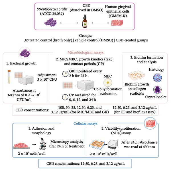
The experimental design included evaluating CBD against S. oralis planktonic growth and biofilm formation as well as assessing its effects on human gingival epithelial cell viability and proliferation. The experimental groups included untreated control, vehicle control, and CBD‐treated groups at different concentrations. The main analyses included bacterial growth inhibition, biofilm formation assessment, histological evaluation, and cell viability analysis. A schematic representation of the study design is presented in Figure 1.
2.2. Outcomes
The primary outcome of this study was the antimicrobial effect of CBD against S. oralis, assessed by measuring planktonic bacterial growth inhibition and determining the minimum inhibitory concentration (MIC) after 24 h of exposure. Bacterial growth was monitored kinetically over 24 h by measuring the optical density (OD) at 600 nm and was additionally assessed at predefined time points.
The secondary outcomes included an evaluation of bactericidal activity, assessed by determining the minimum bactericidal concentration (MBC) after 24 h of CBD exposure, followed by subculture onto brain–heart infusion (BHI) agar. We also evaluated the antibiofilm activity after 3 days of CBD exposure using crystal violet (CV) staining for biofilm biomass quantification and histological analysis for biofilm architecture. Finally, the effect of CBD on human gingival epithelial cell viability and proliferation was assessed after 24 h using cell morphology and adhesion assays and an MTS‐based cell viability/proliferation assay.
2.3. CBD
The CBD used in this study was a pure (purity ≥ 98%) CBD extracted from an isolate of Cannabis sativa [ref. 18]. The CBD powder was dissolved in dimethyl sulfoxide (DMSO) to form a 5 mg/mL stock solution. From this stock, serial dilutions were performed to obtain the desired treatment concentrations. In all assays, a vehicle referring to the same DMSO level as in the highest CBD concentration was included to assess possible solvent effects on bacterial growth.
2.4. Bacterial Strain and Growth Conditions
Growth conditions for S. oralis in this study were adapted from previously published methods for S. mutans [ref. 19]. The S. oralis strains used in this study (ATCC 35,037) were reactivated and grown aerobically at 37°C in a BHI medium supplemented with 0.1% glucose. S. oralis cultures were incubated overnight, collected, and subsequently adjusted to a concentration of 107 CFU/mL based on OD measured at 600 nm using a xMark microplate spectrophotometer (Bio‐Rad, Mississauga, ON, Canada). To convert absorbance to CFU, we used the following formula: Absorbance at 600 nm of 0.2 corresponds to 108 CFU/mL.
2.5. To Evaluate the Effect of CBD on S. oralis Growth Over Time
S. oralis (5 × 10⁴ CFU) was seeded in triplicate in the wells of a 96‐well plate in 200 µL of BHI in the presence of CBD at concentrations ranging from 100 to 3.12 µg/mL. The vehicle control consisted of S. oralis cultured in BHI containing the highest volume of DMSO, at the highest final concentration in the CBD‐treated groups. Eight wells per condition were included. The plates were incubated at 37°C in a Synergy microplate reader (BioTek, Winooski, VT, USA), and growth was monitored every 2 h for 24 h by measuring the absorbance at 600 nm. Four biological replicates were made (n = 4).
2.6. To Evaluate the Growth of S. oralis at Different Contact Periods With CBD
S. oralis (5 × 10⁴ CFU) was seeded in triplicate in the wells of a 96‐well plate in 200 µL of BHI in the presence of CBD at concentrations ranging from 100 to 3.12 µg/mL. Vehicle control consisted of S. oralis being cultured in BHI containing the highest volume of DMSO as the highest final concentration of CBD in the CBD‐treated groups. Plates were incubated for 0, 6, 12, and 24 h at 37°C in a humid atmosphere with 5% CO2. After each incubation period, absorbance was measured at 600 nm using the xMark microplate spectrophotometer (Bio‐Rad, Mississauga, ON, Canada). Results are presented as the mean ± standard deviation (SD) of four biological replicates (n = 4). These experiments allowed the determination of the MIC of CBD. The MIC was defined as the lowest CBD concentration capable of inhibiting detectable bacterial growth after 24 h, as determined by OD measurements [ref. 20].
2.7. Identification of the MBC of CBD
S. oralis (5 × 10⁴ CFU) was seeded in triplicate in the wells of a 96‐well plate in 200 µL of BHI in the presence of CBD at concentrations ranging from 100 to 3.12 µg/mL. Plates were incubated for 24 h at 37°C in a humid atmosphere with 5% CO2. After this incubation period, 5 µL aliquots from each cell suspension of S. oralis, exposed or not to CBD for 24 h, were placed as a drop on the surface of BHI agar and incubated for an additional 24 h under the same growth conditions described above. Macroscopic observations were made to evaluate the absence of bacterial growth (no colony formation). The plates were then scanned and presented, showing the lowest CBD concentration capable of eliminating visible bacterial growth, as indicated by the CBD‐MBC [ref. 20]. Four biological replicates were performed (n = 4).
2.8. Effect of CBD on S. oralis Biofilm Formation
Biofilms of S. oralis were obtained using porous collagen scaffolds (Collatape, Zimmer Dental Inc., Carlsbad, CA, USA), which promote bacterial penetration and adhesion due to the bacterium’s affinity for collagen [ref. 19]. Use of these scaffolds also enables biofilm manipulation with minimal cell loss, thereby preserving its three‐dimensional structure. Biofilm formation was performed using 5 mm × 5 mm samples of the porous scaffolds placed in the wells of a 24‐well plate. The three‐dimensional porous collagen membranes (scaffold) were inoculated with 50 µL of S. oralis suspension (5 × 10⁴ CFU) in triplicate. The plates were incubated for 1 h at 37°C, allowing for initial bacterial adhesion to the scaffold. At this time, S. oralis‐seeded scaffolds were exposed to one of the following CBD concentrations (12.50, 6.25, or 3.12 µg) in 150 µL of BHI medium in triplicate. After 1 h of incubation, to ensure penetration of the lipophilic compound into the collagen membrane and into the adherent bacteria, 950 µL of fresh BHI medium was added to each well, with CBD used at the following concentrations (12.50, 6.25, and 3.12 µg/mL). Negative controls (biofilms without CBD; only DMSO) and positive controls (biofilms treated with 5 µg/mL penicillin/streptomycin) were included in the experiments. Cultures were maintained at 37°C for 3 days, with renewal of the medium and CBD treatment every 24 h. Three biological replicates were performed.
At the end of the third day, collagen membranes were washed three times with phosphate‐buffered saline solution (PBS) and fixed with 4% paraformaldehyde for 24 h, as previously reported [ref. 21]. Samples were then subjected to qualitative (histological) and quantitative (CV staining) analyses.
2.9. Histological Analysis
Histological processing and Masson’s trichrome staining were performed following standard protocols described in the literature [ref. 22–ref. 24]. Briefly, after fixation in paraformaldehyde, biofilms were washed with PBS, dehydrated through a graded ethanol series, cleared in xylene, and embedded in paraffin. Thin sections (5–7 µm) were cut with a microtome and mounted on glass slides. Sections were rehydrated, post‐fixed in Bouin solution, and stained using Weigert’s iron hematoxylin, Biebrich scarlet/acid fuchsin, a phosphotungstic/phosphomolybdic acid solution, and aniline blue, followed by differentiation with 1% acetic acid and graded alcohol dehydration [ref. 23]. After final dehydration and clearing, slides were mounted and analyzed with a high‐resolution digital microscope (Echo Laboratories– Bio‐Rad, San Diego, CA, USA). The system is equipped with high‐resolution optics and an integrated digital camera, both controlled by the proprietary Revolve 4.0 software (Echo Laboratories). Using the software, real‐time adjustments of focus, contrast, brightness, and magnification were performed to ensure the image quality. For each biofilm condition, four glass slides were prepared, each containing two sections. After staining, 10 images were captured per condition, covering distinct and representative areas. Images demonstrating structural definition and histological representativeness were selected for biofilm structural analysis. To minimize assessment bias, histological images were coded before analysis, and biofilm structural evaluation was performed by an investigator blinded to the experimental groups. Group allocation was revealed only after image selection and qualitative assessment were completed.
2.10. Evaluate Biofilm Formation Using the CV Assay
Biofilm biomass was quantified using the CV assay, which stains the biofilm (live cells and the extracellular matrix they secrete), as previously reported [ref. 21]. Briefly, biofilm samples were transferred to a sterile six‐well plate and left to dry for 24 h under a chemical flow hood. They were then layered with 500 µL of 0.1% CV solution and incubated for 15 min at room temperature. S. oralis‐free collagen scaffolds were included as a control. After CV staining, the collagen membranes were thoroughly washed with distilled water until the unbound dye was completely removed and then left to dry for 24 h under a chemical hood. Subsequently, 2.5 mL of 30% acetic acid was added to each well, and the samples were gently agitated to dissolve the bound CV dye. The obtained solutions were transferred (200 µL/well) to the wells of a 96‐well plate in quadruplicate, and the absorbance was measured at 595 nm using the xMark microplate spectrophotometer.
2.11. Evaluate the Interaction of CBD With Human Gingival Epithelial Cells
2.11.1. Cells and Cell Culture Conditions
The gingival epithelial cells used in this study correspond to the non‐cancerous human gingival epithelial cell line GMSM‐K (CVCL_6A82) [ref. 25]. Gingival epithelial cells were cultured in Dulbecco’s Modified Eagle Medium/Ham’s F‐12 (DMEM), supplemented with 10% fetal bovine serum (FBS). The culture medium was renewed three times per week. When cultures reached ~80% confluence, cells were detached using 0.25% trypsin, washed, and resuspended in DMEM supplemented with FBS [ref. 26, ref. 27] and used for our experiments.
2.11.2. Effects of CBD on Gingival Epithelial Cell Adhesion and Cell Morphology
Gingival epithelial cells (2 × 10⁵ cells/well in 1 mL), in triplicate, were seeded in 6‐well tissue culture plates and immediately exposed to CBD at one of the following concentrations (3.12, 6.25, and 12.50 µg/mL), along with a vehicle control (DMSO) and a negative control referring to cells cultured in CBD and DMSO‐free medium. The highest concentration of DMSO in the treated samples was 0.25% (v/v), which, in our studies, was shown to be noncytotoxic to the cells. Cells were cultured in the presence or absence of CBD for 24 h at 37°C in a humidified atmosphere containing 5% CO2. After the incubation period, adherent and nonadherent cells in each well were examined using an inverted light microscope (10x objective) (Nikon Canada, Mississauga, ON, Canada). Photos from experimental conditions (control and CBD‐treated cells) were captured using a Coolpix 950 camera (Nikon Canada).
Cell morphology was evaluated after CV staining of adherent cells in each culture condition (CBD‐exposed and non‐exposed). Briefly, wells were washed with PBS, and then cells were fixed with 4% paraformaldehyde overnight, washed with PBS, and stained with CV, as described above [ref. 21–ref. 23]. The adherent CV‐stained cells were photographed. Four biological replicates were performed (n = 4).
2.11.3. Effects of CBD on Cell Viability and Proliferation
Gingival epithelial cells were seeded at a density of 2 × 10⁴ cells/well in 6‐well plates and incubated for 24 h in a humidified atmosphere containing 5% CO2 at 37°C. After this culture period, the medium was refreshed, and adherent cells were exposed or not to CBD at 3.12, 6.25, and 12.50 µg/mL. As controls, cells were cultured in the presence of 0.25% DMSO (v/v) or in the culture medium. All culture conditions (cells exposed or not to CBD) were incubated in a humidified atmosphere containing 5 % CO2 at 37°C for 24 h. After this period, the medium was refreshed, and cell viability/proliferation was assessed using an MTS‐colorimetric assay (ab197010, Abcam Inc., Toronto, ON, Canada), as previously reported [ref. 27]. Briefly, a volume of 100 µL/mL of CellTiter‐96 Aqueous One Solution reagent was added to each well, and cultures were incubated for 3 h at 37°C in 5% CO2. At the end of the incubation period, 150 µL of the reaction mixture was transferred to a 96‐well plate, and the absorbance was measured at 490 nm using a spectrophotometer. Five biological replicates were performed (n = 5); results were expressed as mean (SD).
2.12. Statistical Analyses
The number of biological replicates was based on previous in vitro studies using similar microbiological and cell viability assays and on experimental feasibility [ref. 18, ref. 19, ref. 21]. No formal a priori sample size calculation was performed. Random allocation and allocation concealment were not applicable because this was an in vitro assay using predefined experimental conditions. When possible, samples and images were analyzed by an investigator independent of the experimental procedures.
The statistical analyses were applied to outcomes related to planktonic growth inhibition (bacteriostatic and bactericidal activities), biofilm biomass, and cell viability. Prior to ANOVA, data distribution and homogeneity of variance were assessed using the Shapiro–Wilk test and the Brown–Forsythe test, respectively. MIC, antibiofilm assays, and cell viability were analyzed by one‐way ANOVA, followed by Dunnett’s multiple comparisons test versus DMSO. The growth kinetics assay was analyzed by two‐way repeated‐measures ANOVA (factors: CBD concentration and time), followed by post hoc multiple comparisons using Dunnett’s test to compare each CBD‐treated condition with the control and, when applicable, Tukey’s test for pairwise comparisons among CBD concentrations. The data were recorded as the mean ± SD. Statistical significance was set at p < 0.05. All analyses were performed in GraphPad Prism version 10 (GraphPad Software, San Diego, CA, USA).
3. Results
3.1. Long‐Term Exposure to CBD Reduced S. oralis Growth
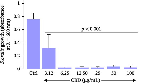
Exposure of S. oralis to CBD for 24 h resulted in a significant decrease in bacterial growth (Figure 2). Indeed, the vehicle (DMSO) used to dissolve CBD did not affect bacterial growth (absorbance = 0.75 ± 0.10). However, all tested CBD concentrations showed growth inhibition compared to the DMSO control ( ∗∗∗ p < 0.0001). The CBD decreased S. oralis growth, even at 3.12 µg/mL, as the absorbance was 0.32 ± 0.27 after 24 h of incubation, compared to that of DMSO (0.75 ± 0.10). However, despite this statistically significant reduction, 3.12 µg/mL did not meet the MIC criterion as detectable bacterial growth persisted after 24 h. Therefore, 6.25 µg/mL, which showed an absorbance of 0.02 ± 0.02, was determined as the MIC. Given this interesting inhibitory effect of CBD on S. oralis growth at 24 h of contact, we evaluate whether this effect occurs at earlier time points.
3.2. Kinetic Inhibition of S. oralis Growth by CBD
The bacteria were cultured in the absence or presence of CBD at different concentrations, with a follow‐up every 2 h. It was observed (Figure 3) that all tested CBD concentrations maintained the S. oralis growth at its lowest rate up to 16 h. At 18 h post contact, only the lowest concentration (3.12 μg/mL) showed growth of S. oralis, which continued increasing with subsequent culture periods (at 20, 22, and 24 h). Interestingly, the statistical analysis showed a significant (p < 0.0001) effect of the CBD on S. oralis growth at all tested times and with all included CBD concentrations above 3.12 μg/mL.
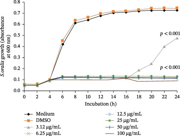
To evaluate the specific efficacy of each chosen CBD concentration relative to the control over the entire 24‐h period, a focused analysis was conducted. Derived from the overarching two‐way ANOVA model, this approach directly compared each treatment group with DMSO at all time points, allowing us to pinpoint significant differences across growth stages. As shown in Figure 3, the 6.25 and 12.50 µg/mL displayed nearly identical kinetic profiles to each other and, importantly, showed statistically significant suppression compared to the DMSO at all tested timepoints, confirming a stable and consistent antimicrobial effect throughout the 24 h. In contrast, the behavior of the 3.12 µg/mL concentration was distinct. While it initially showed significant inhibitory effects compared to the control, this effect was not sustained over time, starting at 16 h post‐exposure when S. oralis began to grow.
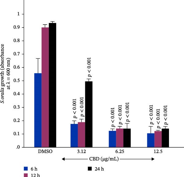
To confirm the inhibitory effect of CBD on bacterial growth, we selected specific exposure times (6, 12, and 24 h). We monitored S. oralis growth after exposure to selected CBD concentrations (3.12, 6.25, and 12.5 µg/mL). The obtained results were subjected to one‐way ANOVA analysis and are presented in Figure 4. It should be noted that even the low concentration (3.12 µg/mL) had significant bacterial growth inhibition at early timepoints (6 and 12 h) but lost this effect later (up to 24 h), while the other concentrations remained efficient in inhibiting the S. oralis growth. Furthermore, considering the effect of CBD on S. oralis growth (Figures 2–4), 6.25 µg/mL was determined as the MIC. With these results, we asked: What would the MBC of CBD be?
3.3. MBC
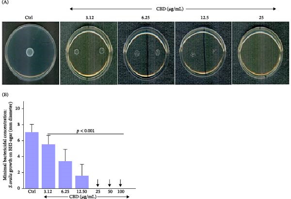
To determine the MBC, we subcultured the bacteria following 24 h of preexposure to CBD using BHI‐Agar plates. Figure 5A shows higher bacterial growth in the control (DMSO), while cells exposed to CBD had a lower growth rate. Indeed, at 3.12 µg/mL CBD, S. oralis showed a smaller bacterial halo than that of the control. It can also be noted that the bacterial growth halo decreases with increased concentrations of CBD. At 12.50 µg/mL, only discrete, isolated colonies are still observable, whereas at 25 µg/mL CBD, no bacterial growth halo was observed. Quantitative measurements of the halo diameter at each CBD concentration confirmed (Figure 5B) the decrease in S. oralis growth due to CBD. In this set of experiments, we demonstrated that 25 µg/mL CBD was the MBC for this product.
3.4. CBD Inhibits S. oralis Biofilm Formation
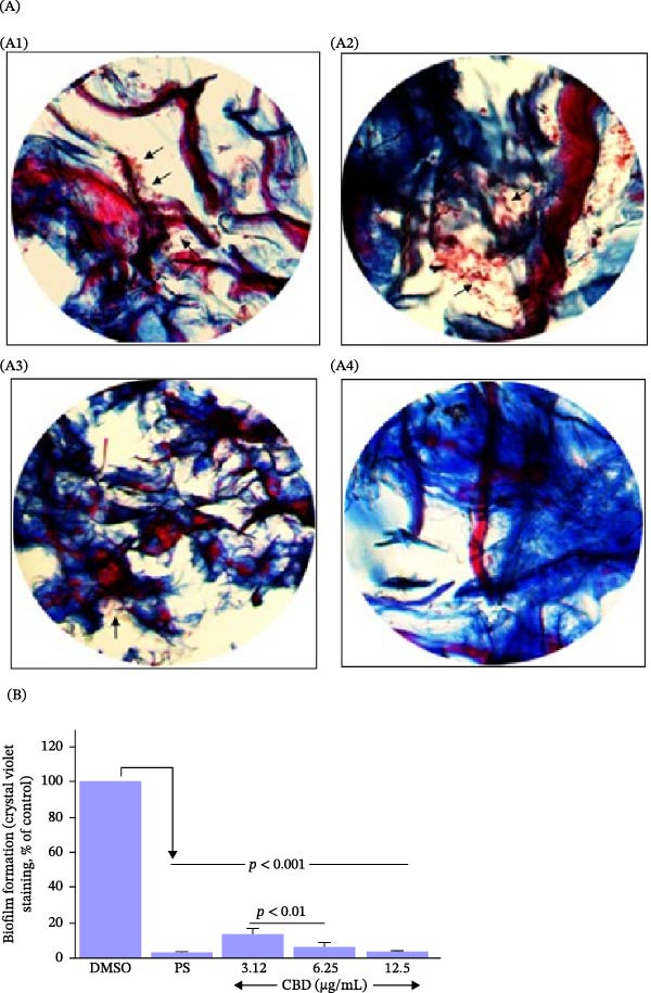
The effect of CBD on S. oralis biofilm formation was evaluated by histological and quantitative assays. Histological analysis (Figure 6A) revealed frequent red‐brown clumps of bacteria within the scaffold in the control group (arrows, Figure 6). Bacterial aggregates were also observable in the 3.12 μg/mL CBD group (arrows, Figure 6A, A2), but they were reduced in the 6.25 μg/mL group (arrows, Figure 6A) and markedly decreased in the 12.5 μg/mL group (Figure 6A, A4). To support these histological observations, biofilm biomass was quantified using CV staining. As shown in Figure 6B, varying concentrations of CBD led to a significant decrease in the S. oralis biofilm formation. Although biofilm formation was still observable at 3.12 (g/mL), in the 6.25 and 12.50 (g/mL) CBD concentrations, we observed a more pronounced reduction in biofilm formation. Interestingly, the effect of CBD on biofilm formation was not statistically different from that observed with the antibiotic (Figure 6B). Overall, histological findings corroborated the CV staining results, demonstrating the effectiveness of CBD in reducing S. oralis biofilm formation.
3.5. Gingival Epithelial Cells Exposed to CBD Showed Shapes and Adhesion Comparable to Those of the Control
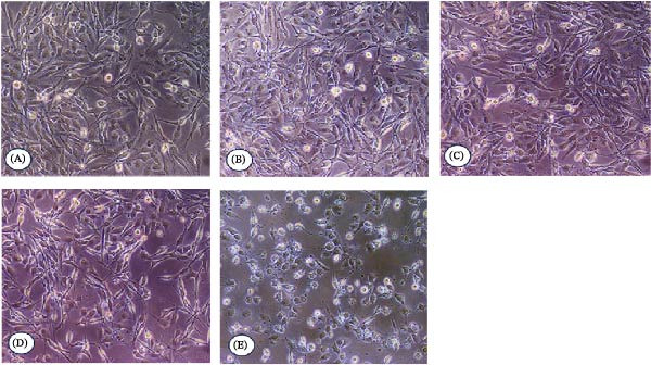
Gingival epithelial cells were cultured for 24 h in the presence of CBD, and the cell morphology and adhesion were evaluated by optical microscopy. Cells exposed to vehicle (DMSO) (Figure 7B) exhibited high adhesion density, with an elongated morphology comparable to the control (cells cultured in medium without CBD or DMSO) (Figure 7A). Similarly, cells exposed to 3.12 µg/mL (Figure 7C) and 6.25 µg/mL (Figure 7D) of CBD showed morphologies and adhesion patterns comparable to those of the control groups (medium alone or with DMSO). Exposure to 12.50 µg/mL showed (Figure 7E) a slight difference in cell morphology as cells did not adopt the elongated shape observed in the control groups. However, based on our qualitative analysis, cell adhesion appeared to be maintained, with similar cell densities adhering to the culture plates (Figure 7E). These findings suggest that CBD concentrations up to 12.5 µg/mL did not markedly alter gingival epithelial cell adhesion or morphology.
3.6. CBD did not Reduce Human Gingival Epithelial Cell Viability/Proliferation
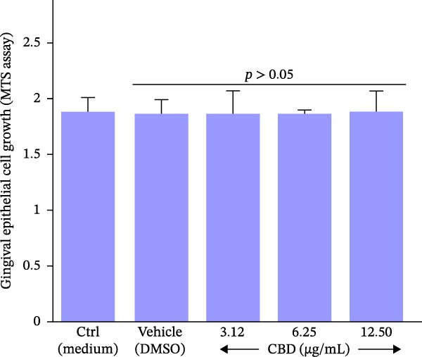
To further assess the compatibility of CBD with human gingival epithelial cells, cell viability/proliferation in CBD‐treated cells was evaluated using the MTS assay. As shown in Figure 8, no significant decrease in gingival epithelial cell viability was observed after 24 h of exposure to CBD. Interestingly, regardless of the CBD concentration (3.12, 6.25, or 12.50 µg/mL), cell viability was the same as that obtained with the control (medium alone) and the vehicle (DMSO). These results demonstrated that CBD concentrations up to 12.5 µg/mL did not reduce cell viability. Notably, this concentration range also showed inhibitory effects against S. oralis growth and biofilm formation.
4. Discussion
This in vitro study demonstrates, for the first time, that CBD affects S. oralis in two physiological states. CBD inhibited planktonic growth in a concentration‐dependent manner and reduced biofilm biomass, while histological analyses revealed a progressively disrupted biofilm architecture following CBD exposure. Given that oral antimicrobials must be effective against biofilm‐associated bacteria while maintaining compatibility with host tissues, we also evaluated gingival epithelial cell responses at the same concentration range. Although the antimicrobial and anti‐inflammatory potential of CBD in oral infections has recently been reviewed [ref. 12–ref. 14], the present findings add a specific experimental contribution by focusing on S. oralis, an early colonizer involved in the initial organization of oral biofilms. This distinction is important because most available studies have focused on overtly pathogenic species or mature disease‐associated biofilms.
In contrast, modulation of early colonizers may represent a preventive strategy for controlling biofilm development. [ref. 14, ref. 15, ref. 28]. Therefore, investigating the effects of CBD on S. oralis, a key early colonizer of the dental biofilm, is highly relevant for understanding its potential to modulate biofilm development and contribute to the control of biofilm‐associated diseases.
Natural plant‐derived compounds have attracted attention as alternative antibacterial agents, with CBD, the main non‐psychoactive cannabinoid of Cannabis sativa, standing out for its diverse biological activities [ref. 29, ref. 30]. Recent studies have demonstrated its antimicrobial potential against Gram‐positive bacteria, including highly resistant species such as Staphylococcus aureus, Streptococcus pneumoniae, and Clostridioides difficile [ref. 14, ref. 29, ref. 31].
In the oral environment, biofilms pose a clinical challenge due to their intrinsic resistance to antimicrobial agents, underscoring the need to investigate new compounds such as CBD to control bacterial colonization and biofilm formation. The antimicrobial effect of CBD appears to involve damage to the bacterial membrane, inhibition of protein, DNA, RNA, and peptidoglycan synthesis, as well as disruption of the plasma membrane of Gram‐positive bacteria, inducing membrane hyperpolarization and impairing ion channel function [ref. 15, ref. 30].
In our study, MIC and MBC analyses revealed that CBD is effective at relatively low concentrations against S. oralis, with an MIC of 6.25 µg/mL (Figures 2–4) and bactericidal activity observed at 25 µg/mL (Figure 5), demonstrating antimicrobial activity in the planktonic state. These findings are consistent with the broader literature, which shows that CBD can exert antimicrobial effects against various bacterial species. However, the concentrations required for inhibition vary considerably depending on the microorganism and experimental conditions. Blaskovich et al. [ref. 30] reported lower MIC values for several Gram‐positive bacteria (1–4 µg/mL) and selected Gram‐negative species (0.25–2 µg/mL), suggesting that S. oralis may be comparatively less susceptible to CBD under the conditions used in the present study. A systematic review focused on Streptococcus and Staphylococcus reported that CBD MICs span a wide range depending on the organism and study design, including values up to 50 µg/mL for Streptococcus pyogenes, suggesting that susceptibility may vary substantially across streptococcal species and experimental settings [ref. 31]. In the oral context, MIC/MBC values in the µg/mL range have also been reported for other streptococci. Recent studies have shown that CBD exhibits antibacterial and antibiofilm activity against oral bacteria, including cannabinoid‐sensitive streptococcal species and periodontopathogens. However, effective concentrations vary according to bacterial species, strain, biofilm model, and assay conditions [ref. 32–ref. 34].
Growth kinetics analysis further showed a concentration‐ and time‐dependent effect: while 3.12 µg/mL did not sustain complete inhibition after 24 h, it delayed bacterial proliferation by ~16 h. This temporal inhibition suggests that sub‐MIC concentrations of CBD may be biologically relevant in the context of oral biofilm development. Sub‐MIC levels of CBD in S. mutans delay growth phases, whereas MIC‐level concentrations sustain growth inhibition without bacterial recovery [ref. 14]. A similar time‐dependent pharmacodynamic profile has been reported for CBD against gram‐positive bacteria in CFU‐based time‐kill assays. Blaskovich et al. [ref. 30] showed that CBD exhibited rapid antibacterial activity against S. aureus within 3 h, with an MBC of 2 µg/mL, and time‐kill curves indicated concentration‐dependent bacterial killing. Importantly, regrowth after initial suppression has also been described in cannabinoid time‐kill settings, including re‐initiation of growth after 8 h, attributed to possible degradation or oxidation of CBD [ref. 35, ref. 36], which supports the interpretation that delayed outgrowth at sub‐MIC can occur even when early inhibition is evident. Together, these results indicate that CBD interferes with bacterial growth dynamics, particularly at lower concentrations, rather than acting solely as an immediate bactericidal agent.
The antibiofilm activity of CBD against S. oralis was demonstrated through both quantitative and structural analyses. CV staining showed a significant reduction in biofilm biomass at all tested CBD concentrations (3.12–12.50 µg/mL), with more pronounced effects at higher concentrations, comparable to those of the control. These results suggest that CBD can be used at concentrations below 12.5 µg/mL to control S. oralis growth for up to 24 h efficiently. The CV assay is widely used for quantitative biofilm assessment, as illustrated by Wieczerza et al. [ref. 37], who used this method to evaluate the effects of cannabinoids on biofilms formed by bacteria associated with endodontic infections. Because CV staining primarily quantifies total biofilm biomass (cells and secreted extracellular matrices) rather than directly assessing bacterial viability, combining this approach with structural imaging strengthens data interpretation. This rationale is supported by previous studies investigating CBD activity against biofilms, in which CV–based minimum biofilm eradication concentration (MBEC)/minimum biofilm inhibitory concentration (MBIC) thresholds (≥70% inhibition relative to the growth control) are used as quantitative proxies for biofilm burden [ref. 29].
Histological analyses of biofilm sections formed on collagen membranes corroborated these findings, revealing a disrupted biofilm architecture and minimal residual bacterial clusters at the highest CBD concentrations. These observations are consistent with those reported by Barak et al. [ref. 14], who showed that CBD interferes with S. mutans biofilm formation, resulting in sparse, poorly organized biofilm structures, as visualized by confocal microscopy. Together, these convergent qualitative patterns support the antibiofilm activity of CBD against oral streptococci and reinforce that CBD compromises biofilm integrity beyond simple biomass reduction. This biomass and structure approach is consistent with prior CBD antibiofilm evidence in Gram‐positive biofilms: CBD was active against established S. aureus biofilms with MBEC ranging from 1 to 2 µg/mL for Methicillin-Susceptible Staphylococcus aureus (MSSA) and from 2 to 4 µg/mL for methicillin‐resistant Staphylococcus aureus (MRSA), and confocal microscopy demonstrated CBD penetration and killing within the biofilm [ref. 30]. It has also been reported that higher CBD concentrations were required to achieve >90% killing (32 µg/mL) than those suggested by CV MBEC values, emphasizing that biomass‐based assays and viability‐oriented imaging can yield different “effective concentrations” due to differences in assay conditions and readouts [ref. 30]. Mechanistically, this is compatible with the broader view that CBD’s lipophilic structure enables membrane interaction, promoting membrane disruption and increased permeability, which can plausibly impact both planktonic cells and biofilm organization [ref. 4]. In streptococcal biofilms specifically, independent antibiofilm literature supports the concept that biofilm architecture and matrix act as barriers to antimicrobial activity and that disrupting matrix components can markedly increase susceptibility, reinforcing why architectural disruption is a meaningful endpoint beyond biomass reduction [ref. 38].
From a translational perspective, the incorporation of CBD into oral formulations has been proposed for mouthrinses, gels, lozenges, and local delivery systems [ref. 15]. The study by Torabi et al. [ref. 39] demonstrated that the use of oral lozenges containing CBD resulted in a significant reduction in the abundance of S. mutans in saliva, suggesting that the effects observed in experimental models may translate into benefits within the complex oral environment. Although the specific mechanisms against S. oralis were not fully elucidated here, the literature suggests that CBD acts by disrupting the cell membrane, reducing extracellular polysaccharides [ref. 14], and inducing cell envelope stress [ref. 30]. This combination of actions explains the double effect observed: the inhibition of growth and the structural disruption of the biofilm, positioning CBD as a modulator of biofilm development rather than a classic bactericidal agent. Notably, cannabinoid‐infused mouthwashes containing CBD or CBG have demonstrated bactericidal efficacy comparable to 0.2% chlorhexidine, the gold standard mouthwash in dentistry, but without the considerable side effects commonly associated with chlorhexidine use [ref. 10, ref. 11, ref. 31]. These findings support the potential of cannabinoids as promising alternatives for oral care applications.
Furthermore, synergistic effects have been reported between CBD and other antimicrobial agents. Avraham et al. [ref. 40] demonstrated that the combination of triclosan and CBD exerted stronger antibacterial and antibiofilm effects than either compound alone. Both agents were shown to induce membrane hyperpolarization and reduce bacterial viability and adhesion. Such combinatorial approaches may be particularly valuable for preventing dental caries and controlling oral inflammation.
The effects of CBD on human gingival epithelial cells were also evaluated for adhesion, morphology, and viability. CBD concentrations up to 12.50 µg/mL were selected for these assays because they corresponded to the concentration range evaluated in the biofilm formation assays, which may be more relevant to the oral environment than the bactericidal concentration determined in the planktonic model. It was observed that at ≤12.50 µg/mL, CBD did not significantly reduce cell adhesion, alter cell morphology, or decrease cell viability after 24 h of exposure. These findings are partially consistent with a recent study by Bahraminia et al. [ref. 18], which investigated the effects of THC and CBD on gingival and skin keratinocytes and found that lower CBD concentrations did not impair cell metabolism, whereas concentrations ≥10 µg/mL were associated with reduced cell viability. In contrast, lower concentrations did not impair the cell metabolism. Since the concentration range evaluated in the present study overlaps with this threshold, the apparent difference between studies should be interpreted cautiously. Such differences may be related to the cell type, exposure conditions, assay methodology, and the concentration range evaluated.
Although the present study demonstrated that CBD inhibited S. oralis growth and biofilm formation under controlled laboratory conditions, these findings should be interpreted within the limitations of an in vitro model. The experiments in this study used a single‐species biofilm model with a single S. oralis strain, which does not fully reflect the strain‐dependent variability in antimicrobial susceptibility. In addition, this model does not reproduce the complexity of the oral cavity, including saliva, multispecies biofilms, host immune responses, tissue interactions, and systemic patient‐related factors. Therefore, further investigations should evaluate the effects of CBD in multispecies biofilm models and in additional oral bacterial species. Moreover, the cellular assays were performed in a single gingival epithelial cell line under in vitro conditions, which may not fully reproduce an in vivo microenvironment. Future ex vivo, in vivo, and clinical studies are necessary to confirm the translational potential of CBD as an antimicrobial agent in oral biofilm management. Despite these limitations, the present findings provide a relevant preliminary foundation for further investigations into the antimicrobial and antibiofilm properties of CBD in oral health contexts.
5. Conclusion
This study demonstrated the effectiveness of CBD at different concentrations in inhibiting the growth of S. oralis at both early and late exposure periods. CBD also had a significant effect on biofilm formation by S. oralis. Low and medium concentrations of CBD, which have antimicrobial effects, did not reduce the viability of human gingival epithelial cells. However, these findings should be interpreted with the limitations of an in vitro model in mind. Overall, this study provides preliminary evidence supporting further investigation of CBD as a potential antimicrobial molecule against S. oralis. Further studies are required to determine its safety, optimal concentration, delivery method, and clinical relevance for oral health applications.
Author Contributions
Celina Cruz Mainardes: conceptualization, methodology, validation, formal analysis, investigation, data curation, writing – original draft, writing – review & editing. Omayma Amri: methodology, validation, investigation, data curation. Denise M. Palomari Spolidorio: writing – review & editing, supervision, project administration. Mahmoud Rouabhia: conceptualization, methodology, validation, formal analysis, investigation, data curation, funding acquisition, writing – original draft, writing – review & editing, supervision, project administration.
Funding
This research was funded by the Natural Sciences and Engineering Research Council of Canada (NSERC) through the NSERC‐Discovery grant to MR (RGPIN‐2019‐04475). Celina Cruz Mainardes benefited from a scholarship from the São Paulo Research Foundation (FAPESP), Grant #2025/04500‐5. NSERC and FAPESP had no role in the study design, data collection and analysis, manuscript preparation, or the decision to publish.
Disclosure
No formal study protocol was registered or publicly available. All authors have read and approved the final version of the manuscript. The authors had full access to all data in this study and took full responsibility for the integrity and accuracy of the data analysis.
Conflicts of Interest
The authors declare no conflicts of interest.
References
- Influence of E-Liquids and Oral Commensal Bacteria on the Growth of Porphyromonas gingivalis Planktonically and in Biofilms,. Dentistry Journal. (, 2026. [DOI | PubMed]
- Biofilm and Antimicrobial Resistance: Mechanisms, Implications, and Emerging Solutions,. Microbiology Research. (, 2025. [DOI]
- A Review on Antimicrobial Strategies in Mitigating Biofilm-Associated Infections on Medical Implants,. Current Research in Microbial Sciences. (, 2024. [DOI | PubMed]
- Bidirectional Effects of Neutrophils on Streptococcus oralis Biofilms in vitro ,. Journal of Oral Microbiology. (, 2025. [DOI | PubMed]
- Oral Streptococci: Modulators of Health and Disease,. Frontiers in Cellular and Infection Microbiology. (, 2024. [DOI | PubMed]
- Characterization of MdpS: An in Depth Analysis of a MUC5B Degrading Protease from, Streptococcus oralis ,. Frontiers in Microbiology. (, 2024. [DOI | PubMed]
- In Vitro Effects of Streptococcus oralis Biofilm on Peri-Implant Soft Tissue Cells,. Cells. (, 2020. [DOI | PubMed]
- Biofilms and Oral Health: Nanotechnology for Biofilm Control,. Discover Nano. (, 2025. [DOI | PubMed]
- Streptococcus oralis Activates the Efg1 Filamentation Pathway in Candida albicans to Promote Cross-Kingdom Interactions and Mucosal Biofilms,. Virulence. (, 2017. [DOI | PubMed]
- Efficacy of Preprocedural Mouth Rinses (Chlorhexidine, Essential Oil, and Hydrogen Peroxide) in Reducing Bacterial Aerosols During Dental Scaling,. BMC Oral Health. (, 2025. [DOI | PubMed]
- Comparison of the Anti-Plaque and Anti-Gingivitis Efficacy of Chlorhexidine and Malva Mouthwashes: Randomized Crossover Clinical Trial,. Journal of Dentistry. (, 2024. [DOI | PubMed]
- Cannabidiol in Dentistry: A Scoping Review,. Dentistry Journal. (, 2022. [DOI | PubMed]
- Phytocannabinoids and Gingival Inflammation: Preclinical Findings and a Placebo-Controlled Double-Blind Randomized Clinical Trial With Cannabidiol,. Journal of Periodontal Research. (, 2024. [DOI | PubMed]
- Anti-Bacterial Effect of Cannabidiol against the Cariogenic streptococcus mutans Bacterium: An In Vitro Study,. International Journal of Molecular Sciences. (, 2022. [DOI | PubMed]
- The Potential of Cannabidiol in the Management of Oral Infections,. Applied Sciences. (, 2025. [DOI]
- The Role and Mechanisms of Gingival Epithelial Cells in Maintaining Periodontal Homeostasis,. Journal of Prevention and Treatment for Stomatological Diseases. (, 2025
- A Review of Cell Adhesion Studies for Biomedical and Biological Applications,. International Journal of Molecular Sciences. (, 2015. [DOI | PubMed]
- Evaluation of the Effects of Tetrahydrocannabinol (THC) and Cannabidiol (CBD) on Gingival and Skin Keratinocyte Growth, Migration, Metabolic Activity, and pro-Inflammatory Cytokine Secretion,. Biomedicines. (, 2025. [DOI | PubMed]
- Electronic Cigarette Vapor Increases Streptococcus mutans Growth, Adhesion, Biofilm Formation, and Expression of the Biofilm-Associated Genes,. Oral Diseases. (, 2021. [DOI | PubMed]
- Methods for In Vitro Evaluating Antimicrobial Activity: A Review,. Journal of Pharmaceutical Analysis. (, 2016. [DOI | PubMed]
- In Vitro Exposure to Vaped Tetrahydrocannabinol Increases Candida albicans (sc5314) Growth, Metabolic Activity, Biofilm Formation, and the Expression of Virulence Genes,. Microorganisms. (, 2025. [DOI | PubMed]
- Electrically Activated Primary Human Fibroblasts Improve In Vitro and In Vivo Skin Regeneration,. Journal of Cellular Physiology. (, 2016. [DOI | PubMed]
- Effect of e-Cigarettes on Nasal Epithelial Cell Growth, Ki-67 Expression, and Pro-Inflammatory Cytokine Secretion,. American Journal of Otolaryngology. (, 2020. [DOI | PubMed]
- Comparative Analysis of Three Staining Techniques for Assessing Liver Fibrosis in Biopsy Samples: A Referral University Hospital,. Biomedical and Biotechnology Research Journal. (, 2025. [DOI]
- Establishment of a Human Polyclonal Oral Epithelial Cell Line. Oral Surg Oral Med Oral Pathol Oral Radiol Endod,. 2000
- Cannabis Smoke Condensate Induces Human Gingival Epithelial Cell Damage through Apoptosis, Autophagy, and Oxidative Stress,. Archives of Oral Biology. (, 2022. [DOI | PubMed]
- Effect of Cannabis Smoke Condensate on Human Nasal Epithelial Cell Adhesion, Growth, and Migration,. American Journal of Otolaryngology. (, 2023. [DOI | PubMed]
- Bacterial Interactions in Dental Biofilm Development,. Journal of Dental Research. (, 2009. [DOI | PubMed]
- Efficacy of Cannabidiol in Reducing Virulence of Listeria monocytogenes ,. International Journal of Molecular Sciences. (, 2026. [DOI | PubMed]
- The Antimicrobial Potential of Cannabidiol,. Communications Biology. (, 2021. [DOI | PubMed]
- Antimicrobial Potential of Cannabinoids: A Scoping Review of the Past 5 Years,. Microorganisms. (, 2025. [DOI | PubMed]
- Identification of Cannabinoid-Sensitive and -Resistant Oral Bacteria,. Frontiers in Microbiology. (, 2026. [DOI | PubMed]
- The Antibacterial and Antibiofilm Role of Cannabidiol Against Periodontopathogenic Bacteria,. Journal of Applied Microbiology. (, 2025. [DOI | PubMed]
- Cannabinoids as Anti-Bacterial Agents: A Systematic and Critical Review of In Vitro Efficacy Against Streptococcus and Staphylococcus ,. Antibiotics. (, 2024. [DOI | PubMed]
- Rapid Antibacterial Activity of Cannabichromenic Acid Against Methicillin-Resistant Staphylococcus aureus ,. Antibiotics. (, 2020. [DOI | PubMed]
- Cannabidiol Is an Effective Helper Compound in Combination With Bacitracin to Kill Gram-Positive Bacteria,. Scientific Reports. (, 2020. [DOI | PubMed]
- Anti-Bacterial Effect of Cannabinoids on Bacteria Associated With Persistent Endodontic Infections,. International Journal of Molecular Sciences. (, 2025. [DOI | PubMed]
- Synergistic Antibiotic Activity Against Planktonic and Biofilm-Embedded Streptococcus agalactiae, Streptococcus pyogenes and Streptococcus oralis ,. Journal of Antimicrobial Chemotherapy. (, 2017. [DOI | PubMed]
- Antimicrobial Effects of Cannabidiol (CBD)-Infused Lozenges Against Streptococcus mutans in Oral Health,. Brazilian Dental Journal. (, 2024. [DOI]
- Improved Anti-Biofilm Effect Against the Oral Cariogenic streptococcus mutans by Combined Triclosan/Cbd Treatment,. Biomedicines. (, 2023. [DOI | PubMed]