
Differential Modulation of Keratinocyte Proliferation, Differentiation, and Barrier Function by Hemp Oil, Blackcurrant Seed Oil, and Vitamin D3 Under Atopic‐Associated Cytokine Stimulation
Abstract
Atopic dermatitis (AD) is a chronic inflammatory skin disease characterized by keratinocyte hyperproliferation, altered differentiation, barrier dysfunction, and oxidative stress. Currently available therapies, including monoclonal antibodies and JAK inhibitors, have improved AD management but do not address all aspects of the disease, supporting the investigation of natural‐product strategies as complementary approaches to long‐term care. An acute in vitro model of canine atopic‐like inflammation was established by exposing canine progenitor epidermal keratinocytes (CPEK) for 24 h to a defined cytokine cocktail (IFN‐γ, IL‐4, IL‐13), followed by 24 h of treatment with hemp oil, blackcurrant seed oil, vitamin D3, or their combination. Proliferation (Ki‐67, cell‐cycle), differentiation (KRT5, KRT10, TGM1, involucrin), tight‐junction organization (CLDN1, TJP1, ZO‐1, TEER), wound closure, nitrosative stress (3‐nitrotyrosine), the NRF2/BACH1/HMOX1 axis, and the secretion of STAT1‐ and NF‐κB‐dependent inflammatory mediators (CXCL9, CXCL10, IL‐8, IL‐6) and the STAT6‐targeted chemokine CCL17 were assessed. The cytokine cocktail induced a coherent AD‐like phenotype: increased Ki‐67, downregulated CLDN1, sustained nitrosative stress, NRF2 elevation paralleled by BACH1 induction, and robust secretion of STAT1‐ and NF‐κB‐dependent mediators. The four treatments modulated this phenotype according to clearly differential, pathway‐specific profiles. Vitamin D3, alone or combined, emerged as the most effective modulator of proliferation and acted preferentially on the downstream redox arm, inducing HMOX1 and reducing BACH1; both vitamin D3‐containing formulations exerted broad anti‐inflammatory activity across the STAT1 and NF‐κB axes. Hemp oil acted preferentially on tight‐junction integrity (CLDN1 recovery), on the upstream NRF2 arm (NFE2L2 induction), on the STAT1 chemokine arm (CXCL9, CXCL10 reduction), and on wound closure. Blackcurrant seed oil acted preferentially on NF‐κB‐dependent IL‐8 and on early wound‐closure dynamics. This differential pharmacological footprint provides a rational basis for further investigation of these formulations as nutraceutical adjuncts in canine AD.
Article type: Research Article
Keywords: atopic dermatitis, blackcurrant seed oil, canine keratinocytes, differentiation, epithelial barrier, hemp oil, nutraceutical adjuvant, oxidative stress, proliferation, vitamin D3
Affiliations: Department of Veterinary Medicine University of Teramo Teramo Italy; European Center of Brain Research/Fondazione Santa Lucia IRCCS Rome Italy; Azienda Sanitaria Locale di Teramo Teramo Italy
License: © 2026 The Author(s). BioFactors published by Wiley Periodicals LLC on behalf of International Union of Biochemistry and Molecular Biology. CC BY 4.0 This is an open access article under the terms of the http://creativecommons.org/licenses/by/4.0/ License, which permits use, distribution and reproduction in any medium, provided the original work is properly cited.
Article links: DOI: 10.1002/biof.70137 | PMC: PMC13457580
Relevance: Core topic: mentioned in title and keywords
Full text: PDF (8.1 MB)
Introduction
Atopic dermatitis (AD) is a multifactorial, chronic inflammatory skin disease affecting both humans (hAD) and dogs (cAD), characterized by dysregulated immune responses, keratinocyte hyperproliferation, abnormal differentiation, barrier dysfunction, and oxidative stress [ref. 1, ref. 2, ref. 3, ref. 4, ref. 5]. A central feature of cAD pathogenesis is the disruption of epidermal barrier integrity. Like hAD, cAD presents reduced expression of superficial adhesion molecules within the stratum corneum, together with alterations involving the basal layer, where tight junction proteins such as claudin‐1 and zonula occludens‐1 (ZO‐1) are critically impaired [ref. 6, ref. 7, ref. 8]. In parallel, changes in epidermal lipid composition [ref. 9] and abnormal keratinocyte differentiation [ref. 10] exacerbate transepidermal water loss (TEWL), facilitate allergen and microbial penetration, and perpetuate chronic inflammatory responses [ref. 11].
These structural alterations are mostly driven by the inflammatory environment acting directly on the keratinocyte, which is now recognized not as a passive target but as an active participant in AD pathology. Th2‐derived cytokines, particularly IL‐4 and IL‐13, signal in keratinocytes mainly through the type II receptor complex (IL‐4Rα/IL‐13Rα1), engaging Janus kinase 1 (JAK1), TYK2, and JAK2 and the downstream transcription factors STAT6 and STAT3 [ref. 12]. Activation of this JAK–STAT6/STAT3 axis suppresses the keratinocyte terminal differentiation program, downregulating filaggrin, loricrin, involucrin, and keratins 1 and 10 in mouse and human [ref. 13, ref. 14, ref. 15]. In addition, IL‐4 and IL‐13 have been reported to reduce the expression of tight‐junction components, including claudin‐1, further weakening epidermal cohesion [ref. 13, ref. 14, ref. 15]. This cytokine‐driven inflammation is not confined to the Th2 arm: the Th1 cytokine IFN‐γ, also detected in atopic skin, activates the JAK1/STAT1 pathway in keratinocytes and cooperates with Th2 signals to sustain a mixed Th1/Th2 inflammatory environment, while the pruritogenic cytokine IL‐31, a key mediator of both hAD and cAD, further perturbs keratinocyte differentiation and barrier homeostasis [ref. 4, ref. 16].
In parallel, oxidative stress plays a central role: reactive oxygen species (ROS) and downstream stress signaling worsen keratinocyte damage, further compromising barrier repair and promoting inflammation [ref. 17, ref. 18, ref. 19]. The cellular response to this redox imbalance is largely orchestrated by the KEAP1–NRF2 system, a thiol‐based sensor–effector apparatus that, upon oxidative challenge, drives the transcription of cytoprotective and antioxidant genes such as heme oxygenase‐1 (HMOX1) [ref. 20, ref. 21]. However, in atopic skin this defensive program is frequently unable to fully counteract the sustained oxidative burden, and the activity of NRF2‐dependent responses in the AD epidermis has been reported to be functionally compromised [ref. 20, ref. 21]. This relative insufficiency of endogenous antioxidant defenses provides a rationale for adjunctive interventions aimed at reinforcing the NRF2 axis.
Current therapies for cAD include topical corticosteroids, immunomodulators, and, more recently, monoclonal antibodies targeting specific cytokines or Janus kinase (JAK) pathway inhibitors [ref. 5, ref. 22, ref. 23, ref. 24]. These biologics are effective but expensive, may have side effects, and do not always fully restore normal barrier function or keratinocyte homeostasis. There is therefore a pressing need for innovative topical approaches based on natural compounds, which are already recognized as safe and represent commercially affordable alternatives to complement current therapies by targeting barrier protection.
There is therefore a pressing need for innovative topical approaches based on natural compounds, already recognized as safe and representing commercially affordable alternatives, to complement current therapies. In this context, vitamin D3, cannabidiol (CBD)‐rich hemp oil, and γ‐linolenic acid (GLA)‐rich blackcurrant seed oil (BCS oil) are of particular interest, as each of them has been reported to act through distinct mechanisms on the inflammatory, redox, and barrier‐related processes that, as outlined above, are central to cAD pathogenesis.
Vitamin D3 (cholecalciferol) is one such promising adjuvant. In a placebo‐controlled, double‐blinded, randomized trial in dogs with AD, systemic administration of vitamin D3 significantly reduced pruritus and lesion severity, and these clinical improvements correlated with increased serum 25‐hydroxyvitamin D levels [ref. 25]. Beyond its well‐established immunomodulatory actions, including the enhancement of antimicrobial peptides, which are often deficient in AD [ref. 26], vitamin D3 also exerts relevant antioxidant and barrier‐supporting effects in the skin. Acting through the vitamin D receptor (VDR), it has been shown to activate the NRF2 pathway and to induce the expression of antioxidant enzymes, including heme oxygenase‐1, while reducing ROS generation in keratinocytes [ref. 27]. In parallel, vitamin D3 reinforces epithelial cohesion by promoting the expression of tight‐junction proteins such as ZO‐1 and occludin, with a measurable increase in transepithelial electrical resistance [ref. 28].
Cannabidiol (CBD), the non‐psychoactive phytocannabinoid derived from Cannabis sativa, and the major bioactive constituents of hemp oil has recently garnered significant interest due to its anti‐inflammatory, antipruritic, and antioxidant properties [ref. 29]. Beyond the canonical CB1/CB2 axis of the cutaneous endocannabinoid system (ECS), CBD engages a broader receptor landscape (e.g., TRP channels, PPARs, adenosine receptors) and can modulate downstream inflammatory signaling (e.g., NF‐κB), pruritus pathways, and redox balance processes central to AD pathophysiology [ref. 29, ref. 30]. Of relevance, CBD has been shown to act directly on the antioxidant machinery of keratinocytes: in primary human keratinocytes it induces several NRF2 target genes, with heme oxygenase‐1 (HMOX1) being the most strongly upregulated, through nuclear export and proteasomal degradation of the transcriptional repressor BACH1 [ref. 31]. Preliminary veterinary data suggest clinically relevant benefits on itch and lesional severity with topical or oral cannabinoid‐based interventions, while safety evaluations generally report good tolerability for CBD in dogs, with mostly mild and self‐limited adverse events [ref. 32, ref. 33].
On the other hand, γ‐linolenic acid (GLA), primarily supplied by BCS oil, has been investigated for its potential benefits in atopic dermatitis. As an omega‐6 fatty acid, GLA bypasses the rate‐limiting Δ6‐desaturase step, an enzymatic activity reported to be deficient in the epidermis, and is readily elongated to dihomo‐γ‐linolenic acid (DGLA) [ref. 34]. DGLA serves as a precursor of anti‐inflammatory eicosanoids rather than of pro‐inflammatory arachidonic acid derivatives, a metabolic feature considered central to the cutaneous effects of GLA [ref. 34]. Consistent with a role in barrier homeostasis, serum levels of GLA and DGLA are negatively correlated with transepidermal water loss in patients with atopic dermatitis [ref. 35]. In cAD, oral administration of BCS oil significantly increased serum levels of GLA and DGLA [ref. 36], although clinical improvements in pruritus and skin lesions were limited and did not reach statistical significance [ref. 36]. In humans, randomized controlled trials have suggested that BCS oil supplementation can reduce the incidence and severity of atopic dermatitis, particularly when administered during pregnancy and early infancy [ref. 37].
Together, these findings indicate that while GLA supplementation may beneficially modulate fatty acid metabolism, its clinical efficacy, especially in canine AD, remains uncertain and warrants further investigation.
Building on this evidence, the present study aimed to investigate whether CBD‐enriched hemp oil, GLA‐enriched BCS oil, and vitamin D3, alone or in combination, can exert protective effects in vitro keratinocyte system based on exposure to cytokines associated with canine atopic dermatitis. Specifically, we assessed their impact on basal keratinocyte proliferation, differentiation, barrier integrity, and oxidative stress under inflammatory conditions. By exploring these key cellular mechanisms, our goal was to evaluate the potential of these natural topical formulations as safe, low‐cost adjuvants to complement current therapeutic options for canine atopic dermatitis.
Materials and Methods
Reagents
Hemp oil and BCS oil were purchased respectively from MH medical hemp GmbH (Wilhelm Kabus Str. 74 D‐10829 Berlin) and NEW INGREDIENTS COMPANY (05018 Orvieto (TR) – ITALIA). Hemp oil was fully profiled for cannabinoid composition. Quantification of major phytocannabinoids revealed 4.22% cannabidiolic acid (CBDA), 6.29% cannabidiol (CBD), 0.08% cannabinol (CBN), 0.04% cannabichromene (CBC), 0.15% Δ9‐tetrahydrocannabinolic acid (THCA), and undetectable levels of Δ9‐tetrahydrocannabinol (THC). Based on these values, total CBD content was 9.99% w/w and total THC content 0.19% w/w. All oils were stored at 4°C in amber containers and used within the period indicated by the manufacturer. BCS oil was analyzed for key oxidative and compositional parameters, showing a free fatty acid content of 0.2% (expressed as oleic acid; AOCS Ca 5a‐40), an iodine value of 176.9 g I2/100 g (AOCS Cd 1‐25), and a peroxide value of 3 meq/kg. The oil contained 17.85% ± 5% γ‐linolenic acid (GLA) relative to total fatty acids, as determined by methods NGD C41‐1976/C42‐1976. Vitamin D3 (cholecalciferol, ≥ 98% purity) was obtained from Sigma‐Aldrich (St. Louis, MO, USA). Canine progenitor epidermal keratinocytes (CPEK) and culture medium kit (CnT‐09) were purchased from CELLnTEC (Bern, Switzerland). Interferon‐γ (IFN‐γ) (Cat: 781‐CG), interleukin‐4 (IL‐4) (Cat: 754‐CL), and interleukin‐13 (IL‐13) (Cat: 5894‐CL) were purchased from R&D Systems Inc. (McKinley Place, MN, USA). Unless otherwise specified, all chemicals were obtained from Sigma‐Aldrich (St. Louis, MO, USA).
Preparation of Compounds
For single treatments, 1 μL of hemp oil or blackcurrant seed oil was initially dissolved in 3 μL of dimethyl sulfoxide (DMSO), and subsequent dilutions were performed in complete culture medium to reach the final concentrations used in the assays. The combined oils stock formulation was prepared at a 5:1 ratio of BCS oil to hemp oil (250 mg BCS to 50 mg hemp oil). To this mixture, vitamin D3 was added at final working concentration of 20 μM. All working dilutions were freshly prepared in complete culture medium, and the final DMSO concentration in cell culture did not exceed 0.1% (v/v), a level considered non‐toxic for keratinocytes.
Cell Culture and AD‐Like Inflammatory Stimuli
CPEK cells were maintained in CnT‐09 Canine Epithelial Proliferation Medium supplemented with L‐Glutamine and 10% fetal bovine serum (FBS) provided with culture medium. Cells were incubated at 37°C, with 5% CO2 in a humidified chamber. To establish a cytokine‐induced inflammatory keratinocyte model reflecting key features of canine atopic dermatitis, CPEK cells were pre‐stimulated for 24 h to a cytokine mixture consisting of interferon‐γ (IFN‐γ, 5 ng/mL), interleukin‐4 (IL‐4, 50 ng/mL), and interleukin‐13 (IL‐13, 50 ng/mL) [ref. 16]. Twenty‐four hours after the inflammatory stimulation, cells were treated for an additional 24 h with either hemp oil, blackcurrant seed oil, vitamin D3, or their combination.
Cell Viability MTT Assay
CPEK cells were seeded at a density of 1 × 10⁴ cells per well in 96‐well plates and allowed to adhere overnight. For single and combined oils testing, a stock solution was prepared by mixing 1 μL of oil with 3 μL of DMSO. From this stock, a 1:1000 dilution was prepared in complete culture medium, which represented the highest concentration tested. Serial 1:2 dilutions were then performed from this starting point, generating a decreasing concentration range for the assay. For vitamin D3, cells were treated with a concentration range of 100, 80, 40, 20, 10, 5, and 2.5 μM. After 24 h of treatment, cells were incubated with MTT solution (0.5 mg/mL) for 30 min at 37°C. Formazan crystals were solubilized in DMSO, and optical densities (OD) were measured at 570 nm using a Varioskan microplate reader (Thermo Scientific, Waltham, MA, USA). Cell viability was expressed as a percentage relative to untreated control cells.
Trypan Blue Exclusion Test
Cell vitality was further assessed by the Trypan Blue exclusion assay. After inflammatory stimulation and subsequent 24 h treatment with hemp oil, blackcurrant seed oil, vitamin D3, or their combination, CPEK cells were harvested by trypsinization and resuspended in complete culture medium. An equal volume of 0.4% Trypan Blue solution was added to the cell suspension, and viable (unstained) and non‐viable (blue‐stained) cells were counted using a hemacytometer under light microscopy. The percentage of viable cells was calculated as the ratio of unstained to total cells.
Flow Cytometric Analysis of Ki‐67 and NRF2
Flow cytometry analysis was performed on 5 × 10⁵ cells per sample, processed in a V‐bottom, untreated 96‐well plate. Additional wells were included for isotype controls, unstained controls, and single‐stained samples with the Live/Dead dye. Except for the unstained control, all wells were stained with LIVE/DEAD Near‐IR (Invitrogen, Thermo Fisher Scientific, Waltham, MA, USA; #L10119) according to the manufacturer’s instructions. Following staining, 100 μL of FACS buffer was added to each well to dilute the dye, and the plate was centrifuged at 300× g for 5 min at room temperature (RT). The cells were then washed by resuspending the pellets in 200 μL of FACS buffer, followed by centrifugation under the same conditions. Cells were fixed with 2% paraformaldehyde at RT for 15 min and washed twice. Permeabilization was performed by adding 200 μL of 1× Permeabilization Buffer (Invitrogen, Thermo Fisher Scientific, Waltham, MA, USA; #00‐8333‐56) to each well, centrifuging at 400–600× g for 5 min at RT, and discarding the supernatant. This step was repeated, and the pellet was resuspended in the residual volume and adjusted to approximately 100 μL with 1× Permeabilization Buffer.
Ki‐67 immunodetection was carried out by incubating the cells with a mouse anti‐Ki‐67 primary antibody (1:50; Invitrogen, Thermo Fisher Scientific, Waltham, MA, USA; Cat: #14‐5699‐82) and with anti‐rabbit NRF2 (C‐20) antibody (1:50; Santa Cruz Biotechnology, Dallas, TX, USA; Cat: SC‐722) diluted in Permeabilization Buffer, for at least 1 h at +4°C, protected from light. After incubation, 200 μL of 1× Permeabilization Buffer was added, and cells were centrifuged and washed as described above. The secondary antibody incubation was performed for 1 h at RT using an Alexa Fluor 647‐conjugated goat anti‐mouse secondary antibody (Invitrogen, Thermo Fisher Scientific, Waltham, MA, USA) diluted 1:200 in Flow Cytometry Staining Buffer for KI‐67 and Goat anti‐Rabbit PE‐conjugated (Invitrogen, Thermo Fisher Scientific, Waltham, MA, USA) diluted 1:200 in the same staining buffer for NRF2. Finally, cells were resuspended in Flow Cytometry Staining Buffer and analyzed using a CytoFLEX flow cytometer (Beckman Coulter, Chaska, MN, USA). Data were processed with the FlowJo software (Tree Star, Ashland, OR, USA). For each experiment, a minimum of 10,000 events was recorded and analyzed. Gating strategy is reported in Figure S1A,B.
Cell Cycle Analysis
Cells were harvested and washed twice with phosphate‐buffered saline (PBS). Approximately, 1 × 10⁶ cells per sample were fixed by adding ice‐cold 70% ethanol dropwise while gently vortexing to prevent cell aggregation. Samples were kept at −20°C for at least 30 min to allow complete fixation. After fixation, cells were centrifuged and washed twice with PBS to remove residual ethanol. The pellets were resuspended in 0.5 mL of FxCycle PI/RNase Staining Solution (Invitrogen, Thermo Fisher Scientific, Waltham, MA, USA; Cat#F10797) and incubated for 30 min at room temperature in the dark. Samples were analyzed without additional washing using a flow cytometer equipped with a 488 nm laser and a 610/20 or 617/20 nm emission filter. DNA content histograms were used to determine the percentage of cells in G 0/G 1, S, and G 2/M phases using FlowJo software. Doublets and aggregates were excluded by gating on forward scatter height (FSC‐H) versus area (FSC‐A). Gating strategy is reported in Figure S1C.
RNA Preparation and Real‐Time RT‐PCR Analysis
Total RNA was isolated from CPEK cells using the ReliaPrep RNA Cell Miniprep System (Promega, Madison, WI, USA), according to the manufacturer’s instructions. RNA quantity and purity were assessed by spectrophotometry (NanoDrop, Thermo Fisher Scientific), and cDNA was synthesized with the SensiFAST cDNA Synthesis Kit (Bioline, London, UK). Quantitative PCR was performed on a StepOne Real‐Time PCR System (Applied Biosystems, Thermo Fisher Scientific) using TaqMan probes (Applied Biosystems). Assay identification numbers are provided in Table 1. Each sample was run in technical duplicate, and three independent biological experiments were performed for each condition. The mean cycle threshold (Ct) value was computed across technical duplicates for each sample. Relative gene expression was calculated with the 2−ΔCt method, using GAPDH as the endogenous reference gene (ΔCt = Ct[target] − Ct[GAPDH]).
TABLE 1: TaqMan gene expression assays.
| Gene | Assay ID |
|---|---|
| GAPDH | Cf004419463_gH |
| KRT5 | Cf02739817_s1 |
| KRT10 | Cf03023136_m1 |
| TGM1 | Cf02690736_m1 |
| CLDN1 | Cf02713195_u1 |
| TJP1 | Cf01552709_m1 |
| NFE2L2 | Cf02665308_g1 |
| BACH1 | Cf02720806_m1 |
| HMOX1 | Cf02626655_m1 |
Note: All probes were purchased from Thermo Fisher Scientific (Waltham, MA, USA). Assay IDs refer to inventoried TaqMan Gene Expression Assays spanning exon boundaries to avoid genomic DNA amplification.
Immunofluorescence
For immunofluorescence analysis, CPEK cells were cultured on glass coverslips until confluence, stimulated with cytokines, and subsequently treated with oils and/or vitamin D3 for 24 h. Cells were rinsed with PBS, fixed in 3% paraformaldehyde for 20 min, permeabilized with 0.1% Triton X‐100 for 15 min, and blocked with 5% BSA for 1 h at room temperature. Primary antibodies against ZO‐1 (1:150, Invitrogen, Cat# 33‐9100) and involucrin (1:200, Invitrogen, Cat# MA5‐11803) were applied overnight at 4°C in a dark, humidified chamber. After washing, cells were incubated for 1 h at room temperature with donkey anti‐rabbit Alexa Fluor 568‐conjugated secondary antibody (1:200, Thermo Fisher Scientific, Cat# A10042). Coverslips were mounted using ProLong Gold Antifade Mountant (Thermo Fisher Scientific). Images were acquired with a confocal fluorescence microscope (LSM 510; Zeiss, Germany) using identical acquisition settings (laser power, gain, exposure time) across all experimental groups within each independent experiment. Image quantification was performed in Fiji (ImageJ, NIH, USA). For each marker, several non‐overlapping fields per condition were analyzed (n = 6 fields per condition for involucrin; n = 5 fields per condition for ZO‐1) from three independent experiments. For involucrin, fluorescence intensity was quantified as the mean gray value of the marker channel within manually defined regions of interest (ROIs) covering the cellular cytoplasm, after subtraction of the local background mean gray value measured in cell‐free areas of the same image. For ZO‐1, intensity was quantified separately in two compartments: cell contours were traced manually based on the ZO‐1 signal, and a peri‐membrane band was defined along the cell boundary using the “Make Band” function in Fiji; the remaining intracellular area within each cell contour was considered the cytoplasmic compartment, and mean gray values were extracted from each compartment after background subtraction. Identical thresholding and segmentation parameters were applied to all images within a given experiment.
Scratch Wound Healing Assay
For wound healing experiments, CPEK cells were seeded in 6‐well plates at a density of 1 × 10⁶ cells/well and cultured until they formed a confluent monolayer. Cells were then exposed to the cytokine cocktail for 24 h. After inflammatory stimulation, a linear scratch was created across the monolayer using a sterile 10 μL pipette tip; detached cells were removed by rinsing with PBS, and cultures were subsequently treated with hemp oil, blackcurrant seed oil, vitamin D3, or their combination in serum‐free medium. To minimize the contribution of cell proliferation to apparent wound closure, cytosine arabinoside (Ara‐C) was added at a final concentration of 10 μM to all conditions at the time of scratching. Wound closure was monitored at 0, 4, 18, 24, and 48 h using a digital microscope (PAULA, Leica Microsystems, Wetzlar, Germany); reference marks drawn on the underside of the wells ensured that the same scratch region was imaged at each timepoint, so that each well was followed longitudinally throughout the experiment. The scratch width was quantified using Leica Application Suite software (version 4.2), and the percentage of wound closure at each timepoint was calculated as follows:
where A 0 is the scratch area at time 0 and A t the scratch area at time t. For each experimental condition, four independent scratches across three independent experiments were analyzed.
Transepithelial Electrical Resistance Measurement
CPEK cells were seeded at a density of 1.8 × 10⁴ cells per insert on 0.4 μm pore‐size transwell membranes (THINCERT, Greiner Bio‐One S.r.l., Rome, Italy) and cultured in complete medium for 3 days until they formed a fully confluent monolayer; cells were maintained at post‐confluence without active induction of terminal differentiation. Transepithelial electrical resistance (TEER) was monitored using a Millicell ERS‐2 voltohmmeter equipped with chopstick electrodes (Millipore, Burlington, MA, USA), and values were corrected for the resistance of a cell‐free, medium‐only insert. Baseline TEER was measured before any treatment to verify monolayer integrity, and only inserts displaying a baseline TEER above 250 Ω cm2 were retained for the experiment. Selected monolayers were then exposed to the cytokine cocktail and TEER was recorded after 24 h of inflammatory stimulation; cells were subsequently treated with hemp oil, BSO, vitamin D3, or their combination, and TEER was measured again after additional 24 h of treatment. Results are expressed as percentage change relative to pre‐stimulation baseline, to account for inter‐insert variability in monolayer resistance prior to treatment.
Enzyme‐Linked Immunosorbent Assay (ELISA)
Secreted analytes related to oxidative protein damage and to the inflammatory response were quantified by ELISA on cell‐culture supernatants. Conditioned media were collected at the end of the treatment period (24 h of cytokine stimulation followed by 24 h of treatment), centrifuged at 500× g for 10 min at 4°C to remove cellular debris, aliquoted, and stored at −80°C until analysis. Each sample was assayed in duplicate, and three independent biological experiments were performed for each condition. All ELISAs were carried out following the manufacturer’s instructions; optical density was read on a Varioskan microplate reader (Thermo Fisher Scientific, Waltham, MA, USA), and analyte concentrations were interpolated from a standard curve generated in parallel on the same plate using four‐parameter logistic (4PL) regression. The commercial kits used in this study are listed in Table 2.
TABLE 2: Commercial ELISA kits.
| Target | Supplier | Catalogue number | Detection range | Assay format |
|---|---|---|---|---|
| 3‐Nitrotyrosine | Antibodies.com | A74164 | 1.56–50 ng/mL | Competitive |
| Interleukin‐8 | MyBioSourse.com | MBS700607 | 0.312–20 ng/mL | Sandwich |
| Interleukin‐6 | MyBioSourse.com | MBS735606 | 1.0–1000 pg/mL | Competitive |
| CXCL10 | MyBioSourse.com | MBS747479 | 1.0–1000 pg/mL | Competitive |
| CXCL9 | MyBioSourse.com | MBS2615990 | 15.6–1000 pg/mL | Sandwich |
| CCL17 | MyBioSourse.com | MBS2884353 | 15.6–1000 pg/mL | Sandwich |
Note: ELISA kits used to quantify the analytes measured in CPEK cell‐culture supernatants: 3‐nitrotyrosine, as a readout of nitrosative/oxidative protein damage, and the panel of inflammatory mediators covering the NF‐κB (IL‐8, IL‐6), STAT1 (CXCL9, CXCL10), and STAT6 (CCL17) signaling axes. For each kit, the target analyte, supplier, catalogue number, detection range, and assay format are reported.
Statistical Analysis
Statistical analyses were performed in Python (version 3.12) using the SciPy (1.17) and pingouin (0.5) libraries. Data are presented as mean ± SD, with individual data points shown for all groups. For each variable, the assumption of normality was assessed using the Shapiro–Wilk test. For single‐timepoint readouts, groups were compared by one‐way ANOVA followed by pairwise Welch’s t‐tests which do not assume equal variances with Šidák correction for multiple comparisons; two separate comparison families were defined per readout, one using CTRL as the reference (to test the effect of the inflammatory stimulus) and one using the Inflamed group as the reference (to test the effect of each treatment). For the wound‐closure and TEER assays, in which the same wells/inserts were followed longitudinally, a mixed two‐way ANOVA was applied, with treatment as the between‐subjects factor and time as the within‐subjects (repeated‐measures) factor, followed by per‐timepoint Welch’s t‐tests with Šidák correction using Inflamed as the reference. Throughout, # denotes a significant difference versus CTRL and * a significant difference versus Inflamed, with p < 0.05 considered statistically significant. Treatments were allocated to experimental replicates in a randomized manner, and imaging and flow‐cytometry acquisitions used identical settings across all groups within each experiment. Image‐based quantifications and flow‐cytometry gating were not performed under blinded conditions, which is acknowledged as a potential source of bias.
Results
Dose–Response Analysis and Selection of Working Concentration
Preliminary assays were conducted to define the optimal experimental conditions and to evaluate the cytotoxicity of the natural compounds in CPEK cells under non‐inflammatory conditions. MTT dose–response experiments (Figure S2A–D) showed that both hemp oil and BCS oil were non‐toxic across the entire range of dilutions tested. Based on these results, the working concentration was set at the fourth 1:2 dilution following the initial 1:1000 dilution, equivalent to roughly 1:8000 of the original stock. The same dilution was applied to all oil formulations, including the mixture of hemp and BCS, to which vitamin D3 was added to reach a final concentration of about 20 μM. For vitamin D3 alone, the dose–response analysis yielded an estimated EC50 of approximately 2.2 × 10⁻⁵ M, and 20 μM was chosen as the working concentration.
To further exclude any cytotoxic effects associated with the inflammatory treatment, Trypan Blue exclusion tests were performed after 24 h of cytokine stimulation followed by 24 h of exposure to the selected compound concentrations. As shown in Figure S3, neither the inflammatory challenge nor the subsequent treatment with the natural formulations caused significant cell death, confirming the suitability of the experimental conditions adopted for all analyses.
Vitamin D3
and Oil Combination With Vitamin Suppress Keratinocyte Hyperproliferation in an Inflamed CPEK Cells
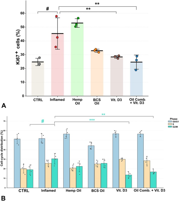
Hyperproliferation of keratinocytes is a hallmark of cAD, contributing to epidermal thickening and impaired barrier function [ref. 38]. Ki‐67 analysis revealed that inflammatory stimulation induced a marked increase in proliferative keratinocytes (CTRL: 24.7 ± 2.7 vs. Inflamed: 45.3 ± 11.4) (Figure 1A). Incidentally, treatment with hemp oil further enhanced Ki‐67 positivity (53.0 ± 3.2), while BCS exerted a partial but not significant reduction (32.9 ± 1.1). Notably, vitamin D3 (28.4 ± 1.1) and the oil combination with vitamin D3 (24.6 ± 5.1) effectively suppressed the inflammatory hyperproliferation, restoring levels close to control cells. Cell cycle analysis showed in Figure 1B corroborated these findings. In inflamed keratinocytes, the proportion of cells in G2/M phase increased markedly (30.5 ± 5.6) compared to CTRL (19.0 ± 5.0), reflecting accelerated proliferation. Both vitamin D3 and the oil combination with vitamin D3 reduced the G2/M fraction (22.5 ± 3.5 and 22.0 ± 3.2, respectively), with a concomitant shift toward G0/G1 (CTRL: 51.3 ± 4.9, Inflamed: 51.8 ± 5.0, vitamin D3: 56.5 ± 3.0, Oil combination with vitamin D3: 56.5 ± 3.0). Together, these data indicate that while hemp oil alone did not attenuate proliferation, vitamin D3 and the oil combination with vitamin D3 strongly counteracted cytokine‐driven hyperproliferation, normalizing both Ki‐67 expression and cell cycle distribution.
Vitamin D3
and Oil Combination Restore Dysregulated Gene Expression of Keratinocyte Differentiation Markers
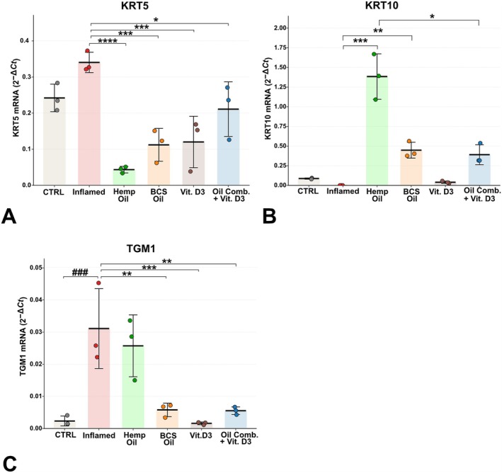
Keratinocyte differentiation is a tightly regulated process essential for maintaining epidermal architecture and barrier function, and its impairment is a hallmark of cAD [ref. 10]. To assess the effects of the treatments on epidermal differentiation, we quantified the gene expression of keratin 5 (KRT5), a cytoskeletal protein expressed in proliferative basal keratinocytes, and keratin 10 (KRT10), a marker of early suprabasal differentiation. Inflammatory stimulation significantly altered basal marker expression, with KRT5 markedly increased compared to control (CTRL: 0.242 ± 0.038; Inflamed: 0.340 ± 0.028), indicating sustained basal proliferation under inflammatory conditions. All treatments reduced KRT5 transcript levels relative to the inflamed group, with the most pronounced decrease observed in hemp oil treated cells (0.043 ± 0.009), followed by BCS oil (0.112 ± 0.046), vitamin D3 (0.120 ± 0.071), and the oil combination with vitamin D3 (0.211 ± 0.076) (Figure 2). In contrast, KRT10, which is typically expressed in suprabasal keratinocytes and inversely correlated with KRT5, was undetectable in inflamed cells, but became clearly detectable in all treatments, suggesting an early activation of the differentiation program rather than restoration of baseline expression. The expression of transglutaminase‐1 (TGM1), an enzyme responsible for cross‐linking structural proteins during cornified envelope assembly, was markedly affected by the inflammatory stimulus. Inflamed keratinocytes displayed a robust induction of TGM1 transcripts compared with control cells (CTRL: 0.0023 ± 0.0015; Inflamed: 0.0311 ± 0.0124), reflecting an abnormal activation of late differentiation pathways under inflammatory stress. Treatment with vitamin D3 significantly reduced TGM1 expression (0.0016 ± 0.0004), restoring values close to those of unstimulated controls. A similar downregulatory effect was observed with the oil combination with vitamin D3 (0.0055 ± 0.0012) and with blackcurrant seed oil (0.0058 ± 0.0021), whereas hemp oil induced only a partial reduction (0.0257 ± 0.0096) (Figure 2).
Vitamin D3, Hemp Oil, and BCS Oil Counteract Inflammation‐Induced Upregulation of Involucrin in CPEK Cells
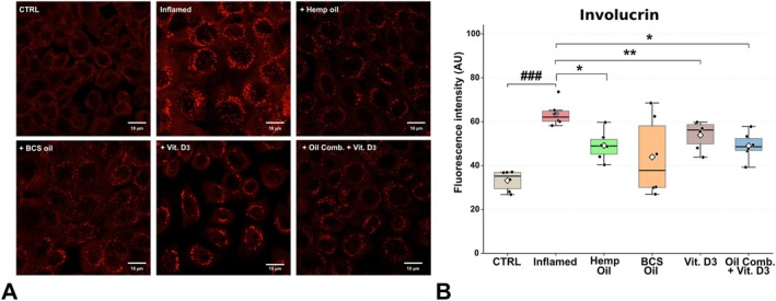
Involucrin (IVL) is a major component of the cornified envelope, and a key structural protein required for terminal keratinocyte differentiation and epidermal barrier integrity [ref. 39]. In atopic dermatitis (AD), epidermal barrier dysfunction is associated with the dysregulated expression of barrier‐related proteins such as filaggrin, loricrin, and involucrin [ref. 13, ref. 14, ref. 15]. Several studies have shown that inflammatory cytokines, particularly IL‐4 and IL‐13, acting through STAT6 and STAT3, downregulate the expression of these differentiation markers, thereby contributing to barrier impairment [ref. 15]. Based on this evidence, we investigated whether the inflammatory environment alters involucrin (IVL) expression in basal keratinocytes and whether treatment with hemp oil, BCS oil, and vitamin D3 could modulate this effect. Immunofluorescence analysis performed 24 h after cytokine stimulation (Figure 3) revealed a marked increase in IVL signal intensity compared with control cells (INFLAMED: 63.54 ± 5.53 vs. CTRL: 33.18 ± 4.67), indicating premature activation of differentiation‐associated pathways in response to inflammation. Treatment with hemp oil (49.11 ± 6.67), BCS oil (43.87 ± 17.9), vitamin D3 (53.86 ± 6.5), or their combination (48.99 ± 6.29) partially normalized involucrin levels. These findings suggest that the treatments, particularly BCS oil and the combined oil + vitamin D3 formulation, suggest that these treatments more effectively counteract inflammation‐induced involucrin overexpression and help preserve basal keratinocyte identity.
Hemp Oil Preferentially Modulates Tight‐Junction Markers, With Limited Effects of the Other Treatments on Barrier Resistance
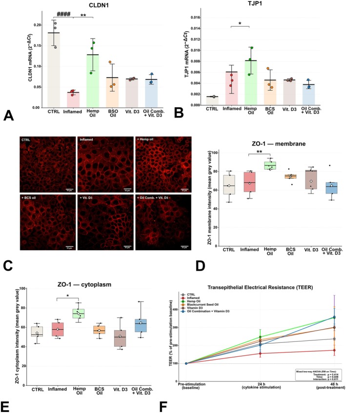
Tight junction proteins are essential components of the epidermal barrier, and their dysregulation represents a key pathogenic feature of cAD [ref. 8, ref. 40]. Consistent with previous evidence showing that Th2 cytokines downregulate claudin‐1 (CLDN1) and alter tight junction protein‐1 (TJP1/ZO‐1) signaling through ERK‐, STAT3‐, and STAT6‐dependent pathways [ref. 41, ref. 42]; inflammatory stimulation of CPEK cells modified the transcriptional profile of these genes (Figure 4A,B). Exposure to pro‐inflammatory cytokines caused a strong downregulation of CLDN1 mRNA (Inflamed: 0.037 ± 0.005 vs. CTRL: 0.181 ± 0.031), confirming barrier‐related gene suppression. Among the tested natural compounds, hemp oil induced the most pronounced recovery of CLDN1 expression (0.128 ± 0.039). In contrast, TJP1 transcription was moderately increased in inflamed cells (Inflamed: 0.004 ± 0.000 vs. CTRL: 0.001 ± 0.000), suggesting a compensatory response to tight junction disruption. Hemp oil further enhanced TJP1 expression (0.008 ± 0.002), while the other treatments did not significantly modify the levels observed under inflammatory conditions. To assess whether transcriptional modulation of TJP1 was accompanied by changes in ZO‐1 abundance and subcellular distribution, fluorescence intensity was quantified separately along the plasma membrane and within the cytoplasmic compartment. In the membrane‐associated fraction, ZO‐1 signal modestly increased under inflammatory conditions (CTRL: 58.90 ± 7.69; Inflamed: 72.22 ± 7.23) and was further elevated following hemp oil treatment (89.64 ± 4.01) (Figure 4C,D). A similar trend was observed in the cytoplasmic compartment (Figure 4C,E), where fluorescence intensity showed a non‐significant increase from control to inflamed cells (CTRL: 52.21 ± 6.25; Inflamed: 62.10 ± 4.55), and a marked enhancement after hemp oil exposure (79.36 ± 3.74) (Figure 4C–E). These data indicate that inflammation increases overall ZO‐1 signal intensity, and hemp oil amplifies this effect, consistent with its transcriptional upregulation of TJP1. To determine whether these molecular and distributional changes translated into functional improvements of the epithelial barrier, transepithelial electrical resistance (TEER) was evaluated. Inflammatory stimulation attenuated the time‐dependent increase in TEER observed in unstimulated monolayers: at 24 h, inflamed cells reached 156% ±37% of their pre‐stimulation baseline compared with 209% ± 31% in CTRL (Welch’s t‐test, p = 0.13) (Figure 4F). At 48 h, all treatments tended to enhance TEER recovery above the inflamed condition, with vitamin D3 (302% ± 33%), the oil combination (360% ± 82%), hemp oil (355% ± 216%), and BCS oil (299%± 204%) all exceeding the inflamed value (174% ± 51%) (Figure 4F); however, none of these comparisons reached statistical significance. A mixed two‐way ANOVA on the post‐stimulation timepoints revealed a significant effect of Time (p = 0.009), but no significant effect of Treatment (p = 0.41) or Treatment × Time interaction (p = 0.61), suggesting that the trends observed are coherent but underpowered at the current sample size. Collectively, these findings demonstrate that hemp oil exerts the strongest protective effect on tight junction integrity in CPEK cells, enhancing CLDN1 and TJP1/ZO‐1 expression and promoting functional recovery of the epithelial barrier.
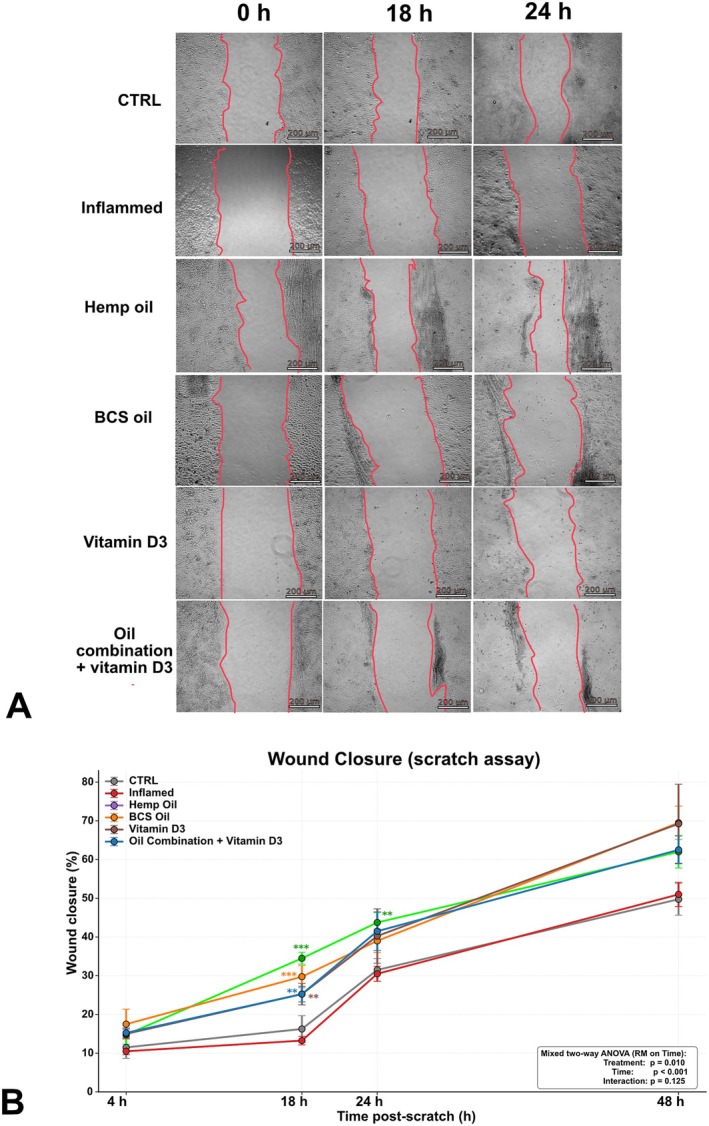
All Treatments Improve Early Keratinocyte Migration, but Only Hemp Oil Sustains Accelerated Scratch Closure Under Inflammatory Conditions
Keratinocyte migration plays a central role in epidermal repair and is strongly impaired in atopic dermatitis (AD), where pruritus‐associated mechanical injury and type‐2 cytokine–driven inflammation delay wound closure and contribute to barrier dysfunction [ref. 43]. Inefficient wound healing in this inflammatory context can promote dysregulated tissue remodeling, favoring excessive fibroblast activation, aberrant collagen deposition, and the development of hypertrophic or keloid‐like scarring in predisposed individuals [ref. 44]. To specifically assess the migratory capacity of CPEK cells under inflammatory conditions and in response to natural compounds, a scratch assay was performed in the presence of 10 μM cytosine arabinoside (Ara‐C), which was used to suppress proliferation and allow evaluation of migration‐dependent wound closure. At early time points (4 h), no significant differences were detected among the experimental conditions, indicating comparable initial motility across groups. Divergence became evident at 18 h (Figure 5), when untreated inflamed cells exhibited reduced wound closure (13.25% ± 2.21%) compared with control CPEK cells (16.25% ± 6.80%). All treatments accelerated wound healing at this time point, with hemp oil showing the strongest effect (34.50% ± 3.10%), followed by blackcurrant seed oil (29.75% ± 5.70%), vitamin D3 (25.20% ± 5.50%), and the oil combination with vitamin D3 (25.25% ± 4.00%) (Figure 5). At 24 h, hemp oil remained the only treatment producing a significant enhancement of wound closure (62.00% ± 8.40%), markedly outperforming both inflamed (30.50% ± 3.80%) and control cells (31.50% ± 5.90%) (Figure 5). By 48 h, no significant differences persisted among groups, consistent with the near‐complete closure of the scratch in all conditions and the intrinsic limitations of the assay at late time points (Figure 5). Overall, these data indicate that inflammation slows keratinocyte migration in this canine AD model, and that natural compounds can partially restore motility, with hemp oil exerting the most pronounced and sustained pro‐migratory activity.
Cytokine Stimulation Triggers Nitrosative Stress and Engages the NRF2/BACH1/HMOX1 Axis, Which Is Differentially Modulated by the Four Treatments
Oxidative stress is a well‐recognized contributor to barrier dysfunction in atopic dermatitis (AD), where type‐2 cytokines and chronic mechanical injury increase ROS and weaken tight junction organization [ref. 45, ref. 46]. To validate the relevance of our in vitro model in this respect, we first quantified 3‐nitrotyrosine (3‐NT), a stable footprint of peroxynitrite‐mediated protein nitration [ref. 47]. 3‐NT levels were strongly increased in inflamed cells compared to CTRL (97.1 ± 3.5 vs. 60.3 ± 9.5 ng/mL), confirming that the cytokine cocktail elicited a robust nitrosative stress response (Figure 6A). None of the treatments reduced 3‐NT relative to the inflamed condition (range 94.7–99.7 ng/mL; all comparisons ns vs. Inflamed). This is consistent with the cumulative nature of the marker: protein nitration occurring during the initial 24 h of cytokine exposure remains in the protein pool sampled at 48 h, and the treatment window (24 h) is shorter than the typical turnover of nitrated proteins. Given this evidence of sustained oxidative challenge, we next interrogated the KEAP1–NRF2 axis, the master regulator of the cytoprotective antioxidant response [ref. 48, ref. 49, ref. 50, ref. 51]. At the transcript level, NFE2L2 mRNA tended to be upregulated in inflamed cells compared with CTRL (0.34 ± 0.08 vs. 0.19 ± 0.03), although this trend did not reach statistical significance (Figure 6B). Hemp oil produced a marked further induction of NFE2L2 mRNA, reaching levels significantly higher than CTRL (0.57 ± 0.11; # vs CTRL), in keeping with previous reports of cannabidiol‐driven NRF2 activation in keratinocytes [ref. 31]; blackcurrant seed oil, vitamin D3 and the combined formulation showed a trend of decline toward control levels, without reaching statistical significance. At the protein level, NRF2 was elevated in inflamed cells compared with CTRL (18.8 ± 5.4 vs. 9.8 ± 4.9 MFI; # vs. CTRL; Figure 6C); hemp oil maintained NRF2 protein at levels comparable to the inflamed condition, whereas blackcurrant seed oil, vitamin D3 and the combined formulation showed intermediate values without reaching statistical significance.
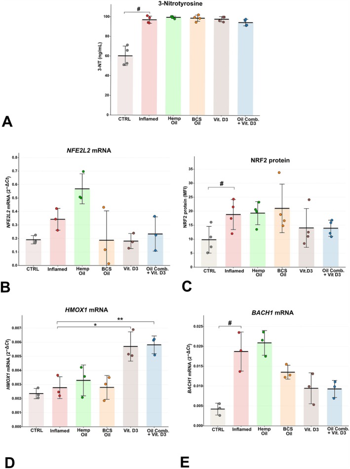
To assess whether the engagement of NRF2 was reflected in the downstream transcriptional output, we measured HMOX1, the prototypical NRF2 target gene, and BACH1, a transcriptional repressor that competes with NRF2 for binding to antioxidant response elements on shared target genes including HMOX1 [ref. 31, ref. 48]. HMOX1 mRNA did not differ significantly between inflamed and control cells (Figure 6D), consistent with the well‐described early‐peak/late‐decline kinetics of this immediate‐early gene, which has typically returned toward basal levels by 24–48 h after stimulation. In contrast, BACH1 mRNA was markedly upregulated by inflammation (0.019 ± 0.005 vs. 0.004 ± 0.001; # vs. CTRL; Figure 6E). The four treatments differentially modulated this axis: hemp oil and blackcurrant seed oil did not significantly affect either HMOX1 or BACH1 mRNA expression relative to the inflamed condition, whereas vitamin D3 and the combined formulation produced a coordinated rebalancing of the axis—vitamin D3 significantly increased HMOX1 expression (* vs. Inflamed; Figure 6D), the combined formulation significantly increased HMOX1 (** vs. Inflamed) and concurrently reduced BACH1 expression (* vs. Inflamed; Figure 6E). Taken together, these results provide a coherent description of the redox state of the inflamed CPEK model at the 48‐h timepoint: the cytokine cocktail elicits a sustained nitrosative stress (3‐NT) that is not reversed by any of the treatments consistent with the cumulative nature of protein nitration over the experimental window and engages the NRF2 transcriptional axis at both the mRNA and protein levels, while concurrently inducing the transcriptional repressor BACH1. The four treatments exhibit a clearly differential effect on this axis: hemp oil and blackcurrant seed oil do not modulate the NRF2/BACH1/HMOX1 pattern at the timepoint assessed, whereas vitamin D3 and especially the combined formulation rebalance the axis by relieving BACH1‐mediated repression and increasing HMOX1 expression, with the combined formulation also producing a coordinated normalization of NRF2 protein levels.
Inflammatory Mediator Secretion Confirms the Engagement of STAT1 and NF‐κB Signaling Axes by the Cytokine Cocktail and Reveals a Pathway‐Specific Modulation by the Tested Compounds
To characterize the inflammatory state of the model and to assess the functional engagement of the major signaling axes activated by the cytokine cocktail, we quantified five cytokines and chemokines in cell‐culture supernatants by ELISA: IL‐8 (CXCL8) and IL‐6 as readouts of NF‐κB‐driven inflammatory signaling, with IL‐6 also functioning as a downstream amplifier of STAT3 through its known autocrine loop; CXCL9 and CXCL10 as IFN‐γ/STAT1‐specific readouts; and CCL17/TARC as a readout of the IL‐4/IL‐13/STAT6 axis. The cytokine cocktail induced a significant secretion of IL‐8 by CPEK keratinocytes (15.1 ± 3.5 vs. 7.8 ± 2.2 ng/mL in CTRL; # vs. CTRL; Figure 7A), confirming the engagement of the NF‐κB inflammatory branch. The four treatments differentially modulated IL‐8 secretion: blackcurrant seed oil, vitamin D3 and the combined formulation significantly reduced IL‐8 relative to the inflamed condition (BCS **, Vit D3 *, Combo *), whereas hemp oil did not. Engagement of the IFN‐γ/STAT1 axis was confirmed by a marked, three‐fold induction of CXCL9 secretion in inflamed cells compared to CTRL (168.0 ± 41.1 vs. 56.6 ± 23.3 ng/mL; # vs. CTRL; Figure 7B). All four treatments significantly reduced CXCL9 relative to the inflamed condition (Hemp Oil **, BCS **, Vit D3 **, Combo **). A second NF‐κB‐dependent cytokine, IL‐6, was also significantly induced by inflammation (25.4 ± 6.7 vs. 10.2 ± 3.0 pg/mL in CTRL; # vs. CTRL; Figure 7C); all treatments modestly reduced IL‐6 secretion, but the differences did not reach statistical significance. The induction of the STAT1 axis was further confirmed by an approximately six‐fold increase in CXCL10 secretion in inflamed cells (122.5 ± 9.0 vs. 21.7 ± 5.0 ng/mL; #*** vs. CTRL; Figure 7D). Hemp oil, vitamin D3 and the combined formulation produced a near‐complete normalization of CXCL10 secretion (all *** vs. Inflamed), whereas blackcurrant seed oil did not significantly reduce CXCL10 levels. CCL17/TARC, included as a readout of the IL‐4/IL‐13/STAT6 axis, was below the lower limit of quantification of the assay in all conditions, including the inflamed group, and the corresponding data are therefore not shown.
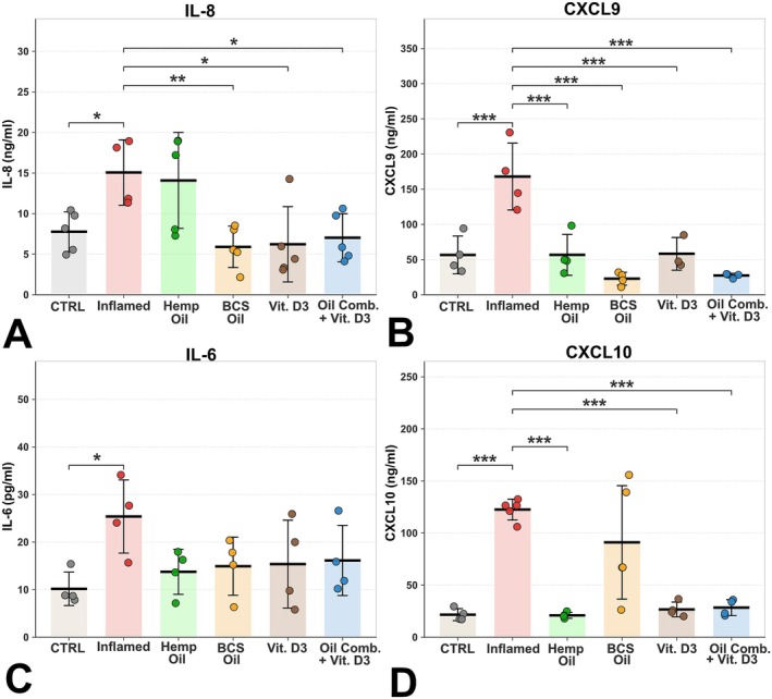
Taken together, these results document a robust engagement of both the NF‐κB and the STAT1 signaling axes in inflamed CPEK keratinocytes with a particularly marked induction of the IFN‐γ/STAT1‐specific chemokines CXCL9 and CXCL10 and reveal a clearly differential pharmacological footprint of the four treatments across these two axes. Hemp oil acts preferentially on the STAT1 chemokine arm (significant reduction of CXCL9 and near‐complete normalization of CXCL10) without significantly affecting NF‐κB‐dependent cytokines. Blackcurrant seed oil acts preferentially on the NF‐κB‐dependent IL‐8 (significant reduction) without normalizing the STAT1‐driven CXCL10. Vitamin D3 and the combined formulation display the broadest action, with significant effects across both signaling axes (CXCL9, CXCL10, IL‐8).
Discussion
This study provides an integrated analysis of the effects of hemp oil, blackcurrant seed oil, vitamin D3, and their combination on cytokine‐stimulated canine progenitor epidermal keratinocytes (CPEK), used here as an acute in vitro model of canine atopic dermatitis (cAD). By examining multiple interconnected features—proliferation, differentiation, tight‐junction organization, barrier function, migration, oxidative stress, and cytokine secretion—we delineate the differential signatures of these four natural formulations under inflammatory conditions: hemp oil acts preferentially on tight‐junction integrity, NRF2 activation, and the STAT1 chemokine arm; BCS oil on NF‐κB‐dependent IL‐8 secretion and wound‐closure dynamics; vitamin D3 on proliferation and on the downstream NRF2/HMOX1/BACH1 transcriptional output; the combined formulation cumulates the broadest action across these axes.
Keratinocyte hyperproliferation is a defining feature of both human AD and cAD, driven by chronic exposure to type‐2 cytokines (IL‐4; IL‐13) and mechanical injury associated with pruritus [ref. 52, ref. 53]. In line with this, cytokine stimulation in our model increased Ki‐67 expression and expanded the G2/M cell‐cycle fraction, reflecting accelerated keratinocyte cycling. Vitamin D3, both alone and in the combined formulation, significantly attenuated this hyperproliferative response, restoring proliferation indices and cell‐cycle distribution toward control levels. These findings are consistent with the established role of vitamin D3 in promoting G0/G1 arrest and coordinating the transition from proliferation to differentiation in keratinocytes [ref. 54, ref. 55]. By contrast, hemp oil did not suppress cytokine‐driven proliferation and was associated with a further increase in Ki‐67 positivity, indicating that its biological activity in this context is not primarily directed toward growth restraint. This observation is consistent with previous evidence [ref. 31] that cannabidiol, the major bioactive constituent of hemp oil, directly stimulates keratinocyte proliferation in vivo, with increased epidermal thickness and upregulation of hyperproliferation and wound repair. This divergence underscores that distinct natural compounds may target different facets of keratinocyte dysregulation in cAD and highlights vitamin D3 as the most effective modulator of proliferation in this model.
Inflammation‐induced disruption of keratinocyte differentiation represents another central component of cAD pathophysiology [ref. 10, ref. 56]. In our model, cytokine stimulation increased expression of the basal marker KRT5, abolished detectable KRT10, and aberrantly induced late differentiation markers such as TGM1 and involucrin in basal keratinocytes, indicative of a distorted differentiation program. This pattern is partly divergent from the canonical signature of human AD lesional skin and of canine atopic biopsies, where TGM1 is typically reduced rather than increased, and likely reflects a specific feature of our experimental system. IFN‐γ, present in the cytokine cocktail, is a well‐characterized inducer of squamous differentiation in primary keratinocytes, driving TGM1 upregulation in parallel with downregulation of cell‐cycle and basal‐compartment genes [ref. 57]; at the 48‐h readout, this pro‐differentiative effect is consistent with the increased TGM1 and the reduced KRT5 and KRT10 observed in inflamed cells. All treatments partially corrected this imbalance, as reflected by reduced KRT5 expression and re‐emergence of KRT10 transcripts, suggesting reactivation of early suprabasal differentiation. Vitamin D3 and the oil combination were particularly effective in normalizing TGM1 gene expression, while BCS oil also exerted a significant attenuating effect. Hemp oil produced a more limited correction of late differentiation markers, suggesting that its principal effects operate through mechanisms other than direct modulation of the differentiation program.
The same pattern was confirmed at the level of involucrin (IVL), a precursor of the cornified envelope and a marker of late keratinocyte differentiation. IVL was significantly upregulated in inflamed cells compared with control, a direction that is opposite to what is consistently reported in human AD lesional skin and in canine atopic biopsies, where IVL is typically downregulated through the IL‐4/IL‐13–JAK–STAT6/STAT3 axis [ref. 15, ref. 58, ref. 59]. As discussed above for TGM1, this suppressive effect of the Th2 component of the cocktail typically requires longer cytokine exposures or three‐dimensional culture systems to fully emerge, and is therefore unlikely to dominate in the acute, monolayer setting used here. The four treatments modulated IVL in a pattern broadly consistent with the one described for TGM1: vitamin D3 and the combined formulation reduced its expression toward control values, hemp oil maintained an inflamed‐like profile, and BCS oil produced an intermediate response. Taken together, the parallel modulation of TGM1 and IVL indicates that vitamin D3‐containing formulations exert a coordinated restraint of the squamous‐differentiation program engaged in our acute model.
Disruption of tight junctions and impaired epidermal barrier function are key pathogenic features of cAD [ref. 1, ref. 6, ref. 11]. Consistent with previous reports [ref. 1, ref. 32], cytokine stimulation markedly downregulated claudin‐1 expression, reproducing one of the canonical hallmarks of atopic skin and providing indirect evidence for the engagement of the IL‐4/IL‐13–JAK–STAT6 axis [ref. 15]. In contrast, TJP1 mRNA and membrane‐associated ZO‐1 protein were not significantly modified, while the apparent increase in cytoplasmic ZO‐1 staining should be interpreted with caution. Cytokine‐stimulated CPEK cells exhibit a hypertrophic morphology, consistent with the well‐documented cell‐enlargement effect of cytokines acting through STAT3, most notably IL‐6, which is produced autocrinously by inflamed keratinocytes, described by Niehues and colleagues as a hypertrophy‐inducing input on keratinocytes independent of hyperproliferation [ref. 60]. The increased per‐cell cytoplasmic ZO‐1 signal measured by immunofluorescence in inflamed cells is therefore most parsimoniously explained as a morphometric consequence of this cellular enlargement, rather than as a genuine increase in the intracellular pool of ZO‐1 protein, an interpretation supported by the unchanged TJP1 mRNA levels, which indicate no transcriptional upregulation of the gene. Two aspects of the tight‐junction readout in our model deserve a specific note: the cytoplasmic rather than membranous accumulation of ZO‐1, and the absence of significant TEER changes. Marsella and colleagues have shown that CPEK cells grown in monolayer display, already at baseline, a predominantly cytoplasmic localisation of ZO‐1 and a markedly lower trans‐epithelial electrical resistance (~150 Ohms/cm) than primary canine keratinocytes from skin biopsies (~2000 Ohms/cm), concluding that monolayer CPEKs are not fully representative of normal canine keratinocytes for permeability studies [ref. 61]. The further increase in cytoplasmic ZO‐1 signal observed in inflamed cells therefore overlays a baseline already shifted toward this distribution and is compatible with the cytokine‐induced cellular hypertrophy discussed above, while the unchanged TEER reflects the intrinsically narrow dynamic range of this readout in CPEK monolayers.
Keratinocyte migration is tightly linked to epidermal repair and is often impaired in atopic dermatitis [ref. 62]. By performing scratch assays under inflammatory conditions while pharmacologically suppressing proliferation, we specifically assessed migration‐dependent wound closure. Inflammation modestly reduced early migration, consistent with impaired motility observed in atopic keratinocytes [ref. 63]. All treatments enhanced migration at intermediate time points, indicating a general capacity to support re‐epithelialization under inflammatory stress. However, only hemp oil sustained a significant pro‐migratory effect at 24 h, resulting in markedly accelerated wound closure compared with both inflamed and control cells.
This sustained migratory response aligns with the pronounced barrier‐restorative effects of hemp oil and suggests coordinated regulation of junctional plasticity and cell motility [ref. 64, ref. 65]. The more transient effects observed with BCS oil and vitamin D3 are consistent with previous evidence supporting their involvement in early phases of wound repair [ref. 66, ref. 67, ref. 68, ref. 69, ref. 70, ref. 71] but suggest limited capacity to counteract prolonged inflammatory inhibition of migration. The accelerated wound closure observed in treated cells is unlikely to reflect the cytokine‐induced cellular hypertrophy discussed earlier, since wound closure was quantified as percent area covered relative to the initial scratch within each individual well, a metric that is intrinsically independent of individual cell size and reflects the net advancement of the wound edge over time.
Oxidative and nitrosative stress play a recognized pathogenic role in atopic dermatitis, where increased reactive oxygen and nitrogen species coexist with a functionally insufficient endogenous antioxidant response, contributing to barrier dysfunction and to the chronic engagement of inflammatory signaling [ref. 17, ref. 45, ref. 72]. In keeping with this background, the cytokine cocktail produced in our model a marked accumulation of 3‐nitrotyrosine in inflamed cells, confirming a sustained nitrosative protein damage at the 48‐h timepoint and providing a functional readout of the redox imbalance generated by the inflammatory stimulus. The choice to focus subsequently on the NRF2 transcriptional axis was based on the central role of this pathway in coordinating the cytoprotective antioxidant response in keratinocytes and on direct evidence that its activity is reduced in the epidermis of AD patients [ref. 20, ref. 21]. Additional rationale was provided by previous reports describing the modulation of this axis by natural compounds with cytoprotective activity in keratinocytes, including cannabidiol [ref. 31]. The four treatments produced a clearly differential pattern of modulation along the NRF2/BACH1/HMOX1 axis. Hemp oil acted primarily on the upstream/activator arm, with a significant induction of NFE2L2 mRNA above control levels and maintained NRF2 protein, whereas the downstream output of the pathway, HMOX1 mRNA was not further increased and the transcriptional repressor BACH1 was not modulated. The data therefore document, in our acute monolayer setting, a partial engagement of the cannabidiol–NRF2 link previously described by Casares and colleagues in primary human keratinocytes [ref. 31, ref. 51]: NRF2 activation was present, but BACH1 degradation and HMOX1 induction were not observed. This divergence likely reflects the different experimental matrix (canine immortalized CPEK monolayer vs. primary human keratinocytes), the acute cytokine challenge, and the 48‐h timepoint, at which the early peak of HMOX1 induction described in vitro may already have declined. Vitamin D3 and the combined formulation, in contrast, acted preferentially on the downstream arm of the axis, with significant induction of HMOX1 and a marked reduction of BACH1 approaching the conventional significance threshold, in line with the recognized role of VDR signaling in relieving transcriptional repression and enhancing the antioxidant output of keratinocytes. BCS oil did not significantly modulate any component of the axis. Two observations deserve specific framing: the persistence of 3‐nitrotyrosine and the maintenance or, for hemp oil, the further increase of NRF2 protein in treated cells. Neither finding should be read as evidence that the treatments worsen oxidative damage. 3‐NT is a cumulative footprint of protein nitration generated during the initial 24 h of cytokine exposure, with a protein‐turnover‐limited persistence longer than the 24 h treatment window; the dataset informs on the reversal of established nitration at the late timepoint, not on whether the treatments limit ongoing nitration. The sustained NRF2 protein elevation, similarly, is a canonical response to oxidative challenge and reflects an antioxidant program actively engaged in response to the inflammatory stimulus rather than compound‐induced toxicity. For hemp oil specifically, the parallel induction of NFE2L2 mRNA above control values indicates a direct activator role on the NRF2 axis rather than a stress‐driven compensatory response.
The ELISA‐based quantification of secreted mediators provides functional evidence that the cytokine cocktail engages the major signaling axes operative in CPEK cells and reveals, at the same time, that the model recapitulates a clinically relevant inflammatory output. The robust induction of CXCL9 (three‐fold) and CXCL10 (six‐fold) in inflamed cells reflects the engagement of the IFN‐γ/STAT1 axis and is consistent with the well‐documented role of these CXCR3‐binding chemokines in recruiting Th1 and cytotoxic CD8+ T cells to the lesional skin, particularly in the chronic phase of AD and in intrinsic AD endotypes [ref. 15, ref. 73]. In keratinocytes specifically, IL‐4 has been shown to potentiate the action of IFN‐γ in inducing these CXCR3 ligands, providing a mechanistic link between the Th2‐skewed initiation phase and the Th1‐amplified chronicisation of the disease, a process that our acute model captures by combining IFN‐γ, IL‐4, and IL‐13 within the same cocktail. The NF‐κB‐dependent branch, captured by IL‐8 (CXCL8) and IL‐6 secretion, was also significantly engaged, although with a smaller amplitude: IL‐8 is a recognized mediator of neutrophil recruitment in inflammatory skin diseases and is among the chemokines that contribute to the innate inflammatory component of AD, while IL‐6 produced autocrinously by keratinocytes sustains epidermal hyperplasia and acts as a downstream amplifier of STAT3 signaling, contributing both to disease chronicisation and to the keratinocyte hypertrophy discussed in previous sections [ref. 15]. CCL17/TARC, included as a STAT6‐specific readout, was below the lower limit of quantification of the assay in all conditions; the engagement of STAT6 by the IL‐4/IL‐13 components of the cocktail is, however, indirectly supported by the marked downregulation of CLDN1, a gene consistently suppressed by the IL‐4/IL‐13–JAK‐STAT6 axis in keratinocytes [ref. 74].
The pattern of treatment‐induced modulation maps onto a clearly pathway‐specific footprint, which translates directly into distinct potential immunological consequences. Hemp oil reduced CXCL9 and CXCL10 secretion but did not significantly modify IL‐8 or IL‐6, indicating a preferential action on the STAT1 chemokine arm and, by extension, a potential to limit the IFN‐γ/CXCR3‐driven recruitment of Th1 and CD8+ cells without significant suppression of the NF‐κB‐dependent innate inflammatory branch. BCS oil produced a mirror‐image profile, significantly reducing IL‐8 without normalizing the STAT1‐driven CXCL10, consistent with a γ‐linolenic acid‐mediated suppression of NF‐κB‐dependent pathways and with a potential preferential effect on the innate, neutrophil‐recruiting component of the response. Vitamin D3 and the combined formulation acted broadly across both axes, reducing CXCL9, CXCL10, and IL‐8, in line with the well‐documented broad‐spectrum anti‐inflammatory action of VDR signaling on multiple keratinocyte pathways and with the potential to dampen both the adaptive Th1 and the innate components of the inflammatory output. The convergence of these results with the proliferation, differentiation, barrier, and redox readouts discussed above reinforces the picture of four natural formulations modulating distinct but partially complementary nodes of the inflamed keratinocyte phenotype.
Limitations and Future Directions
Several limitations should be acknowledged when interpreting these findings. First, this study relied on an immortalized keratinocyte line and an in vitro cytokine‐based inflammatory model, which cannot fully recapitulate the cellular heterogeneity, immune–epithelial interactions, and chronic environmental influences present in canine atopic skin in vivo. While CPEK cells reproduce many hallmark features of cAD [ref. 75, ref. 76, ref. 77, ref. 78], disease‐specific behaviors of primary keratinocytes [ref. 79] and contributions from immune cells, fibroblasts, and the microbiome remain unaddressed. Second, although the combined oil and vitamin D3 formulation demonstrated beneficial effects, the study was not designed to formally distinguish additive from synergistic interactions or to optimize formulation ratios. Third, the extension of the redox characterization to BACH1 quantification and to additional NRF2 transcriptional targets (NQO1, GCLC, GCLM) would strengthen the mechanistic interpretation of the differential modulation of the NRF2/BACH1/HMOX1 axis observed here.
Future studies should therefore extend these findings to primary keratinocytes derived from atopic dogs, incorporate co‐culture systems with immune cells, and validate barrier and wound‐healing effects in ex vivo or in vivo models. Detailed mechanistic analyses dissecting the contributions of specific hemp oil constituents, including cannabidiol and lipid components, will be essential to refine formulation strategies. Ultimately, controlled clinical studies will be required to determine whether the barrier‐supportive and antioxidant effects observed here translate into meaningful therapeutic benefits for dogs with atopic dermatitis.
Funding
This work was supported by NBF Lanes.
References
- Advances in Our Understanding of Canine Atopic Dermatitis,”. Veterinary Dermatology, December 2021. [DOI | PubMed]
- Update on the Role of Genetic Factors, Environmental Factors and Allergens in Canine Atopic Dermatitis,”. Veterinary Dermatology, February 2024. [DOI | PubMed]
- Update on the Skin Barrier, Cutaneous Microbiome and Host Defence Peptides in Canine Atopic Dermatitis,”. Veterinary Dermatology, February 2024. [DOI]
- Update on the Role of Cytokines and Chemokines in Canine Atopic Dermatitis,”. Veterinary Dermatology, February 2024. [DOI | PubMed]
- Updated Insights Into the Molecular Pathogenesis of Canine Atopic Dermatitis,”. Veterinary Dermatology, August 2025. [DOI | PubMed]
- Tight Junction Proteins in the Canine Epidermis: A Pilot Study on Their Distribution in Normal and in High IgE‐Producing Canines,”. Canadian Journal of Veterinary Research, January 2015. [PubMed]
- Expression Patterns of Superficial Epidermal Adhesion Molecules in an Experimental Dog Model of Acute Atopic Dermatitis Skin Lesions,”. Veterinary Dermatology, February 2015. [DOI | PubMed]
- A Comparative Study of Epidermal Tight Junction Proteins in a Dog Model of Atopic Dermatitis,”. Veterinary Dermatology, February 2016. [DOI | PubMed]
- Altered Lipid Properties of the Stratum Corneum in Canine Atopic Dermatitis,”. Biochimica et Biophysica Acta, Biomembranes, February 2018. [DOI | PubMed]
- Alterations of Keratins, Involucrin and Filaggrin Gene Expression in Canine Atopic Dermatitis,”. Research in Veterinary Science, December 2012. [DOI | PubMed]
- Pilot Study Using Five Methods to Evaluate Skin Barrier Function in Healthy Dogs and in Dogs With Atopic Dermatitis,”. Veterinary Dermatology, January 2019. [DOI]
- JAK–STAT Signaling Pathway in the Pathogenesis of Atopic Dermatitis: An Updated Review,”. Frontiers in Immunology, 2022. [DOI | PubMed]
- In Vitro Assessment of IL‐4‐ or IL‐13‐Mediated Changes in the Structural Components of Keratinocytes in Mice and Humans,”. Journal of Investigative Dermatology, May 2014. [DOI | PubMed]
- Interleukin‐13: Targeting an Underestimated Cytokine in Atopic Dermatitis,”. Allergy, January 2020. [DOI | PubMed]
- Regulation of Filaggrin, Loricrin, and Involucrin by IL‐4, IL‐13, IL‐17A, IL‐22, AHR, and NRF2: Pathogenic Implications in Atopic Dermatitis,”. International Journal of Molecular Sciences, July 2020. [DOI | PubMed]
- Polyphenols and Cannabidiol Modulate Transcriptional Regulation of Th1/Th2 Inflammatory Genes Related to Canine Atopic Dermatitis,”. Frontiers in Veterinary Science, March 2021. [DOI | PubMed]
- Oxidative Stress Markers in Canine Atopic Dermatitis,”. Research in Veterinary Science, June 2012. [DOI | PubMed]
- Selected Serum Oxidative Stress Biomarkers in Dogs With Non‐Food‐Induced and Food‐Induced Atopic Dermatitis,”. Veterinary Dermatology, June 2018. [DOI | PubMed]
- Investigation on the In Vitro Effects of Resveratrol on Peripheral Blood Mononuclear Cells Harvested From Healthy and Atopic Dogs,”. Research in Veterinary Science, November 2024. [DOI | PubMed]
- The Role of NRF2 Transcription Factor in Inflammatory Skin Diseases,”. BioFactors, March–April 2025. [DOI | PubMed]
- AHR and NRF2 in Skin Homeostasis and Atopic Dermatitis,”. Antioxidants, January 2022. [DOI | PubMed]
- Evaluation of Oclacitinib Maleate and Prednisolone Combined Therapy for the Control of Atopic Dermatitis in Dogs: A Controlled Clinical Trial,”. Veterinary Dermatology, April 2025. [DOI | PubMed]
- A Systematic Review of Allergen Immunotherapy, a Successful Therapy for Canine Atopic Dermatitis and Feline Atopic Skin Syndrome,”. Journal of the American Veterinary Medical Association, March 2023. [DOI | PubMed]
- Current Review of Monoclonal Antibody Therapeutics in Small Animal Medicine,”. Animals, February 2025. [DOI | PubMed]
- Vitamin D Shows In Vivo Efficacy in a Placebo‐Controlled, Double‐Blinded, Randomised Clinical Trial on Canine Atopic Dermatitis,”. Veterinary Record, April 2018. [DOI | PubMed]
- Is Vitamin D3 a Worthy Supplement Protecting Against Secondary Infections in Dogs With Atopic Dermatitis?,”. Pathogens, January 2023. [DOI | PubMed]
- Protective Effects of Novel Derivatives of Vitamin D3 and Lumisterol Against UVB‐Induced Damage in Human Keratinocytes Involve Activation of Nrf2 and p53 Defense Mechanisms,”. Redox Biology, 2019. [PubMed]
- Vitamin D and the Aging Skin: Insights Into Oxidative Stress, Inflammation, and Barrier Function,”. Immunity & Ageing, November 2025. [DOI | PubMed]
- The Potential of Cannabidiol for Treating Canine Atopic Dermatitis,”. Veterinary Sciences, February 2025. [DOI | PubMed]
- Therapeutic Potential of Cannabinoids for Treating Atopic Dermatitis,”. Journal of Cannabis Research, August 2025. [DOI | PubMed]
- Cannabidiol Induces Antioxidant Pathways in Keratinocytes by Targeting BACH1,”. Redox Biology, January 2020. [DOI | PubMed]
- Effects of Cannabidiol Without Delta‐9‐Tetrahydrocannabinol on Canine Atopic Dermatitis: A Retrospective Assessment of 8 Cases,”. Canadian Veterinary Journal, April 2022
- Dermatological Evaluation in Dogs With Atopic Dermatitis Treated With Full‐Spectrum High Cannabidiol Oil: A Pre Study Part 1,”. Frontiers in Veterinary Science, October 2023. [DOI | PubMed]
- The Role of Linoleic Acid in Skin and Hair Health: A Review,”. International Journal of Molecular Sciences, December 2024. [DOI | PubMed]
- Linoleic Acid Metabolite Levels and Transepidermal Water Loss in Children With Atopic Dermatitis,”. Annals of Allergy, Asthma & Immunology, January 2008. [DOI]
- Conjugated Linoleic Acid and Black Currant Seed Oil in the Treatment of Canine Atopic Dermatitis: A Preliminary Report,”. Veterinary Journal, March 2007. [DOI | PubMed]
- Effect of Omega‐3 Polyunsaturated Fatty Acid Supplementation on Clinical Outcome of Atopic Dermatitis in Children,”. Nutrients, August 2024. [DOI | PubMed]
- Missing C‐Terminal Filaggrin Expression, NFkappaB Activation and Hyperproliferation Identify the Dog as a Putative Model to Study Epidermal Dysfunction in Atopic Dermatitis,”. Experimental Dermatology, August 2010. [DOI | PubMed]
- Involucrin—Structure and Role in Envelope Assembly,”. Journal of Investigative Dermatology, May 1993. [DOI | PubMed]
- Expression of ZO‐1 and Claudin‐1 in a 3D Epidermal Equivalent Using Canine Progenitor Epidermal Keratinocytes,”. Veterinary Dermatology, May 2018. [DOI]
- IL‐33 Down‐Regulates CLDN1 Expression Through the ERK/STAT3 Pathway in Keratinocytes,”. Journal of Dermatological Science, June 2018. [DOI | PubMed]
- Benvitimod Inhibits IL‐4‐ and IL‐13‐Induced Tight Junction Impairment by Activating AHR/ARNT Pathway and Inhibiting STAT6 Phosphorylation in Human Keratinocytes,”. Journal of Investigative Dermatology, March 2024. [DOI | PubMed]
- Atopic Dermatitis Itch: Scratching for an Explanation,”. Journal of Investigative Dermatology, 2024. [DOI | PubMed]
- Abnormal Wound Healing: Keloids,”. Clinics in Dermatology, January–February 2007. [DOI | PubMed]
- Oxidative Stress Products and Managements in Atopic Dermatitis,”. Frontiers in Medicine (Lausanne), May 2025. [DOI]
- Role of Oxidative Stress and Antioxidants in the Course of Atopic Dermatitis,”. International Journal of Molecular Sciences, April 2025. [DOI | PubMed]
- The Antioxidant Barrier, Oxidative/Nitrosative Stress, and Protein Glycation in Allergy: From Basic Research to Clinical Practice,”. Frontiers in Immunology, December 2024. [DOI | PubMed]
- The Role of KEAP1‐NRF2 System in Atopic Dermatitis and Psoriasis,”. Antioxidants, July 2022. [DOI | PubMed]
- Cardamonin Inhibits Oxazolone‐Induced Atopic Dermatitis by the Induction of NRF2 and the Inhibition of Th2 Cytokine Production,”. Antioxidants, September 2020. [DOI | PubMed]
- Role of Phytochemicals in Skin Photoprotection via Regulation of Nrf2,”. Frontiers in Pharmacology, May 2022. [DOI | PubMed]
- Therapeutic Potentials of Cannabidiol: Focus on the Nrf2 Signaling Pathway,”. Biomedicine & Pharmacotherapy, October 2023. [DOI | PubMed]
- Identification of Keratinocyte Mitogens: Implications for Hyperproliferation in Psoriasis and Atopic Dermatitis,”. JID Innovations, October 2021. [DOI | PubMed]
- Proliferative Processes in the Epidermis of Patients With Atopic Dermatitis Treated With Thymodepressin,”. Bulletin of Experimental Biology and Medicine, May 2002. [DOI | PubMed]
- Reciprocal Role of Vitamin D Receptor on β‐Catenin Regulated Keratinocyte Proliferation and Differentiation,”. Journal of Steroid Biochemistry and Molecular Biology, October 2014. [DOI | PubMed]
- The Transcriptional Coactivator DRIP/Mediator Complex Is Involved in Vitamin D Receptor Function and Regulates Keratinocyte Proliferation and Differentiation,”. Journal of Investigative Dermatology, October 2010. [DOI | PubMed]
- An Innovative Three‐Dimensional Skin Model for Advancing Canine Atopic Dermatitis Research,”. Veterinary Journal, November 2025. [DOI | PubMed]
- Control of Growth Regulatory and Differentiation‐Specific Genes in Human Epidermal Keratinocytes by Interferon Gamma. Antagonism by Retinoic Acid and Transforming Growth Factor Beta 1,”. Journal of Biological Chemistry, January 1994. [PubMed]
- Scratching Counteracts IL‐13 Signaling by Upregulating the Decoy Receptor IL‐13Rα2 in Keratinocytes,”. International Journal of Molecular Sciences, July 2019. [DOI | PubMed]
- An Interleukin‐4 and Interleukin‐13 Induced Atopic Dermatitis Human Skin Equivalent Model by a Skin‐on‐a‐Chip,”. International Journal of Molecular Sciences, February 2022. [DOI | PubMed]
- Identification of Keratinocyte Mitogens: Implications for Hyperproliferation in Psoriasis and Atopic Dermatitis,”. JID Innovations, October 2021. [DOI | PubMed]
- Canine Epidermal Keratinocytes (CPEK) Grown in Monolayer Are Not Representative of Normal Canine Keratinocytes for Permeability Studies: Pilot Studies,”. Veterinary Sciences, January 2022. [DOI | PubMed]
- Aberrant Wound Healing in an Epidermal Interleukin‐4 Transgenic Mouse Model of Atopic Dermatitis,”. PLoS One, January 2016. [DOI | PubMed]
- Glycomacropeptide Protects Against Inflammation and Oxidative Stress, and Promotes Wound Healing in an Atopic Dermatitis Model of Human Keratinocytes,”. Food, May 2023. [DOI]
- Anti‐Inflammation and Gingival Wound Healing Activities of Cannabis sativa L. Subsp. Sativa (Hemp) Extract and Cannabidiol: An In Vitro Study,”. Archives of Oral Biology, August 2022. [DOI | PubMed]
- Cannabidiol Nanoemulsion for Eye Treatment—Anti‐Inflammatory, Wound Healing Activity and Its Bioavailability Using In Vitro Human Corneal Substitute,”. International Journal of Pharmaceutics, August 2023. [DOI | PubMed]
- Oral Administration of Oleic or Linoleic Acid Accelerates the Inflammatory Phase of Wound Healing,”. Journal of Investigative Dermatology, January 2012. [DOI | PubMed]
- The Impact of Vitamin D on Skin Aging,”. International Journal of Molecular Sciences, August 2021. [DOI | PubMed]
- Effects of Vitamin D Status on Cutaneous Wound Healing Through Modulation of EMT and ECM,”. Journal of Nutritional Biochemistry, December 2024. [DOI | PubMed]
- Vitamin D Promotes Wound Healing in Aged Skin by Modulating Inflammation, Angiogenesis, and EMT via the Hippo Pathway,”. Journal of Steroid Biochemistry and Molecular Biology, September 2025. [DOI | PubMed]
- The Role of Vitamin D on the Wound Healing Process: A Case Series,”. International Medical Case Reports Journal, April 2023. [DOI | PubMed]
- 1α,25‐Dihydroxyvitamin D3 Accelerates Skin Wound Re‐Epithelialization by Promoting Epidermal Stem Cell Proliferation and Differentiation Through PI3K Activation: An In Vitro and In Vivo Study,”. Brazilian Journal of Medical and Biological Research, March 2025. [DOI | PubMed]
- Oxidative Stress and Atopic Dermatitis,”. Antioxidants, February 2020. [DOI | PubMed]
- Multiple Roles for Cytokines in Atopic Dermatitis: From Pathogenic Mediators to Endotype‐Specific Biomarkers to Therapeutic Targets,”. International Journal of Molecular Sciences, 2022. [DOI | PubMed]
- Tight Junction Defects in Patients With Atopic Dermatitis,”. Journal of Allergy and Clinical Immunology, March 2011. [DOI | PubMed]
- Identification of the Signaling Pathway of TNF‐α‐Induced CCL17/TARC Transcription in a Canine Keratinocyte Cell Line,”. Veterinary Immunology and Immunopathology, February 2011. [DOI | PubMed]
- Phenotypic Analysis for a Cell Line of Canine Epidermal Keratinocytes,”. Journal of Veterinary Medical Science, August 2008. [DOI | PubMed]
- Toll‐Like Receptor 2 Activation Induces C‐C Motif Chemokine Ligand 5 Production in Canine Keratinocytes,”. Veterinary Dermatology, April 2025. [DOI | PubMed]
- Tumour Necrosis Factor‐α Induces C‐C Motif Chemokine Ligand 5 Production in Canine Keratinocytes,”. Veterinary Dermatology, April 2024. [DOI | PubMed]
- Differences in Behaviour Between Normal and Atopic Keratinocytes in Culture: Pilot Studies,”. Veterinary Sciences, June 2022. [DOI | PubMed]