
Brain reward function in people with moderate-to-severe cannabis use disorder who tried to cut down or quit: an fMRI study
Abstract
Supplementary Information:
The online version contains supplementary material available at 10.1038/s41598-026-50870-y.
Article type: Research Article
Keywords: Diseases, Neuroscience
Affiliations: https://ror.org/04cxm4j25grid.411958.00000 0001 2194 1270Neuroscience of Addiction and Mental Health Program, Healthy Brain and Mind Research Centre, School of Behavioural and Health Sciences, Faculty of Health Sciences, Australian Catholic University, Level 5 Daniel Mannix Building, 115 Victoria Parade, Fitzroy, VIC Australia; https://ror.org/0220mzb33grid.13097.3c0000 0001 2322 6764Department of Social, Genetic & Developmental Psychiatry Centre (SGDP), Institute of Psychiatry, Psychology & Neuroscience, King’s College London, London, UK; https://ror.org/04cxm4j25grid.411958.00000 0001 2194 1270Mary MacKillop Institute for Health Research, Australian Catholic University, Melbourne, VIC Australia; Braincast Neurotechnologies, Melbourne, VIC Australia; https://ror.org/00rqy9422grid.1003.20000 0000 9320 7537Centre of Advanced Imaging, Australia Institute for Bioengineering and Nanotechnology (AIBN), University of Queensland, Brisbane, Australia; https://ror.org/02bfwt286grid.1002.30000 0004 1936 7857Urner Institute for Brain and Mental Health, School of Psychological Sciences, Monash University, Clayton, VIC Australia; https://ror.org/04cxm4j25grid.411958.00000 0001 2194 1270School of Behavioural & Health Sciences, Australian Catholic University, Fitzroy, VIC Australia; https://ror.org/00rqy9422grid.1003.20000 0000 9320 7537School of Psychology, University of Queensland, Brisbane, QLD Australia; https://ror.org/02bfwt286grid.1002.30000 0004 1936 7857Turning Point, Eastern Health, Monash University, Melbourne, VIC Australia; OIMR Berghofer Institute of Medical Research, Queensland Brisbane, Australia; https://ror.org/02jx3x895grid.83440.3b0000 0001 2190 1201 Clinical Psychopharmacology Unit, Research Department of Clinical, Educational & Health Psychology, University College London, London, United Kingdom
License: © The Author(s) 2026 CC BY 4.0 Open Access This article is licensed under a Creative Commons Attribution 4.0 International License, which permits use, sharing, adaptation, distribution and reproduction in any medium or format, as long as you give appropriate credit to the original author(s) and the source, provide a link to the Creative Commons licence, and indicate if changes were made. The images or other third party material in this article are included in the article’s Creative Commons licence, unless indicated otherwise in a credit line to the material. If material is not included in the article’s Creative Commons licence and your intended use is not permitted by statutory regulation or exceeds the permitted use, you will need to obtain permission directly from the copyright holder. To view a copy of this licence, visit http://creativecommons.org/licenses/by/4.0/.
Article links: DOI: 10.1038/s41598-026-50870-y | PubMed: 42086744 | PMC: PMC13333791
Relevance: Moderate: mentioned 3+ times in text
Full text: PDF (1.9 MB)
Introduction
Cannabis Use Disorder (CUD) is reported in 22% of people who use cannabis1, therefore affecting ~ 50 million individuals worldwide2. Symptoms of CUD include an inability to control consumption and unsuccessful attempts to cut down or quit using cannabis3 despite the experience of adverse outcomes such as mental ill-health (e.g., mood, anxiety, and psychotic disorders4); impaired cognition (e.g., learning and memory5); and affective flattening (e.g., greater levels of apathy6). The global burden of disease for CUD (measured by disability-adjusted life years [DALYs]) increased by 21.2% between 1990 and 20197, reflecting a rising public health concern affecting healthcare (e.g., hospitals and treatment facilities8), public safety (e.g., driving while intoxicated and motor vehicle collisions9), and economic productivity (e.g., poor work attendance10). Therefore, elucidating the mechanisms of CUD is essential to inform the identification of effective treatment targets.
Most neuroscientific theories of addiction suggest the reward system plays a central role, with typically heightened responses to drug rewards and blunted responses to non-drug rewards11–15. This notion is confirmed by consistent evidence in Substance Use Disorders other than CUD (SUDs; e.g., alcohol, cocaine, nicotine16), and by emerging mixed evidence in cannabis users and CUD. Further, prominent theories, including the incentive-sensitisation theory of addiction14, postulate that distinct brain systems underscore distinct phases of reward processing. For example, the ‘anticipation of rewards’ would reflect the incentive motivation (“wanting”) for addictive substances/other rewards, supported by the mesolimbic dopamine system, which can undergo long-lasting sensitisation with chronic consumption14. Meanwhile, the ‘receipt of rewards’ would reflect the hedonic impact (“liking”) of consuming substances/other rewards14, and in some individuals, it would not change with long-term consumption of substances. Importantly, understanding if CUD is associated with changes in specific reward systems could prove useful to advance a mechanistic understanding of CUD and to inform the development of treatments that target the neurobiology underlying CUD.
Some studies using functional magnetic resonance imaging (fMRI), which measures brain function of cannabis users in vivo, suggest that brain reward function is altered in people who use cannabis compared to controls6,17. Specifically, cannabis users have been shown to exhibit altered activity during the anticipation of monetary rewards in the nucleus accumbens (NAcc)/ventral striatum18–21, a brain area central to reinforcement learning and anticipatory reward processing22,23, with a few findings highlighting additional regions (e.g., caudate and whole/dorsal putamen20,21). Further, some fMRI studies have found that cannabis vs. control groups show altered brain function during the receipt of monetary rewards6,18. Yet, findings vary in direction (e.g., greater, lower, and no difference in activity/connectivity) and location of brain functional differences across studies6,17. For example, Skumlien et al.24 reported greater activity in the inferior parietal cortex and frontal pole in cannabis use vs. controls. Similarly, Van Hall et al.20 reported increased activity in the dorsal striatum during the receipt of monetary rewards in cannabis users vs. controls. In contrast, other studies have reported no significant group difference during the receipt of monetary rewards25 or have primarily focused on loss outcomes rather than reward receipt26,27.
Preliminary evidence also suggests that brain reward function is associated with cannabis use patterns. For example, the OFC and ACC have been found to correlate with withdrawal symptom scores and the ventral striatum/caudate with cannabis quantity17. However, yet again, the direction and significance of the correlations are inconsistent, with both positive, negative, and non-significant correlations17. The inconsistent fMRI results on reward processing in cannabis users may be explained in part by methodological limitations. First, the evidence is largely based on samples where CUD status is not confirmed17,28,29. Therefore, it is unclear if brain reward function is altered in CUD. Importantly, there are reports that as much as ~ 46% of cannabis users endorse failed attempts to cut down or quit30. Thus, it is essential to elucidate the underlying neurobiology in those with unsuccessful quit attempts. Second, only a few studies have examined whether brain reward function in cannabis users is associated with cannabis use patterns17 and apathy, which is characterised by reduced motivation and has been ascribed to alterations in the brain’s reward system31. Therefore, the role of cannabis use patterns and apathy in the association between cannabis use and brain reward function is unclear. Third, previous fMRI analyses have inconsistently accounted for key demographic variables that affect brain function independently of or in interaction with cannabis use, including age32 and sex33. As such, age and sex should be accounted for to better elucidate the potential effect of CUD.
This study aimed to investigate how brain reward function is affected during distinct phases of reward processing postulated to be relevant for addiction14 – anticipation/’wanting’ and receipt/’liking’ – for the first time in a sample of people with moderate-to-severe CUD who tried to cut down or quit their use in the past two years, compared to controls. In line with the incentive-sensitisation theory of addiction14 and emerging fMRI evidence on reward processing in addiction14, it was hypothesised that the CUD group vs. controls would show lower activity in the region-of-interest (ROI) ventral striatum, while anticipating rewards. Additionally, we explored if brain activity during both anticipation and receipt phases was altered in other key ROIs implicated in prominent neuroscientific theories of addiction14 and with known relevance for reward processing34,35: the dorsal striatum (i.e., dorsal caudate and putamen; habit formation), insula (interoception), and the anterior cingulate and orbitofrontal cortices (i.e., inhibitory control and motivation).
Finally, we explored whether brain activity within the CUD group, in the regions that significantly differed between individuals with CUD and controls, was associated with cannabis use patterns (i.e., Cannabis Use Disorder Identification Test [CUDIT], cannabis grams/past month, years of regular use) and levels of apathy, as measured by the Apathy Evaluation Scale (AES).
Methods
This cross-sectional study was nested within a larger pre-registered project (http://www.isrctn.com/ISRCTN76056942) and received ethical approval from the Australian Catholic University Human Research Ethics Committee (HREC ID: 2019-71H). All methods were carried out in accordance with relevant guidelines and regulations. All participants provided written informed consent before participating in the study.
Participants
Participants were neurologically healthy adults recruited from the general community in the Melbourne metropolitan area via online advertisements (e.g., Facebook) and local flyers. The CUD and control groups were recruited to be matched on age and sex. Key inclusion criteria for the CUD group were: i) used cannabis daily or almost for ≥ 12 months; ii) attempt to quit cannabis use at least once in the last 24 months; iii) endorse DSM-5 criteria for moderate-to-severe CUD, defined as four or more symptoms, confirmed by the Structured Clinical Interview for DSM-5 (SCID-5-RV36). Key exclusion criteria for all participants included i) illicit substance use (other than tobacco/alcohol) above recreational levels (i.e., 50-lifetime episodes or > weekly use over a 3-month period, other than cannabis in the CUD group); and ii) significant alcohol use defined as scoring ≥ 13 on the Alcohol Use Identification Test scores (AUDIT;37. A detailed description of the study’s inclusion and exclusion criteria, recruitment procedure, and testing measures is outlined in Supplementary S1.1—S1.3.
Face-to-face testing procedure
Participants’ face-to-face assessment for socio-demographic data, cognition, substance use, and mental health lasted ~5 hours with breaks being provided throughout (Supplementary S1.3). Assessments were conducted at the Monash Biomedical Imaging facility in Clayton, Victoria, Australia. All participants were required to provide a urine sample to confirm the level of cannabis metabolites, specifically 11-nor-delta-9-tetrahydrocannabinol-9-carboxylic acid: creatinine (THC-COOH: creatinine), to corroborate self-reported cannabis use in the CUD group and to confirm the lack of cannabis use in controls. Testing started with a study description and written informed consent. Upon testing completion, controls and CUD participants were reimbursed A$100 and A$150 vouchers, respectively, and given a 2D image of their brain. A full description of the face-to-face testing procedure and testing measures is outlined in Supplementary S1.3–1.4
Imaging methods
All participants were scanned with a 3 T Skyra MRI at Monash Biomedical Imaging in Melbourne between 2019 and 2022. The MRI data acquisition parameters are given in Supplementary S1.5.
Monetary incentive delay (MID) fMRI task
The MID fMRI task was administered using a modified version of the original38 to analyse changes while anticipating and receiving rewards separately34. The instructions and figure of the schematic representation of the MID are located in Supplementary S1.6, Figure S1.
fMRI task design
The fMRI task ran for 11 min and 54 s. Participants underwent 20 practice trials to confirm their understanding of the task and to adjust the task to each participant’s mean reaction times (RTs). This system allowed participants to press the button fast enough to win approximately 50% of the trials, which were presented in randomised order (i.e., 15 reward trials out of 30, same for the neutral trials). During the 30 neutral trials, participants were able to win if they hit the button fast enough, but they did not receive any money. Participants completed 30 reward trials (represented by a smiley face cue) and 30 neutral trials (represented by a neutral face). Each cue was presented for 750 ms. Then, a star symbol appeared for a mean of 3,286 ms (range: 779–6,729 ms), with an inter-trial mean duration of 3,535 ms (range: 1029–6,979 ms), indicating the task’s anticipatory phase. Then, participants saw an exclamation mark, which indicated that they needed to press a button as quickly as possible. Finally, participants received feedback on their performance on the screen, including whether they won money and their cumulative total. Participants could win a total of A$15. This was virtual money; participants understood they would not receive this sum in actual A$.
Imaging data processing
The data was pre-processed and quality-checked using fMRI prep https://fmriprep.org/en/stable/39. Details of pre-processing are outlined in Supplementary S1.7.1.
First-level analysis
We examined three contrasts: i) anticipation of monetary cues vs. anticipation of neutral cues, ii) receipt of monetary wins vs. receipt of neutral wins, and iii) receipt of monetary wins vs. receipt of missed wins (see Supplementary S1.7.2 for other details).
Second-level analysis
We investigated whole-brain voxel-wise group differences on the contrasts of interest, using independent sample t-tests, with age and sex as covariates. Multiple comparisons correction was performed using a cluster-level FDR correction p < 0.05, with an initial uncorrected p-value < 0.001 and k > 5040.
Region of interest
ROI analyses were performed in a priori ROIs selected based on Hoogendam et al.34, which include the bilateral ventral striatum, dorsal caudate, putamen, insula, and cingulate and orbitofrontal cortices. ROIs were defined using the Anatomic Automatic Labelling (AAL) atlas41 and generated using the WFU PickAtlas Toolbox implemented in SPM (version 12). Specifically, the ventral striatum and dorsal caudate were defined as parts of the caudate nucleus located above and below the Z = 0 mm plane. The orbitofrontal cortex ROI included the orbital sections of both the middle and superior frontal gyri. The cingulate cortex ROI was made up of the anterior and medial parts of the cingulate cortex. All other ROIs matched the anatomical regions defined in the AAL atlas. All ROIs were visualised on representative MNI slices at y = 8 (coronal) and z = -8 (axial).
Activation (beta values) within each ROI were extracted by overlapping the ROI masks with the three contrast-of-interest maps generated at the first-level analysis. We adjusted for age and sex by residualising them from the beta values before the analyses. These beta values were entered into a non-parametric Mann–Whitney U test (additional data handling is provided in Supplementary S1.7.3).
Statistical analysis of behavioural data
Descriptive variables
All non-imaging data used in the manuscript were quality-checked via boxplots to inspect for outliers. Data normality tests were performed via the Shapiro–Wilk test and visual inspections, including histograms and quantile–quantile plots. Chi-square tests were used to compare groups on categorical variables (i.e., sex and handedness). Independent sample t-test were used to compare groups on normally distributed quantitative variables (i.e., IQ [WASI-II], education years, Perceived Stress Scale [PSS], MID neutral reaction times). Mann–Whitney U tests were used for non-normally distributed variables (i.e., age, Beck Depression Index—II [BDI-II], Community Assessment of Psychic Experiences [CAPE], State-Trait Anxiety Inventory [STAI], Apahty Evaluation Scale [AES], AUDIT, alcohol days/past month, standard drinks/past month, Fagerström Test for Nicotine Dependence [FTND], and MID smile reaction times).
Brain-behaviour correlations
We conducted exploratory Spearman’s rank correlations within the CUD group to examine associations between brain activity in the ROIs that significantly differed in the CUD vs. control group and cannabis use patterns (e.g., CUDIT scores, age onset of regular use, grams/past month, and cannabis withdrawal), apathy scores (AES), alcohol problem severity (AUDIT), nicotine severity (FNTD), and MID smile/neutral reaction times. We adjusted for age and sex by residualising them from the beta values before analyses (see Supplementary S1.8). All statistical analyses for behavioural data were conducted using IBM SPSS Statistics version 30, with significance determined as p < 0.05.
Results
Sample characteristics
Table 1 summarises sample descriptives. The sample comprised 92 people, with a mean age of 28 years, of which 57 (19 female) individuals endorsed a moderate-to-severe CUD with attempts to cut down or quit their cannabis consumption, and 35 (15 female) were controls.
Table 1: Overview of sample characteristics.
| Variable | CUD | HC | Group difference | ||||
|---|---|---|---|---|---|---|---|
| M (SD) | Min–Max | M (SD) | Min–Max | Z /ta/ χb | p | ||
| Age, yrs | 27.23 (8.19) | 18–56 | 28.51 (9.96) | 18–55 | − 0.46 | .649 | |
| N, total [F] | 57[19] | – | 35[15] | – | 0.84b | .358 | |
| Handedness [Right] | 57[42] | – | 33[31] | – | 0.03b | .861 | |
| Education, yrs | 15.28 (2.88) | 10–23 | 15.85 (3.85) | 7–25 | − 0.81a | .211 | |
| IQ (WASI-II) | 106.58 (9.71) | 90–129 | 108.68 (13.66) | 84–135 | − 0.81a | .202 | |
| Mental health | |||||||
| Depression (BDI-II) | 10.84 (6.78) | 2–27 | 7.11 (8.58) | 0–37 | − 3.21 | < .001** | |
| CAPE | Depressive symptoms | 23.17 (8.18) | 8–45 | 20.06 (7.37) | 10.47 | − 1.88 | .060 |
| Positive psychotic symptoms | 38.78 (12.16) | 20–76 | 32.49 (8.04) | 22–56 | − 2.82 | .005* | |
| Negative symptoms | 39.67 (12.27) | 21–80 | 33.20 (11.80) | 14–53 | − 2.12 | .034* | |
| State anxiety (STAI-Y) | 32.86 (8.61) | 20–60 | 30.66 (8.39) | 20–53 | − 1.23 | .219 | |
| Perceived stress (PSS) | 15.96 (7.43) | 1–33 | 13.54 (7.54) | 1–29 | 1.50a | .138 | |
| Apathy (AES) | 32.15 (7.38) | 20–51 | 28.97 (7.71) | 18–47 | − 1.98 | .048* | |
| Substance use | |||||||
| Alcohol | AUDIT | 6.93 (4.55) | 0–17 | 2.89 (2.77) | 0–13 | − 4.39 | < .001** |
| Alcohol days/pm (TLFB) | 6.02 (6.57) | 0–30 | 2.74 (4.41) | 0–25 | − 3.05 | .002* | |
| Standard drinks/pm (TLFB) | 31.23 (48.57) | 0–206 | 10.03 (17.27) | 0–81 | − 3.22 | < .001** | |
| Nicotine | FTND | 1.15 (1.77) | 0–6 | .00 | .00 | .00 | .00 |
| Days/pm | 13.65 (13.83) | 0–30 | .00 | .00 | .00 | − .00 | |
| Cigarettes/pm | 84.58 (143.29) | 0–600 | .00 | .00 | .00 | − .00 | |
| Cannabis | CUD symptoms, N | 7.11 (1.86) | 4–11 | – | – | – | – |
| CUDIT | 16.18 (5.14) | 7 – 30 | |||||
| Days /past month | 25.75 (4.98) | 14–30 | – | – | – | – | |
| Grams/past month | 26.85 (21.71) | 1 – 84 | – | – | – | – | |
| Age of first use, years | 16.5 (2.92) | 13 – 32 | – | – | – | – | |
| Age of regular use, years | 18.54 (3.50) | 14 – 32 | – | – | – | – | |
| Hours from last use | 20.71 (11.92) | 12 – 73 | – | – | – | – | |
| Withdrawal (CWS) | 33.02 (27.72) | 0 – 118 | – | – | – | – | |
| THC-COOH:creatinine ng/mg | 216.42 (215.74) | 5–909 | .00 | .00 | .00 | .00 | |
| MID | Smile trials, RT | 240.44 (21.10) | 191–292 | 256.43 (47.33) | 197–417 | − 0.911 | .362 |
| Neutral trials, RT | 247.32 (33.81) | 184–386 | 260.18 (31.07) | 207–328 | − 2.05a | .041* | |
a, parametric t-test. b, nonparametric chi-square test. Z, nonparametric Mann–Whitney U test; M, mean; SD, standard deviation; F = Female; pm, past month; N, number, RT, reaction times; CUD, Cannabis User Disorder; HC, Healthy Controls; WASI-II, Wechsler Abbreviated Scale of Intelligence, 2nd Edition; BDI-II, Beck Depression Index – II; CAPE, Community Assessment of Psychic Experiences; STAI-Y, Spielberger State-Trait Anxiety Index – Y; PSS, Perceived Stress Scale; AUDIT, Alcohol Use Identification Test; Age of Onset and Age of Regular Use was measured by Cannabis Use Intervention [CUI], Cannabis/nicotine/alcohol days/grams per month measured by Timeline Follow-Back [TLFB]; FTND, Fagerström Test for Nicotine Dependence, CUD symptoms measured by Structured Clinical Interview for DSM-5 Research Version [SCID-5-RV]; CWS, Cannabis Withdrawal Scale; THC-COOH, 11-nor-delta-9-tetrahydrocannabinol-9-carboxylic acid; ng/mg, nanograms per milligram; MID, Monetary Incentive Delay. For Z /t a/ χ negative sign signifies CUD > HC, a positive sign signifies CUD < HC. *p < .05. **p < .001.
Groups did not significantly differ in years of education completed, IQ, CAPE depressive symptoms, state anxiety, and perceived stress. Compared to controls, the CUD group endorsed significantly higher levels of positive and negative psychotic symptoms, BDI depressive symptom scores, and AES scores.
Substance use
Compared to controls, the CUD group had significantly higher drinking levels (i.e., AUDIT, standard drinks and consumption days/past month), and higher nicotine dependence scores. In total, 10 of the 58 individuals with a CUD endorsed nicotine dependence (FTND score ≥ 3). No control participant had consumed nicotine in the past month.
Cannabis use
A severe CUD was endorsed by 43 participants, followed by a moderate CUD endorsed by 14 participants. The mean age at which individuals with a CUD reported first trying cannabis was about 17 years old, and the mean age for regularly using cannabis (at least 3 times weekly) was 19 years. All participants reported consuming cannabis via smoking joints or bongs, and only 10 of 57 participants additionally reported occasional use of non-smoked products, including edibles (M = 3.67 days of 30) or vapes (M = 4.33 days of 30). On average, the CUD group reported using almost one gram of cannabis daily to almost daily for an average of 26 of 30 days. Additionally, they abstained from using cannabis on average for 20.7 h before testing and reported low levels of withdrawal. We collected urine specimens from all participants to measure urinary levels of THC metabolites (i.e., 11-nor-delta-9-tetrahydrocannabinol-9-carboxylic acid [THC-COOH]), adjusting for creatinine concentration to confirm current cannabis use status in the CUD group and lack of cannabis use in the control group.
Group differences in RTs during the MID fMRI task
Groups did not significantly differ in reaction times (RTs) for monetary reward trials during the smiley cues. The CUD group were faster than controls during the neutral cues (see Supplementary Figure S2, Section S2.1).
Neuroimaging results from whole-brain analyses
There were no significant group differences in brain activity for the voxel-wise whole-brain analysis, for any of the contrasts (Supplementary S2.2).
Neuroimaging results from ROI analysis
There were significant group differences in selected ROIs for receipt outcomes. See below for significant results; additional results are located in Supplementary Table S4, Section S2.3.1.
Anticipating monetary cues vs. neutral cues
There were no significant group differences in ROIs activity while anticipating monetary cues vs. neutral cues (see supplementary S2.3.2 and Figure S6).
Receipt of monetary wins vs. neutral wins
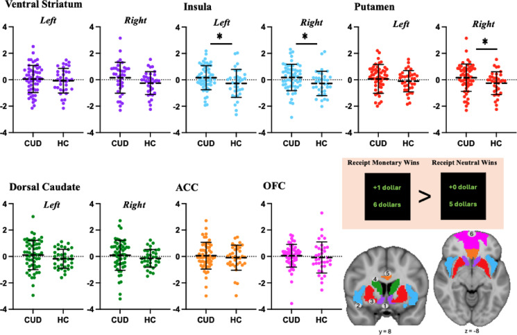
Individuals in the CUD group compared to controls, showed greater activity during the receipt of monetary wins vs. neutral wins in select ROIs, with moderate effect sizes (Fig. 1): insula (left, p < 0.05; d = 0.42; right p < 0.05; d = 0.47) and putamen (right: p < 0.05, d = 0.43).
Receipts of monetary wins vs. missed wins
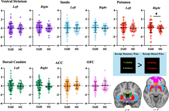
For the contrast receipt of monetary wins vs. missed wins, the CUD group showed greater activity than the control group in the putamen (right: p < 0.05; d = 0.28; small effect size; see Fig. 2).
Brain-behaviour correlations
No correlations between brain activity and other variables survived correction for multiple comparisons (Bonferroni corrections: 0.05/33 comparisons; adjusted p ≤ 0.00152). See Supplementary 2.4, Figure S7 for uncorrected brain-behaviour correlations.
Discussion
This fMRI study examined brain reward function for the first time in participants with a moderate-to-severe CUD who have attempted to quit or cut down their cannabis use. The individuals with CUD, compared to controls, showed greater activity bilaterally in the insula and right putamen while receiving monetary wins vs. neutral wins. The CUD vs. the control group also showed greater right putamen activity while receiving monetary wins vs. missed wins. Contrary to our hypothesis, there was no significant group difference while anticipating monetary cues vs. anticipating neutral cues. Overall, our findings suggest that alteration of reward processing in CUD may be specific to receiving rewards (and not to anticipating rewards). The findings partly align with prominent neuroscientific theories of addiction that postulate altered activity in habit-related (e.g., dorsal striatum) and interoceptive (e.g., insula) pathways during the ‘liking’/receipt phase of rewards11,15; but do not support neuroadaptations during the ‘wanting’ phase of reward processing in this sample.
Receipt of monetary wins and cannabis use
We found that the CUD vs. control group showed greater activity in the putamen and insula during the ‘liking’ phase of reward processing measured during the receipt of monetary wins. Specifically, greater putamen activity was found across both receipt contrasts (receiving monetary wins vs. neutral wins and missed wins), consistent with a previous study that found increased putamen activity in cannabis users vs. controls while receiving monetary wins20. Notably, the putamen (part of the dorsal striatum) is implicated in habit formation and automated/compulsive substance use11,43,44. Therefore, increased putamen activity during the receipt of monetary wins may reflect reinforcement processes that contribute to the development of habitual substance use.
We also found greater insula activity in the CUD group vs. controls during the receipt of monetary wins vs. the receipt of neutral wins. Although some previous studies have reported greater insula activity in cannabis users vs. controls during other reward/loss conditions21,26, no study has found a significant group difference in insula activity during the receipt of monetary wins (e.g.,20,24,25). Interestingly, the insula is implicated in arousal and interoception45, and is often altered in SUDs46. Therefore, greater insula activity while receiving monetary rewards in the CUD group may reflect increased awareness of bodily states or greater salience attribution. However, further research is needed to understand the role of insula activity while receiving monetary rewards in people with CUD.
Anticipation of monetary cues and cannabis use
No significant group differences were found in brain activity during the ‘wanting’ phase of reward processing, measured while anticipating monetary cues vs. neutral cues. This aligns with several previous studies24–27,47, but needs to be interpreted with caution. Indeed, brain activity was not robust within the control group for this contrast, suggesting that this contrast might not have robustly recruited neural pathways related to anticipation, possibly due to the lack of real monetary rewards.
At the behaviour level, we also found no differences in RTs when responding to monetary cues, but faster RTs towards neutral cues, suggesting that at a behavioural level, there might have been a blunting of anticipation of rewards; and a dissociation between the behavioural and neural correlates of reward anticipation. Our findings, in conjunction with existing evidence, demonstrate inconsistent alterations of the neural correlates of reward anticipation in CUD.
Strengths and limitations
A main strength of the current study is the novel sample of individuals with a moderate-to-severe CUD who have tried to cut down or quit using cannabis in the past two years. By understanding how those with more severe forms of CUD process rewards, treatment targets can be better informed.
Nevertheless, several important methodological limitations must be considered when interpreting these findings. First, the cross-sectional design limits conclusions about directionality; we cannot determine whether greater putamen and insula activity during the receipt phase of rewards reflects neuroadaptations from chronic cannabis use or pre-existing traits that increase vulnerability to CUD. Future longitudinal studies are needed to track how these patterns change over time and how they relate to changes in cannabis use.
Second, while providing a robust methodology to measure the activity of the brain’s reward neurocircuitry, the controlled fMRI environment, where participants performed the task, and the structure of the MID may not accurately reflect how people process rewards in real-life situations48.
Third, the results might reflect, instead of cannabis use-related effects on brain integrity, the effects of nicotine and alcohol use, and related problems, or other measures of hedonic tone (e.g., anhedonia). These factors may influence brain function independently and in interaction with cannabis16. Indeed, nicotine was consumed by most participants in the CUD group but by none in the control group; alcohol was consumed in greater amounts and on more days by participants in the CUD group than by controls; and we did not measure anhedonia. Further, we did not measure nicotine withdrawal because we did not ask participants to abstain from nicotine use, and we allowed participants to consume nicotine during scheduled breaks. Yet, we could not confirm that nicotine withdrawal in some participants might have affected brain reward function. Future work is required to unpack the independent and interactive effects of CUD, cannabis, alcohol, and nicotine consumption and related problems, and of distinct metrics of hedonic tone, on the neurocircuitry of reward processing. Fourth, the current study did not examine loss-related contrasts. Previous studies using the MID reported blunted neural responses to loss outcomes in cannabis users25,27. As such, future research should incorporate loss-related contrast (e.g., losing monetary rewards) to clarify whether CUD is associated with altered reward processing of negative outcomes.
Fifth, the strict exclusion criteria used during recruitment may have resulted in a sample that is not fully representative of individuals who endorse a CUD. While the exclusion criteria were designed to meet the objectives of the experiments, future research should include cannabis users with polydrug use and comorbid psychiatric disorders to better understand their impact on brain reward function and improve generalisability. Sixth, while the study employed robust metrics of cannabis consumption (e.g., TLFB49), reliance on self-report measures introduces the potential for underreporting biases due to recall, stigma, and lack of awareness50. Although urinary THC metabolites screening was included, which provides an objective indicator of recent cannabis use, future studies should incorporate international standardised quantification metrics, such as the iCannToolkit51, the ‘roll a joint paradigm’52,53, and tools that measure the amount of THC in participants’ cannabis products (e.g., Standard THC units42) to improve accuracy. Finally, consistent with prior work investigating reward processing using the MID fMRI task34, corrections for multiple comparisons were not applied to the exploratory ROI analyses. As such, results should be interpreted with caution.
Conclusions
We examined brain reward function for the first time in a sample of participants with a moderate-to-severe CUD with previous attempts to quit or cut down their cannabis use. The findings suggest that the CUD group showed greater activity in the putamen and insula during the receipt phase of rewards compared to controls. These current findings are partly in line with prominent neuroscientific theories of addiction that postulate altered activity in key brain reward regions during the receipt phase of monetary reward11,15. Future longitudinal neuroimaging studies are required to determine whether changes in brain reward function predate or follow the onset of CUD.
Supplementary Materials
References
- J Leung, GCK Chan, L Hides, WD Hall. What is the prevalence and risk of cannabis use disorders among people who use cannabis? A systematic review and meta-analysis. Addict. Behav., 2020. [DOI | PubMed]
- 2.United Nations Office on Drugs and Crime. World drug report 2024. Vienna, Austria: United Nations Office on Drugs and Crime, 2024.
- 3.American Psychiatric Association. Diagnostic and statistical manual of mental disorders. Arlington, VA (optional for online version: American Psychiatric Association; 2013.
- E Hoch, ND Volkow, CM Friemel, V Lorenzetti, TP Freeman, W Hall. Cannabis, cannabinoids and health: A review of evidence on risks and medical benefits. Eur. Arch. Psychiatry Clin. Neurosci., 2025. [DOI | PubMed]
- A Selamoglu, C Langley, R Crean, G Savulich, F Cormack, BJ Sahakian. Neuropsychological performance in young adults with cannabis use disorder. J. Psychopharmacol., 2021. [DOI | PubMed]
- M Skumlien, C Langley, W Lawn, V Voon, HV Curran, JP Roiser. The acute and non-acute effects of cannabis on reward processing: A systematic review. Neurosci. Biobehav. Rev., 2021. [DOI | PubMed]
- H Shao, H Du, Q Gan, D Ye, Z Chen, Y Zhu. Trends of the global burden of disease attributable to cannabis use disorder in 204 countries and territories, 1990–2019: Results from the disease burden study 2019. Int. J. Neuropsychopharmacol., 2023
- DT Myran, M Pugliese, AJ McDonald, J Xiao, B Fischer, Y Finkelstein. Cannabis use disorder emergency department visits and hospitalizations and 5-year mortality. JAMA Netw. Open, 2025. [DOI | PubMed]
- UW Preuss, MA Huestis, M Schneider, D Hermann, B Lutz, A Hasan. Cannabis use and car cashes: A review. Front. Psych., 2021. [DOI]
- CG Imhoff. Long-term outcomes of chronic, recreational marijuana use. Corinthian, 2020
- GF Koob, ND Volkow. Neurobiology of addiction: A neurocircuitry analysis. Lancet Psychiatry, 2016. [DOI | PubMed]
- K Blum, ER Braverman, JM Holder, JF Lubar, VJ Monastra, D Miller, DE Comings. The reward deficiency syndrome: A biogenetic model for the diagnosis and treatment of impulsive, addictive and compulsive behaviors. J. Psychoactive Drugs, 2000. [DOI]
- P Anselme. The effect of exposure to drugs on the processing of natural rewards. Neurosci. Biobehav. Rev., 2009. [DOI | PubMed]
- TE Robinson, KC Berridge. The incentive-sensitization theory of addiction 30 years on. Annu. Rev. Psychol., 2025. [DOI | PubMed]
- BJ Everitt, TW Robbins. Drug addiction: Updating actions to habits to compulsions ten years on. Annu. Rev. Psychol., 2016. [DOI | PubMed]
- IM Balodis, MN Potenza. Anticipatory reward processing in addicted populations: A focus on the monetary incentive delay task. Biol. Psychiatry, 2015. [DOI | PubMed]
- E Beyer, G Poudel, S Antonopoulos, H Thomson, V Lorenzetti. Brain reward function in people who use cannabis: A systematic review. Front. Behav. Neurosci., 2024. [DOI | PubMed]
- ME Martz, EM Trucco, LM Cope, JE Hardee, JM Jester, RA Zucker. Association of marijuana use with blunted nucleus accumbens response to reward anticipation. JAMA Psychiat., 2016. [DOI]
- HC Karoly, AD Bryan, BJ Weiland, A Mayer, A Dodd, SW Feldstein Ewing. Does incentive-elicited nucleus accumbens activation differ by substance of abuse? An examination with adolescents. Dev. Cogn. Neurosci., 2015. [DOI | PubMed]
- HH van Hell, M Vink, L Ossewaarde, G Jager, RS Kahn, NF Ramsey. Chronic effects of cannabis use on the human reward system: An fMRI study. Eur. Neuropsychopharmacol., 2010. [DOI | PubMed]
- L Nestor, R Hester, H Garavan. Increased ventral striatal BOLD activity during non-drug reward anticipation in cannabis users. Neuroimage, 2010. [DOI | PubMed]
- KC Berridge, TE Robinson. Parsing reward. Trends Neurosci., 2003. [DOI | PubMed]
- B Knutson, JC Cooper. Functional magnetic resonance imaging of reward prediction. Curr. Opin. Neurol., 2005. [DOI | PubMed]
- M Skumlien, C Mokrysz, TP Freeman, MB Wall, M Bloomfield, R Lees. Neural responses to reward anticipation and feedback in adult and adolescent cannabis users and controls. Neuropsychopharmacology, 2022. [DOI | PubMed]
- B Enzi, S Lissek, MA Edel, M Tegenthoff, V Nicolas, N Scherbaum. Alterations of monetary reward and punishment processing in chronic cannabis users: an FMRI study. PLoS ONE, 2015. [DOI | PubMed]
- FM Filbey, J Dunlop, US Myers. Neural effects of positive and negative incentives during marijuana withdrawal. PLoS ONE, 2013. [DOI | PubMed]
- SW Yip, EE DeVito, H Kober, PD Worhunsky, KM Carroll, MN Potenza. Pretreatment measures of brain structure and reward-processing brain function in cannabis dependence: An exploratory study of relationships with abstinence during behavioral treatment. Drug Alcohol Depend., 2014. [DOI | PubMed]
- V Lorenzetti, A Batalla, J Cousijn. Cannabis use disorders and altered brain morphology: Where is the evidence?. Curr. Addict. Rep., 2016. [DOI]
- V Lorenzetti, J Cousijn, N Solowij, H Garavan, C Suo, M Yucel. The neurobiology of cannabis use disorders: A call for evidence. Front. Behav. Neurosci., 2016. [DOI | PubMed]
- JR Hughes, S Naud, AJ Budney, JR Fingar, PW Callas. Attempts to stop or reduce daily cannabis use: An intensive natural history study. Psychol. Addict. Behav., 2016. [DOI | PubMed]
- MA Bloomfield, CJ Morgan, S Kapur, HV Curran, OD Howes. The link between dopamine function and apathy in cannabis users: an [18F]-DOPA PET imaging study. Psychopharmacology, 2014. [DOI | PubMed]
- C Gorey, L Kuhns, E Smaragdi, E Kroon, J Cousijn. Age-related differences in the impact of cannabis use on the brain and cognition: a systematic review. Eur. Arch. Psychiatry Clin. Neurosci., 2019. [DOI | PubMed]
- AM Francis, JN Bissonnette, SE MacNeil, CE Crocker, PG Tibbo, DJ Fisher. Interaction of sex and cannabis in adult in vivo brain imaging studies: A systematic review. Brain Neurosci. Adv., 2022. [DOI | PubMed]
- JM Hoogendam, RS Kahn, MH Hillegers, M van Buuren, M Vink. Different developmental trajectories for anticipation and receipt of reward during adolescence. Dev. Cogn. Neurosci., 2013. [DOI | PubMed]
- S Oldham, C Murawski, A Fornito, G Youssef, M Yucel, V Lorenzetti. The anticipation and outcome phases of reward and loss processing: A neuroimaging meta-analysis of the monetary incentive delay task. Hum. Brain Mapp., 2018. [DOI | PubMed]
- 36.First MB, Williams JB, Karg RS, Spitzer RL. SCID-5-RV. 2015.
- 37.Babor TF, Higgins-Biddle JC, Saunders JB, Monteiro MG, Organization WH. AUDIT: the alcohol use disorders identification test: guidelines for use in primary health care. World Health Organization, 2001.
- B Knutson, CM Adams, GW Fong, D Hommer. Anticipation of increasing monetary reward selectively recruits nucleus accumbens. J. Neurosci., 2001. [DOI | PubMed]
- O Esteban, CJ Markiewicz, RW Blair, CA Moodie, AI Isik, A Erramuzpe. fMRIPrep: A robust preprocessing pipeline for functional MRI. Nat. Methods, 2019. [DOI | PubMed]
- R Heller, D Stanley, D Yekutieli, N Rubin, Y Benjamini. Cluster-based analysis of fMRI data. Neuroimage, 2006. [DOI | PubMed]
- N Tzourio-Mazoyer, B Landeau, D Papathanassiou, F Crivello, O Etard, N Delcroix. Automated anatomical labeling of activations in SPM using a macroscopic anatomical parcellation of the MNI MRI single-subject brain. Neuroimage, 2002. [DOI | PubMed]
- TP Freeman, V Lorenzetti. ‘Standard THC units’: A proposal to standardize dose across all cannabis products and methods of administration. Addiction, 2020. [DOI | PubMed]
- 43.Ghandili M, Munakomi S. Neuroanatomy, Putamen. StatPearls. Treasure Island (FL): StatPearls Publishing; 2024.
- CM Gillan, TW Robbins, BJ Sahakian, OA van den Heuvel, G van Wingen. The role of habit in compulsivity. Eur. Neuropsychopharmacol., 2016. [DOI | PubMed]
- V Droutman, SJ Read, A Bechara. Revisiting the role of the insula in addiction. Trends Cogn. Sci., 2015. [DOI | PubMed]
- AM Herman. Interoception within the context of impulsivity and addiction. Curr. Addict. Rep., 2023. [DOI | PubMed]
- G Jager, RI Block, M Luijten, NF Ramsey. Tentative evidence for striatal hyperactivity in adolescent cannabis-using boys: A cross-sectional multicenter fMRI study. J. Psychoactive Drugs, 2013. [DOI | PubMed]
- ML Elliott, AR Knodt, AR Hariri. Striving toward translation: Strategies for reliable fMRI measurement. Trends Cogn. Sci., 2021. [DOI | PubMed]
- LC Sobell, MB Sobell. Timeline Follow-Back. Measuring Alcohol Consumption, 1992
- JJ Palamar, A Le. Self-correction of unreported marijuana use by participants taking a street intercept survey. Am. J. Drug Alcohol Abuse, 2020. [DOI | PubMed]
- V Lorenzetti, C Hindocha, K Petrilli, P Griffiths, J Brown, A Castillo-Carniglia. The international cannabis toolkit (iCannToolkit): A multidisciplinary expert consensus on minimum standards for measuring cannabis use. Addiction, 2022. [DOI | PubMed]
- D Dawson, D Stjepanovic, V Lorenzetti, WD Hall, J Leung. How much cannabis is used in a joint in Australia? An experimental investigation into use by potency and frequency. Drug Alcohol Rev., 2024. [DOI | PubMed]
- LJ Nestor, B Behan, J Suckling, H Garavan. Cannabis-dependent adolescents show differences in global reward-associated network topology: A functional connectomics approach. Addict. Biol., 2020. [DOI | PubMed]