
Cannabidiol inhibits both human KV7.1 and KV7.1/KCNE1 channels through distinct sites
Abstract
Several essential physiological systems express voltage-gated potassium channels within the KV7 family (comprising KV7.1–7.5), sometimes also co-assembled with auxiliary subunits in the KCNE family (comprising KCNE1–5). An ongoing challenge to KV7 drug development is creating subtype-selective compounds to limit adverse effects. Prior work has shown that the antiepileptic cannabidiol (CBD), a pan-KV7 modulator, inhibits cardiac- and epithelia-associated KV7.1 and KV7.1/KCNE1 channels, while activating neuronal KV7 subtypes (KV7.2–7.5). However, little is known about the binding sites through which CBD mediates inhibitory effects on KV7.1 and KV7.1/KCNE1, limiting insight towards the development of selective KV7 modulators. To address this knowledge gap, we used a combination of the Chai-1 artificial intelligence model (to generate CBD binding site predictions in human KV7.1 and KV7.1/KCNE1 channels), site-directed mutagenesis and electrophysiology of these channels expressed in Xenopus laevis oocytes (to corroborate CBD binding site predictions), and molecular dynamics simulations (to study the biophysical mechanisms underlying CBD binding). We found that CBD binds to two unique sites within KV7.1 and KV7.1/KCNE1. In KV7.1 alone, CBD was bound to an intrasubunit S5–S6 pore domain binding site; referred to as the S5–S6 site. In KV7.1/KCNE1, the addition of the KCNE1 subunit created a novel binding site for CBD, sandwiched between two KV7.1 subunits and one KCNE1 subunit; referred to here as the S6–S5’–E1 site. Molecular dynamics simulations showed that CBD binding to the S6–S5’–E1 KV7.1/KCNE1 site closes off the KV7.1 S5–S6 site. A sequence comparison between KV7 channels revealed key amino acid differences at both the S5–S6 and S6–S5’–E1 sites relative to neuronal KV7s. These support the notion that CBD binds differently in KV7.1 and KV7.1/KCNE1 channels in accordance with its unique inhibitory pharmacological effects on these channels compared to the activating effect in neuronal KV7s. Thus, we provide support for KV7.1 and KV7.1/KCNE1 being inhibited by CBD via distinct binding sites, which can guide future research focused on the rational development of drugs that avoid inhibitory effects on KV7.1 and KV7.1/KCNE1 channels or utilize these sites to modulate channel activity.
Article type: Research Article
Keywords: electrophysiology, molecular modelling, binding site, cannabidiol, KCNQ1, KCNE1
Affiliations: https://ror.org/05ynxx418grid.5640.70000 0001 2162 9922Department of Biomedical and Clinical Sciences, Linköping University, SE-581 85 Linköping, Sweden; https://ror.org/026vcq606grid.5037.10000 0001 2158 1746Department of Applied Physics, Science for Life Laboratory, KTH Royal Institute of Technology, Stockholm, Sweden
License: © The Author(s) 2026 CC BY 4.0 Open Access This article is licensed under a Creative Commons Attribution 4.0 International License, which permits use, sharing, adaptation, distribution and reproduction in any medium or format, as long as you give appropriate credit to the original author(s) and the source, provide a link to the Creative Commons licence, and indicate if changes were made. The images or other third party material in this article are included in the article’s Creative Commons licence, unless indicated otherwise in a credit line to the material. If material is not included in the article’s Creative Commons licence and your intended use is not permitted by statutory regulation or exceeds the permitted use, you will need to obtain permission directly from the copyright holder. To view a copy of this licence, visit http://creativecommons.org/licenses/by/4.0/.
Article links: DOI: 10.1038/s41401-025-01742-0 | PubMed: 41776084 | PMC: PMC13279787
Relevance: Relevant: mentioned in keywords or abstract
Full text: PDF (5.9 MB)
Introduction
The KV7.1/KCNE1 channel, generating the slow-delayed component of the outward potassium current (IKs), plays a crucial role in cardiac repolarisation [ref. 1, ref. 2]. Drug-induced inhibition of the KV7.1/KCNE1 channel may result in proarrhythmic cardiac action potential prolongation. Indeed, clinically used drugs like the anti-inflammatory celecoxib and the anti-malarial quinidine, which are known to inhibit KV7.1/KCNE1 channels, increase the risk of adverse cardiac events [ref. 3, ref. 4]. In contrast, KV7.1/KCNE1 channel activators have been put forward as promising compounds for treating cardiac arrhythmias caused by impaired KV7.1/KCNE1 channel function or arrhythmias related to delayed cardiac repolarisation in general [ref. 5–ref. 7]. Besides the heart, KV7.1 is expressed in other tissues like epithelia, either co-assembled with other KCNE subunits or potentially as KV7.1 alone [ref. 8, ref. 9]. Evidently, detailed knowledge of drug binding sites in KV7.1 and KV7.1/KCNE1 is vital to avoid adverse effects and drive rational drug development initiatives.
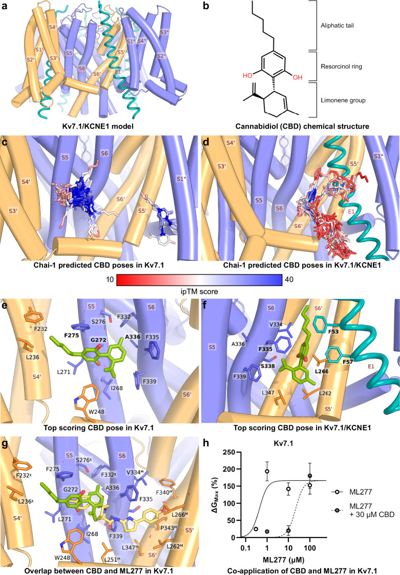
The KV7.1/KCNE1 channel is co-assembled from the voltage-gated potassium (KV) channel α-subunit KV7.1, encoded by the KCNQ1 gene, and the auxiliary subunit KCNE1, encoded by the KCNE1 gene. The KV7.1 subunits are arranged as a tetramer in an interleaved domain-swapped conformation. This creates interfaces between several KV7 subunits; therefore, we label the helices from subunits 2 and 3 with the suffixes ‘ and “, respectively, to differentiate them from subunit 1 in all figures (exemplified in Fig. 1a). Each subunit contains six transmembrane helices (S1–S6), the S1–S4 helices comprise the voltage-sensing domain (VSD) while the S5–S6 helices form the ion-conducting pore domain (PD) [ref. 10]. KV7.1 functions as a KV channel by itself, however, co-assembly with up to four KCNE1 subunits (Fig. 1a), confers biophysical properties characteristic of the native IKs (like slow activation kinetics and shifted voltage-dependence of channel activation) [ref. 2].
The challenge in developing drugs targeting KV7 channels, including KV7.1 and KV7.1/KCNE1, lies in the difficulty of making channel subtype-selective compounds. This is highlighted by the anti-epileptic drug retigabine, which was withdrawn from the market due to adverse effects, like urinary retention [ref. 11]. These adverse effects are thought to occur because retigabine binds to KV7.2–KV7.5 channels rather than being selective for the neuronal KV7.2/7.3 heteromeric channels [ref. 11]. The retigabine binding site has been extensively characterised, and was ultimately resolved in the KV7.2-bound retigabine structures [ref. 12]. Ligands at the site resolved in complex with KV7.2 include: retigabine, CBD, HN37, Ebio1, Ebio2 and Ebio3 [ref. 12–ref. 15]. This detailed binding site characterisation has enabled rational drug development initiatives leading to the discovery of more efficacious subtype-selective compounds than retigabine [ref. 14, ref. 16]. In contrast, to date, KV7.1 structures have been resolved with only one activator, ML277 [ref. 17, ref. 18], and there are no KV7.1/KCNE1 structures with ligands (although two studies with apo structures were published at the time of writing) [ref. 19, ref. 20], further adding to the challenge of rational drug development initiatives.
A compound with notable parallels to retigabine is the phytocannabinoid cannabidiol (CBD, Fig. 1b). Both have anti-epileptic effects that are thought to result from their activation of the KV7.2/7.3 channels [ref. 21–ref. 23]. We have previously shown that, like retigabine, CBD affects the KV7.2–KV7.5 channels differently compared to KV7.1 and KV7.1/KCNE1. Specifically, CBD activates KV7.2 and KV7.3 by shifting the voltage-dependence of activation to more negative potentials (−ΔV50) and activates KV7.4 and KV7.5 by increasing maximum channel conductance (+ΔGMax). In a previous study of human KV7.1 or KV7.1/KCNE1 channels expressed in Xenopus laevis oocytes, we found that CBD reduced the GMax of KV7.1/KCNE1 in a concentration-dependent manner with a concentration causing half-maximal inhibition (IC50) of 14 µM, and more potently inhibited the KV7.1 channel (IC50 = 6 µM) [ref. 24]. Given CBD’s distinct pharmacological effects on KV7.1 and KV7.1/KCNE1 channels compared to KV7.2–7.5, a detailed binding site characterisation may offer key molecular insights and guide the development of subtype-selective KV7-targeted drugs.
In KV7.2, recent cryo-EM structures have resolved that CBD binds at two adjacent binding sites in the PD [ref. 13]. One CBD molecule binds in the same site as retigabine, while another one binds just above it (when viewing from the cross-section of the membrane). No structures of CBD bound to KV7.1 are currently available. However, we have previously narrowed down the binding site to the PD of KV7.1, but were unable to differentiate between several putative binding sites at the time [ref. 24]. Moreover, the binding site for CBD in KV7.1/KCNE1 remains uncharacterised. To address this, we conducted a thorough binding site investigation using a combination of computational molecular modelling and electrophysiology-based methods. This included using recent innovations in artificial intelligence (AI)–based structural prediction methods to make initial binding site predictions. Then we refined our prediction through molecular docking to a KV7.1 structure and a KV7.1/KCNE1 homology model and used molecular dynamics (MD) simulations to identify the most stable binding modes among our predictions. To validate our predictions, we utilised site-directed mutagenesis, electrophysiological measurements of human KV7.1 and KV7.1/KCNE1 channels expressed in Xenopus laevis oocytes, and pharmacological co-application experiments. We found that CBD binds in an S5–S6 intrasubunit site in KV7.1, and that KCNE1 co-assembly creates a novel binding site for CBD located at the intersubunit interface of S5 and S6 of adjacent KV7.1 subunits and the KCNE1 subunit. Intriguingly, this binding site in KV7.1/KCNE1 overlaps the canonical retigabine binding site in the KV7.2–7.5 channels. These previously uncharacterised CBD binding sites in the KV7.1 and KV7.1/KCNE1 channels provide insights into the inhibiting effect of CBD on these channels and should aid future efforts to design KV7 subtype-selective drugs.
Materials and methods
Molecular biology
Xenopus laevis frog oocytes were harvested, prepared, and selected in accordance with published procedures [ref. 25] and established permits (#1941 and #14515) from the regional ethics board of Linköping, Sweden, or purchased from EcoCyte Bioscience, Germany. Plasmids containing human KCNE1 and KCNQ1 were linearized, purified, and in vitro transcribed (T7 mMessage mMachine, Invitrogen) to RNA. For channel mutants, plasmids were mutated via site-directed mutagenesis (QuickChange II XL, Agilent) with mismatched primers (Invitrogen), and then amplified in a commercial E. coli bacterial line, XL10-Gold Ultracompetent (Agilent). Nucleic acid concentrations were determined using spectrophotometry (NanoDrop 2000c, Thermo Scientific). Plasmids were sequenced to confirm mutagenesis via Sanger sequencing at the Linköping University Core Facility. RNA was injected into defolliculated oocytes using a microinjector either with 50 ng of KV7.1 or a KCNE1-saturating mixture of 25 ng:8 ng KV7.1 with KCNE1. For mutants with low current expression, RNA concentrations were doubled. RNA for KV7.1 A336G/KCNE1 injection was diluted to a KV7.1/KCNE1 ratio of 0.5 ng:8 ng due to high expression after 2 days. Oocytes expressing KV7.1 or KV7.1/KCNE1 were incubated for 1–3 days at 16 °C, and measured either directly afterwards or kept at 8 °C.
Electrophysiology
The two-electrode voltage clamp technique was applied to oocytes expressing KV7.1 or KV7.1/KCNE1 channels after an acclimatising time of 15 min at room temperature in the control extracellular solution. Cells were penetrated with 0.2–1.6 MΩ borosilicate glass tipped Ag/AgCl-electrodes backfilled with 3 M KCl. Signals were amplified with a TEC-10CX (NPI Electronic) amplifier and filtered with a 1 kHz Bessel low-pass filter. Stimulation protocols via Clampex 11.2 software (Molecular Devices) for establishing current traces at differing voltages for KV7.1 consisted of a holding voltage (−80 mV), pre-pulse (−100 mV, 2 s), test pulse (−100 mV to +70 mV, +10 mV with each repeat, 3 s) and tail voltage (−30 mV, 1 s), with 17 repeats (15 s sweep-to-sweep interval). For KV7.1/KCNE1, the protocol was: holding voltage (−80 mV), test pulse (−80 mV to +80 mV, +10 mV with each repeat, 5 s), and tail voltage (−30 mV, 5 s), with 17 repeats (30 s sweep-to-sweep). For select mutants with shifted voltage dependence, the standard protocol was extended to more positive test pulse voltages (up to +130 mV). The extracellular control solution contained (in mM): 88 NaCl, 1 KCl, 0.4 CaCl2, 0.8 MgCl2, 15 HEPES, with pH 7.4 set by NaOH. Extracellular control solution was perfused continuously during recordings (1 mL/min). Control solution supplemented with CBD (commercially obtained from Chiron/Sigma-Aldrich or synthesized in-house according to a previous protocol [ref. 26] with >98% purity) and/or ML277 (Tocris) was applied using a repeated voltage-step protocol (KV7.1: −20 mV for 3 s, KV7.1/KCNE1: +40 mV for 5 s) with a holding voltage of −80 mV (sweep-to-sweep: 10 s). CBD and ML277 were dissolved in a 25 mM DMSO stock solution and stored at −20 °C until use. Dilutions of CBD and/or ML277 in control solutions were used immediately after dilution. The application was continued until changes to the current morphology were stabilized or after a maximum of 7 min if no change was observed (time point chosen based on CBD’s application time course). To study CBD effects in each oocyte, a stimulation protocol was applied to determine baseline channel behaviour before the solution exchange, and a stimulation protocol was applied after the solution exchange. Changes to recording conditions are stated where applicable.
Experimental analysis
Leak compensation was used, or cells were excluded from measurement if the leak was >600 nA. Tail currents (currents at the beginning of the tail pulse, but after the transient spike) were plotted against their preceding test pulse step voltage to establish a tail current vs voltage relationship, as an approximation of the conductance vs voltage (G(V)) relationship. Data points were fitted with a sigmoidal Boltzmann curve:
G\left(V\right)={G}_{{Min}}+\frac{({G}_{{Max}}-{G}_{{Min}})}{[1+{e}^{\frac{{V}_{50}-V}{S}}]}
\]
where GMin is the minimum conductance, GMax is the maximum conductance, V50 is the voltage generating half maximal conductance, and s is the slope of the curve (intrinsic properties of mutants in SI Tables 1, 2). Changes to GMax and V50 were determined for each cell individually from the analysis of baseline tail currents (before CBD exposure) and tail currents after CBD exposure. ΔGMax is calculated as % of the current increase/decrease in baseline GMax, where 0% denotes no change. ΔV50 is calculated as baseline V50 subtracted by CBD-exposed V50, where 0 mV denotes no change. Likewise, Δs denotes a subtraction of the slope measured in the presence of CBD from the slope s of the baseline recording.
Concentration-response relationships for CBD or ML277 effects on GMax were fitted using a Hill equation:
\Delta {G}_{{Max}}=\frac{{E}_{{Max}}}{1+\,{\left(\frac{{C}_{50}}{C}\right)}^{{{{\rm{H}}}}}}
\]
where EMax is the maximum observed response (i.e. maximal reduction in GMax for CBD or maximal increase in GMax for ML277, respectively), C50 is the concentration at which 50% of the response is reached (corresponds to IC50 for CBD and EC50 for ML277, respectively), C is the applied concentration, and H indicates the Hill slope.
For the L266W mutant with and without KCNE1, which showed linearly time-dependent (CBD-independent) current run-up, this was corrected for as previously described [ref. 24]. For mutants that showed non-linear time-dependent (CBD-independent) current run-up that may influence the interpretation of CBD effects, putative CBD effects were compared to time-matched control experiments in the absence of CBD and referred to in those cases.
Statistical analysis
We used Welch’s ANOVA and Dunnett’s T3 to test and compare the effects of CBD on mutant channels relative to wild-type (WT) channels. Student’s t-test with Welch’s correction was used for comparison of two experimental conditions. The symbol abbreviations used within the manuscript designating statistical significance are ns: P > 0.05, *P < 0.05, **P < 0.01, ***P < 0.001, ****P < 0.0001. n denotes the number of oocytes, which was ≥5 for each data set. Every mutant and experimental condition included oocytes from at least two separate batches of oocytes. Experimental data are shown as mean ± SEM if not stated otherwise. The selected representative examples have parameters within one SD of V50, ΔGMax, and ΔV50 of the sample mean.
Prediction of CBD binding sites in KV7 and KV7.1/KCNE1
The Chai-1 tool [ref. 27] was used to predict models of human KV7.1 and KV7.1/KCNE1 in complex with four CBD molecules. For the KV7.1 subunit, the prediction encompassed residues T104 to Q361, reflecting the first resolved N-terminal residue to the end of the S6 helix in the KV7.1 structures [ref. 10]. For the KCNE1 subunit, the prediction encompassed residues P35–K69, reflecting the helical portion predicted to interact with the transmembrane domain of KV7.1. For KV7.1 and KV7.1/KCNE1 in complex with CBD, 100 models were generated (20 seeds with 5 model predictions each) and ranked by the interface predicted template (ipTM) score [ref. 28] of the CBD molecules. The top-scoring complexes were then selected for simulations.
Molecular docking
In addition to the CBD-bound models predicted using Chai-1, we generated docking poses for CBD in KV7.1 and KV7.1/KCNE1. Here, we used the KV7.1 structure in the closed pore conformation (PDB: 6UZZ) [ref. 10] and constructed a model of KV7.1/KCNE1 based on the KV7.1/KCNE3 structure in the closed pore conformation (PDB: 6V01) [ref. 10]. In brief, 100 models of KV7.1/KCNE1 were created and ranked by the intersection of the discrete optimised protein energy (DOPE) and molecular probability density function (MolPDF) scores using the Modeller program [ref. 29], and the top-ranking model was selected for docking. The DOPE score is calculated from a statistical potential based on interatomic distances; meanwhile, the MolPDF score is an energy-based metric derived from the sum of all restraints in each model. Both provide a means to assess the quality of a protein model. The sequence lengths of the KV7.1 structure and KV7.1/KCNE1 model were truncated to match the Chai-1 predictions. A comparison between the KV7.1 structure and KV7.1/KCNE1 model and their respective Chai-1 predictions is provided (Figure S1). The Kv7.1 structure and KV7.1/KCNE1 model were prepared for docking using the protein preparation wizard implemented in Maestro (Schrödinger Release 2022-2: Maestro, Schrödinger, LLC, New York, NY, 2022). This involved adding missing hydrogen atoms and assigning the tautomeric states of titratable residues at pH = 7.0. The CBD molecule was docked using the induced fit docking protocol [ref. 30] with the extended sampling option that generates up to 80 docking poses. The docking sites were based on the top-scoring poses from the Chai-1 prediction.
Molecular dynamics simulations
The top-scoring docked, and Chai-1 poses for CBD in KV7.1 and KV7.1/KCNE1 were prepared for molecular dynamics simulations. Additionally, we prepared the apo KV7.1 structure (PDB: 6UZZ) and KV7.1/KCNE1 model based on the KV7.1/KCNE3 structure (PDB: 6V00). Each complex and apo protein was placed in a simulation box 140 Å× 140 Å× 100 Å in size using the CHARMM-GUI webserver [ref. 31]. This comprised a palmitoyl-2-oleoyl-sn-glycero-3-phosphocholine (POPC) lipid bilayer and TIP3P water [ref. 32] with 0.15 M KCl solution and excess Cl ions to neutralise the overall charge. The system was described using the CHARMM36M [ref. 33] and CHARMM36 (lipids) forcefields; parameters for CBD were generated using CgenFF [ref. 34]. Simulations were conducted using the GROMACS program [ref. 35], simulation parameters for non-bonded interactions, pressure, and temperature during both equilibration and production followed the CHARMM-GUI defaults [ref. 36]. The default CHARMM-GUI equilibration protocol involved gradually withdrawing positional restraints on protein, lipid, and ligand atoms. In the last step, where 50 kJ· mol−1 ·nm−1 restraints are applied to the protein backbone and ligand heavy atoms, the simulation was extended from 0.5 to 10 ns. Following equilibration, production simulations were conducted for 500 ns in triplicate using a 2 fs timestep.
Simulation analysis
The root mean square deviation (RMSD) and distance measurement analyses were conducted using the MDAnalysis tool [ref. 37]. Clustering analyses were conducted using the “gromos” method [ref. 38] implemented in GROMACS, an RMSD cutoff of 2.0 Å was selected for forming clusters. This method works by counting the number of neighbours for each structure using a cut-off distance, selecting the structure with the highest number of neighbours and removing this structure together with its neighbours from the pool of structures. This is repeated until all structures are sorted into clusters; the structure with the highest number of neighbours from each cluster is called the “central structure”. Molecular interactions of CBD within the clusters were calculated using the ProLIF tool [ref. 39]. The definitions for counting H-bonds, π-stacking interactions, hydrophobic interactions, and van der Waals (vdW) contacts are provided in Fig. S2. To calculate binding free energies, molecular mechanics/Poisson–Boltzmann surface area (MM/PBSA) calculations were performed using the gmx_MMPBSA tool [ref. 40]. Illustrations were made using the PyMOL program (The PyMOL Molecular Graphics System, Version 2.5 Schrödinger LLC). Values reported for analyses of unclustered MD simulations reflect mean ± SD from each of the 4 binding sites of the 3 simulation replicas (n = 12).
Results
CBD is predicted to bind to the PD of KV7.1 and KV7.1/KCNE1
To predict potential binding sites for CBD in KV7.1 and KV7.1/KCNE1, we generated models of CBD in complex with the two proteins using the Chai-1 tool [ref. 27]. The top 50 models from a total of 100 were ranked by the ipTM score of the CBD molecules in KV7.1 and KV7.1/KCNE1 (Fig. 1c, d). When predicted with KV7.1 alone, CBD was bound primarily adjacent to the S5–S6 helices of each subunit, which we refer to as the “S5–S6 site”. When predicted with the KV7.1/KCNE1 complex, CBD bound primarily at the interface of the KCNE1 subunit and two KV7.1 subunits. This site is formed by the S6 helix of the left subunit, the S5’ helix of the right subunit, and the KCNE1 subunit. We refer to this as the “S6–S5’–E1 site”. The top-scoring poses for CBD in complex with KV7.1 show that the methyl group of the limonene moiety was positioned between G272 from S5 and A336 from S6, forming hydrophobic interactions with both. The resorcinol ring rested against G272 and formed π-stacking with F275 (Fig. 1e). Furthermore, hydrophobic interactions were observed between the limonene group and F335/F339 and between the hydrophobic tail of CBD and F232/L236. The top-scoring pose for CBD in complex with KV7.1/KCNE1 exhibited hydrophobic interactions between the limonene moiety and L266 and F335 from KV7.1, and between the resorcinol ring and F53 and F57 from KCNE1. Furthermore, the hydroxy group oxygen of the resorcinol is 4.6 Å from the hydroxy oxygen of S338, suggesting that a small shift in the position of CBD would enable H-bond interactions (Fig. 1f).
The structure of KV7.1 has been resolved in complex with the activator ML277, which binds at a PD-spanning site [ref. 17]. The availability of ML277-bound KV7.1 structures enables comparison to the proposed CBD binding mode in KV7.1. This revealed that the predicted CBD binding mode partially overlapped with ML277 in KV7.1 (Fig. 1g). To test whether CBD and ML277 indeed use the same binding pocket, we conducted competition experiments between CBD and ML277 (Fig. 1h). For ML277 alone, clear effects were seen at 1 µM as an increase of GMax by 193% ± 28%. In contrast, for cells pretreated and co-exposed with 30 µM CBD, GMax was only increased by 18% ± 3% by 1 µM ML277 (Fig. 1h). In the presence of 30 µM CBD, the EC50 of ML277 was shifted by about two orders of magnitude, suggesting that CBD and ML277 compete for the same binding site, in accordance with our predictions.
KV7.1 mutagenesis corroborates CBD-interacting residues in the S5–S6 site
To test the validity of the proposed binding site in KV7.1, we mutated residues predicted to interact with CBD in the S5–S6 site. Given that CBD was predicted to interact with two opposing residues between the S5 and S6 helices, G272 and A336, respectively, we mutated these residues. We hypothesised that changing G272 to a bulkier residue, cysteine (G272C), may reduce the space available for CBD to bind. G272C had previously been reported to abolish ML277 effects [ref. 18, ref. 41]. In line with this, the G272C mutation decreased the GMax reduction induced by 30 µM CBD, from −57% ± 2% for WT to −39% ± 2% for G272C (Fig. 2a, Figure S3 for representative examples of CBD effects on mutants), due to a decrease in CBD’s efficacy for G272C (Fig. S4). For A336, we hypothesized that a smaller residue, glycine (A336G), may increase the space for CBD to bind. Strikingly, we observed a complete removal of the inhibitory effect of CBD on GMax for 30 µM CBD (ΔGMax = +16% ± 5%, Fig. 2a, Fig. 2c, d for a representative example of CBD effect on WT and the A336G mutant), and a smaller inhibition than WT for 100 µM CBD (highest concentration tested, WT: ΔGMax = −62% ± 14% vs. A336G: ΔGMax = −18% ± 6%). The small apparent increase in GMax for 30 µM was not significantly different from time-matched control experiments (Fig. S5).
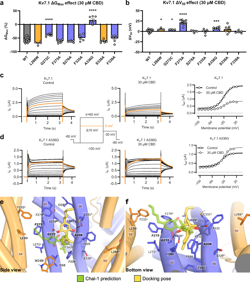
We also generated mutants of other residues in the S5–S6 site. This included mutating F275, predicted to form π-stacking interactions with CBD, to alanine (F275A). This mutation did not change the GMax effect of 30 µM CBD (ΔGMax = −58 ± 3%), but induced a clear shift in the voltage dependence of channel activation (V50), which was not seen in WT (from ΔV50 = +1.4 ± 0.8 mV for WT to +21.6 ± 1.9 mV for F275A, Fig. 2b). Adjacent to F275, we tested whether the hydroxy sidechain of S276 could form an H-bond with CBD by mutating it to S276A. The mutation was not found to impact the CBD response compared to WT (Fig. 2a, b). Adjacent to A336 is F335, which formed hydrophobic interactions to CBD. Mutating F335 did not change the CBD response compared to the WT when mutated to alanine (Fig. 2a, b). The WT-like response of F335A was in contrast to our previous study [ref. 24], which is likely a result of varied CBD loss in different experimental systems (see Discussion for further details). Additionally, we tested F339, located one helical turn below F335 and adjacent to the limonene group of CBD. Mutating this residue to alanine did not appear to alter CBD response compared to WT.
To verify the absence of CBD binding to the S6–S5’–E1 site in KV7.1 alone, we assessed the CBD effect on S338A (S6) and L266W (S5). Neither mutant changed the GMax effect of 30 µM CBD (Fig. 2a). In summary, the site-directed mutagenesis experiments suggest that several residues in the S5–S6 site (G272, F275, A336) are important for CBD binding or effects on KV7.1 alone, while residues in the S6–S5’–E1 site are not.
After validating that the S5–S6 site is important for binding CBD in KV7.1, we generated alternative docking poses for CBD at this site using an induced fit docking protocol. This protocol samples alternative residue conformations in the binding site and generates docking poses not otherwise accessible with the original residue conformations. Here, we used the KV7.1 structure in the closed pore conformation (PDB: 6UZZ) [ref. 10]. The top-scoring docking pose shows that CBD is able to bind deeper into the binding pocket than predicted by the Chai-1 tool (Fig. 2e, f). Like what we concluded after using Chai-1 prediction, the predicted CBD docking pose is only compatible with the absence of ML277 in the binding site (Fig. S6). The docking pose of CBD is oriented vertically, lining the groove between the S5 and S6 helices, while in the Chai-1 prediction, CBD is perpendicular to the helices. The vertical CBD binding pose predicted through flexible docking is supported by H-bond interactions from the hydroxy groups of the resorcinol to the backbone carbonyl oxygens of G272 and C331. The limonene group of CBD penetrates deeper into the pocket between G272 and A336 (Fig. 2e, f). Both in the Chai-1 and docking poses, the limonene group of CBD forms hydrophobic interactions with F335/F339. This alternative binding pose shifts the aliphatic tail away from F232, L236, and F275 compared to the Chai-1 prediction. We explore the implications of this through MD simulations in a subsequent section.
KV7.1/KCNE1 mutagenesis corroborates CBD interacting primarily with residues in the S6–S5’–E1 site
As with KV7.1, we experimentally tested the binding site prediction for CBD in KV7.1/KCNE1. Firstly, we examined S338, which is near CBD in the KV7.1/KCNE1 model and was observed to have no impact on the CBD effect in KV7.1 alone. Unlike in KV7.1, in KV7.1/KCNE1 the S338A mutant yielded a channel that was more strongly inhibited by 30 µM CBD than WT (ΔGMax = −89% ± 12% for S338A compared to −39% ± 4% for WT, Fig. 3a, Figure S7 for representative examples of CBD effects on mutants) and responded to CBD with a more pronounced V50 shift (ΔV50 = −11.1 ± 1.8 mV for S338A compared to +5.4 ± 0.8 mV for WT, Fig. 3b). We hypothesised that a much bulkier residue at position 338, tryptophan (S338W), would displace CBD from the site. In line with this, the S338W mutant was not inhibited by 30 µM CBD (ΔGMax = +43% ± 6%, Fig. 3a, Fig. 3c, d for a representative example of CBD effect on WT and the S338W mutant) or 100 µM CBD (ΔGMax = +20% ± 11%). The apparent increase in GMax was not significantly different from time-matched control experiments (Fig. S5).
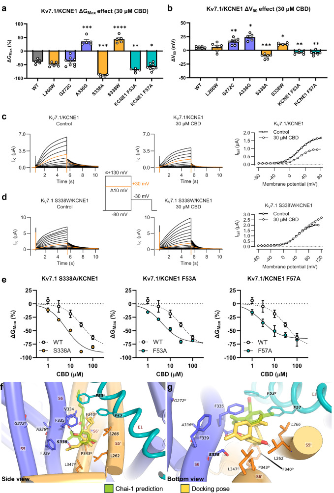
To assess if KCNE1 contributes to the S6–S5’–E1 CBD binding site, we tested the KCNE1 mutants F53A and F57A, both predicted to form hydrophobic interactions to CBD. The F53A mutant was more strongly inhibited by 30 µM CBD than WT (ΔGMax = −70% ± 3%, Fig. 3a). Likewise, the F57A mutant was more strongly inhibited by 30 µM CBD than WT (ΔGMax = −58% ± 5%, Fig. 3a). Like for the S338A mutant, 30 µM CBD shifted V50 to more negative voltages for the F53A and F57A mutants (ΔV50 = −4.4 ± 1.5 mV, and −5.4 ± 0.8 mV, respectively, Fig. 3b). Despite the proximity of CBD to L266 in the S6–S5’–E1 site, the bulkier L266W mutant, which was intended to prevent CBD binding, did not alter the CBD response in KV7.1/KCNE1 (Fig. 3a, b). For Kv7.1/KCNE1 mutants with enhanced response to 30 µM CBD, we assessed CBD effects across a broader range of concentrations to get further insights into how the mutants alter the pharmacology. All alanine mutants in the S6–S5’–E1 CBD binding site clearly increased CBD’s potency by about one order of magnitude (Fig. 3e).
To verify that CBD did not bind at the S5–S6 site in KV7.1/KCNE1, we assessed the CBD effect on G272C co-expressed with KCNE1. The G272C mutant did not change the GMax effect of 30 µM CBD in KV7.1/KCNE1 (Fig. 3a); however, a V50 response to CBD was observed (ΔV50 = +16.1 ± 1.7 mV, Fig. 3b). Hence, in KV7.1/KCNE1, the G272C mutant preserved the GMax response to CBD and displayed a small, additional V50 effect. This suggests that CBD may exhibit some binding to the S5–S6 site in addition to the S6–S5’–E1 site. Interestingly, the A336G mutant, when co-expressed with KCNE1, resulted in a complete abolishment of the GMax reduction normally induced by 30 µM CBD (ΔGMax = +36% ± 7%, Fig. 3a, which was a significant increase in GMax compared to time-matched experiments, Fig. S5) and induced a more pronounced V50 shift (ΔV50 = +24.0 ± 2.9 mV, Fig. 3b). Moreover, there was no GMax reduction induced by 100 µM CBD for the A336G mutant (ΔGMax = +5.6% ± 8.0%). We note that A336 is only two residues away from S338 in the S6 helix, indicating that A336 may contribute to both the S5–S6 site and the S6–S5’–E1 site. In summary, the site-directed mutagenesis experiments suggest that some residues in the S6–S5’–E1 site (S338 on KV7.1 and F53 and F57 in KCNE1) are important for CBD binding or effects in KV7.1/KCNE1. However, unlike KV7.1, where mutations to residues in the S6–S5’–E1 site did not affect the CBD response, in KV7.1/KCNE1, mutation of G272 at the S5–S6 site did impact the CBD response.
As with KV7.1, we generated alternative docking poses for CBD binding in KV7.1/KCNE1 using an induced fit docking protocol (Fig. 3f, g). We constructed a model of KV7.1/KCNE1 based on the KV7.1/KCNE3 structure in the closed pore conformation (PDB: 6V00) [ref. 10]. The docking pose of CBD is deeper into the S6–S5’–E1 pocket compared to the Chai-1 prediction, similar to what we observed in KV7.1. As such, additional hydrophobic interactions are established to S6’ helix residues, between the hydrophobic tail and F340, between the resorcinol ring and P343, and between the limonene group and L347. Relative to the Chai-1 prediction, the resorcinol ring is rotated by ~90°, meanwhile the limonene group is flipped by 180°. This prediction enables CBD to pack closer to S338 and enables F335 and KCNE1 residue F53 to come closer to each other and form a “seal” around the aliphatic tail of CBD.
MD simulations illustrate the stability of CBD poses
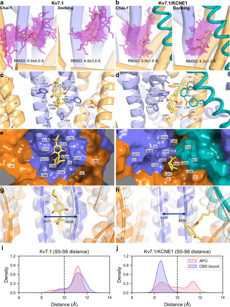
To test the stability of the CBD poses predicted from Chai-1 and docking, we conducted triplicate MD simulations lasting for 500 ns in each binding site. First, the stability of the protein was assessed using the RMSD of Cα atoms relative to the initial protein conformation. The plots demonstrate that the protein Cα atom positions were stable by 300 ns with an overall deviation of around 3.0 Å from the initial structure, except in the Chai-1 predicted KV7.1/KCNE1 protein structure, which was shifted by around 4.0 Å (Figure S8). We therefore present binding pose and interactions for CBD from 300–500 ns to avoid frames where the protein was still equilibrating. The stability of CBD was quantified by average RMSD and visually, by overlaying simulation frames on the initial protein conformations (Fig. 4a, b). Both RMSD and the simulation frames show that CBD had more stable binding modes for simulations initiated from docking poses compared to the Chai-1 predicted poses. In KV7.1, the simulation frames show that CBD simulations initiated from the Chai-1 prediction sampled a variety of poses that spanned the PD (RMSD = 6.9 ± 4.0 Å); meanwhile, the simulation initiated from the docking pose sampled more uniform poses (RMSD = 4.8 ± 3.4 Å), mostly around the S5 and S6 helices. In KV7.1/KCNE1, the simulation initiated from the Chai-1 prediction shows that CBD drifted further from the binding pocket under the KCNE1 subunit (RMSD = 5.9 ± 1.6 Å) compared to the simulation initiated from the docking pose (RMSD = 4.2 ± 1.2 Å).
We tracked the distance between CBD and residues known to influence its effect in KV7.1 (G272, F275, A336) and KV7.1/KCNE1 (S338, and KCNE1 residues F53 and F57) (Fig. S9). In KV7.1, for the majority of the simulation, CBD remained overall closer to G272, F275, and A336 in the simulation initiated from the docking pose compared to the Chai-1 pose. The reason is that throughout the simulation initiated from the Chai-1 prediction, CBD drifts away from the three binding site residues (distance >= 10 Å). We note, however, that for the simulation initiated from the docking pose, CBD leaves the binding site in one simulation replica around 400 ns. In contrast to the shallower S5–S6 site of KV7.1, in the deeper S6–S5’–E1 KV7.1/KCNE1 site, CBD did not leave the binding site in any simulation replicas. We observed that in the Chai-1–initiated pose, CBD moved away from S338 but remained close to F53 across most simulation replicas, while the docked pose showed the opposite trend—drifting from F53 but staying near S338. Both poses stayed similarly close to F57. Because S338 lies at the base of the S6–S5’–E1 site pocket, the Chai-1–initiated pose shifted away from the base of the pocket, but in doing so came closer to F53 than the docked pose.
To investigate how the docked and Chai-1 CBD poses affected binding free energies, we performed MM/PBSA calculations. Interestingly, we found that CBD had similar binding free energy in KV7.1, regardless of whether the simulation was initiated from the docked or Chai-1 predicted pose (−24.3 ± 4.0 and −21.2 ± 4.5 kcal/mol, respectively). A greater difference was observed in KV7.1/KCNE1 where the simulations initiated from the docked pose had a more favourable energy of −27.5 ± 3.2 kcal/mol compared to the one from Chai-1 at −22.9 ± 2.0 kcal/mol.
In summary, we have compared the differences in CBD binding in terms of RMSD, distance to binding site residues, and binding free energy in KV7.1 and KV7.1/KCNE1. We found that in KV7.1, the shallower S5–S6 site enabled CBD to drift away from the site. Despite the pose initiated from docking being overall more stable in terms of RMSD and distance to binding site residues, this did not translate into a meaningful difference in binding free energy between the Chai-1 and docked poses. In contrast, in the deeper S6–S5’–E1 site of KV7.1/KCNE1, the pose initiated from docking, which remained closer to the base of the site (S338), had a more favourable binding free energy compared to the pose initiated from the Chai-1 prediction, which drifted away from the base of the site.
MD simulations illustrate CBD molecular interactions and binding pocket dynamics
We performed clustering analysis to find the most representative binding pose of CBD in KV7.1 and KV7.1/KCNE1 from simulations initiated from the docked conformations (Figs. 4c and 4d, respectively). We note that the simulation frames illustrated in Fig. 4 are based on the “central structure” (see Methods), which is the most representative of each cluster [ref. 38], but a spectrum of simulation frames is provided in Fig. S10 to highlight the distribution of conformations. The most populated simulation cluster in KV7.1 resembled the initial docked pose and enabled the resorcinol ring of CBD to remain within the gap between S5 (G272) and S6 (A336). However, the resorcinol ring could only maintain the H-bond interaction with the backbone carbonyl of C331 and not G272 (Fig. 4c). We quantified the percentage of cluster frames where this H-bond interaction was present, showing that CBD interacted with G272 and C331 in ~2% and ~93% of cluster frames, respectively. Analysis of other prominent molecular interactions (present in > 33% of cluster frames) revealed that the resorcinol group of CBD formed vdW/hydrophobic interactions with G272 and F275 as well as π-stacking interactions with F332. Further, the limonene group formed hydrophobic interactions with A336, in addition to I268, F335, and F339, see Fig. S2 for details. The proposed binding mode of CBD in KV7.1 is supported by agreement between the simulation and site-directed mutagenesis findings, suggesting a role for G272, F275, and A336 in CBD binding. However, given prominent interactions with F335 and F339, it is unclear why mutating these residues to alanine did not affect response to CBD.
Similarly, in KV7.1/KCNE1, the most populated simulation cluster for CBD resembled the initial docking pose. However, the resorcinol ring had shifted to break the H-bond with the backbone carbonyl of V334 and instead formed interactions with the sidechain hydroxy group of S338 (Fig. 4d). This interaction was present in ~95% of the cluster frames. Analysis of other prominent interactions (present in > 33% of cluster frames) revealed that in addition to KCNE1 residues F53 and F57, F335 formed hydrophobic interactions with the resorcinol ring of CBD. The limonene group formed hydrophobic interactions with L262, F339, P343, and L347. Furthermore, the CBD aliphatic tail formed other hydrophobic interactions, including with L266 (Fig. S2). The proposed binding mode for CBD in KV7.1/KCNE1 is supported by agreement between the simulation and site-directed mutagenesis findings, suggesting a role for S338 and KCNE1 residues F53 and F57 in CBD binding. However, given the proximity to L266, it is unclear why mutating it to the bulkier tryptophan did not affect response to CBD.
A surface representation of the most populated clusters is provided to further characterise the S5–S6 site of KV7.1 and the S6–S5’–E1 site of KV7.1/KCNE1. In KV7.1, we observe that G272 and A336 form a shallow pocket that houses the limonene group of CBD (Fig. 4e). F275 partially forms a cover over the resorcinol ring of CBD; this conformation was present in 92% of cluster frames (see Fig. S10 for the spectrum of simulation frames). Meanwhile, in KV7.1/KCNE1, CBD is inside a pocket behind the KCNE1 subunit; the residue F335 and the KCNE1 residue F53 can interact, forming a “cover” over the resorcinol ring of CBD (Fig. 4f). We note, however, that F53 exhibited a variety of conformations such that CBD was “covered” in around 66% of cluster frames (Figure S10b). In addition to contributing potentially favourable molecular interactions to CBD, the KCNE1 subunit may stabilise the conformations of binding site residues in the S6–S5’–E1 site of KV7.1/KCNE1. Specifically, KCNE1 subunit residues F53 and F57 seemed to stabilise the conformations of residues F270 and L266, in KV7.1/KCNE1 (see Fig. S10c for details).
We investigated whether the binding of CBD to the S6–S5’–E1 site influences the S5–S6 site. To achieve this, we measured the distance between the helical cores of the S5 and S6 helices centred around G272 and A336, respectively (Fig. 4g–h). In KV7.1, the distance distribution was similar regardless of the presence of CBD (Fig. 4i). The pocket remained open with an average interhelical distance of 10.8 ± 0.7 Å without CBD and 11.1 ± 0.7 Å with CBD. By contrast, in KV7.1/KCNE1, without CBD, the S5–S6 helical distance varied (Fig. 4j), sampling closed and open conformations of the pocket (interhelical distance of 9.9 ± 1.3 Å on average). With CBD bound to the S6–S5’–E1 site, the helices are mainly packed together and close the pocket (interhelical distance of 8.6 ± 0.3 Å on average). The opening distance from each replica/binding site is compared to the initial opening distance in Fig. S11. We observe that only with CBD in KV7.1/KCNE1, all replicas shifted towards a smaller, more closed interhelical distance.
In summary, we obtained the most prominent binding poses of CBD in KV7.1 and KV7.1/KCNE1 through clustering analyses of the simulations. Consistent with site-directed mutagenesis, CBD interacted with residues critical for its activity: G272, F275, and A336 in KV7.1, and S338, along with KCNE1 residues F53 and F57 in KV7.1/KCNE1. CBD also interacted with residues whose mutation did not alter activity, including F335 and F339 in KV7.1 and L266 in KV7.1/KCNE1. Surface representations showed that the S6–S5’–E1 binding site of KV7.1/KCNE1 forms a deep pocket sealed by F335 and KCNE1 residue F53, whereas the S5–S6 site in KV7.1 is comparatively shallow and open. The KCNE1 subunit not only contributes to the S6–S5’–E1 pocket but also influences S5–S6 site dynamics, reducing its accessibility, particularly when CBD is bound to KV7.1/KCNE1.
Sequence and structure comparison of CBD pockets elucidates KV7 selectivity
The recently published KV7.2 structure in complex with CBD [ref. 13] enables a comparison of the binding modes proposed in different isoforms. The CBD-bound KV7.2 structure in the closed pore state (PDB: 8J00) [ref. 13] displays two bound CBD molecules (Fig. 5a). Except for KV7.2 residues L232 and S303, only residues that are different between KV7.2 and KV7.1 are displayed. The lower CBD molecule (dark green) is positioned below the KV7.2 residue W236, which corresponds to L266 in KV7.1. The KV7.2 residue I300 forms hydrophobic interactions with the limonene group of both CBD molecules and corresponds to the bulkier KV7.1 residue F335. Superimposing the CBD molecules bound in the KV7.2 structure on our CBD-bound KV7.1 prediction reveals that in KV7.1, L266 and F335 sterically hinder the lower binding mode of CBD in KV7.2 (Fig. 5b). On the other hand, the upper CBD molecule (light green) forms H-bonds through the resorcinol hydroxyls with W236 and T296 in KV7.2 (KV7.2 residue T296 corresponds to C331 in KV7.1). Further, CBD, through its limonene group, forms hydrophobic interactions with KV7.2 residue F104 (which corresponds to L134 in KV7.1). Therefore, these residue differences, which remove important molecular interactions, rationalise why CBD likely does not exhibit the upper binding pose in KV7.1 that is seen in the KV7.2 structure.
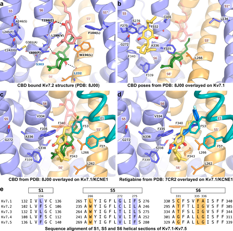
On the other hand, there are several differences around the S5–S6 pocket in KV7.2 compared to KV7.1. In this pocket, all three KV7.1 residues (G272, F275 and A336) shown to be important for the effect of CBD are different in KV7.2 (C242, L245, and G301, respectively). We have shown that the mutants G272C and A336G, mirroring the KV7.2 substitutions, reduce the CBD response of KV7.1; therefore, CBD is unlikely to bind in a similar pose in KV7.2.
Alignment of the CBD-bound KV7.2 structure to the binding site prediction for KV7.1/KCNE1 shows that the CBD pose in the S6–S5’–E1 site resembles the lower CBD binding pose in KV7.2 (Fig. 5c). This contrasts with the KV7.1 structure, where L266 and F335 sterically hinder CBD from binding. In addition to CBD in KV7.2, CBD at the S6–S5’–E1 site also overlaps with retigabine from KV7 structures, displayed as an overlay from the KV7.2 structure (PDB: 7CR2 [ref. 12], Fig. 5d). This suggests that the addition of KCNE1, specifically KCNE1 residue F53, creates a retigabine-like site in KV7.1/KCNE1.
Expanding the sequence comparison of regions involved in CBD binding to all other KV7 subtypes (Fig. 5e) shows that KV7.1 residue L134 is homologous to KV7.3 residue L134, and KV7.1 residue F275 is homologous to KV7.4 residue F251 and KV7.5 residue F279. For the other highlighted residues, KV7.1 is unique among the KV7 family. In this sense, the S5–S6 site may be unique to KV7.1 as it is the only channel in the family with a glycine (S5) and alanine (S6) in the positions G272 and A336. Furthermore, KV7.1’s unique residues L266 and F335 seem to occlude access to the retigabine/CBD binding site of other KV7 channels. However, our findings suggest that inclusion of the KCNE1 subunit creates a binding mode where CBD partly overlaps the retigabine/CBD binding modes found in other KV7 channels.
Discussion
The molecule CBD uniquely inhibits the KV7.1 and KV7.1/KCNE1 channels while having activating effects on neuronal KV7.2–KV7.5 channels [ref. 24]. As such, investigating the CBD binding mode that underlies these effects is important from the perspective of developing subtype-selective drug compounds. In this study, using a combination of computational and experimental approaches, we propose that CBD binds to sites that are not only different between the KV7.1 and KV7.1/KCNE1 channels but also different compared to the neuronal KV7 channels due to key amino acid differences at the binding sites.
Our electrophysiology data support the idea that CBD primarily derives its effects in KV7.1 from binding at the S5–S6 site. This is because the activity of CBD was affected by mutations to S5–S6 site residues G272, A336, and F275. On the other hand, mutations of S338 from the S6–S5’–E1 site, which altered CBD response in KV7.1/KCNE1, had no effect on the CBD response in KV7.1. This supports our second proposal that the S6–S5’–E1 site can only exist in the presence of the KCNE1 subunit. Unlike KV7.1, however, mutations in both the S5–S6 and S6–S5’–E1 sites impacted the CBD response in KV7.1/KCNE1, in which both the G272C and A336G mutants altered CBD response. It can be argued that A336 is only two residues away from S338, which is important for the CBD response in KV7.1/KCNE1, but G272 is one helix away from the S6–S5’–E1 binding site. In contrast to WT KV7.1/KCNE1, the G272C mutant did not display a CBD-induced reduction in GMax (however, a V50 response to CBD was observed for this mutant, which was not seen in WT KV7.1/KCNE1). In addition to S338, mutations to F53 and F57 of KCNE1 also influenced response to CBD, supporting the presence of the S6–S5’–E1 binding site. Therefore, we propose that in KV7.1/KCNE1, CBD primarily derives its response from binding at the S6–S5’–E1 site, but a portion of its response comes from CBD binding at the S5–S6 site, which exists regardless of the presence of the KCNE1 subunit.
MD simulations provided a means to explain this nuanced data for the S6–S5’–E1 binding site. We observed that the S6–S5’–E1 binding site constitutes a deeper pocket compared to the relatively shallow S5–S6 site (compare Fig. 4e and f). The implication of this is that CBD exited the S5–S6 site in a few simulation replicas but did not do so in any simulation replicas at the S6–S5’–E1 binding site (Fig. S9). The differences between the two pockets also seemed to manifest in the binding free energies of CBD. Specifically, CBD binding deep into the S6–S5’–E1 binding site conferred the most favourable binding free energy of the poses investigated. In essence, the presence of the KCNE1 subunit in KV7.1/KCNE1 creates a deep pocket for CBD to bind, which is not present in KV7.1 alone.
A striking observation from our electrophysiology experiments is that mutation of S338 and KCNE1 residues F53 and F57 to alanine comparably improved the potency of CBD in KV7.1/KCNE1. These residues formed favourable molecular interactions with CBD, with F53 having the additional role of forming a cover over the CBD molecule together with F335. Furthermore, we observe that KCNE1 residues F53 and F57 may stabilise binding site residues F270 and L266, respectively (Fig. S10), though it is unclear if this stabilisation persists when mutated to the smaller alanine residue. Evidently, if S338 and KCNE1 residues F53 and F57 play a role in forming favourable molecular or stabilising interactions, it is surprising that alanine mutations at these residues improve CBD potency in experiments. A plausible explanation is provided by the consistency of the potency shift, which suggests a shared trend whereby mutation to the smaller alanine residue favourably expands the space available within the S6–S5’–E1 binding site. In essence, we suspect that despite their favourable contacts, these residues may sterically constrain CBD within the pocket, and a more favourable/stable binding mode may be achieved by alleviating this strain.
Beyond forming part of the S6–S5’–E1 binding site, the KCNE1 subunit seemed to modulate the accessibility of the S5–S6 pocket. Even without CBD present, the KCNE1 subunit seemed to push the S6 helix, reducing the gap between the S5 and S6 helices. This gap, located between G272 and A336, is needed to allow CBD binding in KV7.1 by housing its limonene group. When CBD is bound in the S6–S5’–E1 site, this seemed to further push the S6 helix to the point where the S5–S6 binding site gap disappeared. These observations are consistent with our proposal that CBD in KV7.1/KCNE1 derives its effect primarily from binding to the S6–S5’–E1 site.
Based on sequence comparisons, the described S5–S6 binding site for CBD in KV7.1 is likely not present in neuronal KV7s, which have bulkier residues in place of G272 and a glycine in place of A336. However, this site appears to be part of a modulatory hub for other KV7.1 selective compounds, including the channel activators ML277 and R-L3. Structures of ML277 bound to Xenopus and human KV7.1 [ref. 13, ref. 18] show that ML277 overlaps with the S5–S6 CBD site proposed here (see also Fig. S6), which aligns with our experimental data, suggesting that CBD and ML277 use the same site for binding. Mutations of residues in this pocket have been previously described to impact the effect of R-L3 and ML277 [ref. 41, ref. 42] (see ref. [ref. 24] for a comparison of important residues). Although mutagenesis of specific residues lining the S5–S6 binding site in KV7.1 shows differences in how they impact the effect of ML277, R-L3, and CBD [ref. 18, ref. 41, ref. 42], the geometry of the space allotted by A336 and G272 appears to be critical for all three compounds: ML277 [ref. 41], R-L3 [ref. 42], and CBD (this study). Of note, both ML277 and R-L3 lose their effect with saturating levels of KCNE1 [ref. 43]. Given their overlapping binding sites with our S5–S6 CBD site, it is tempting to speculate that the reduced gap between the S5 and S6 helices induced by KCNE1 may contribute to this phenomenon.
On the other hand, the S6–S5’–E1 binding site for CBD in KV7.1/KCNE1 overlaps with the lower CBD molecule in the KV7.2 structure and with the retigabine binding site of neuronal KV7 channels. Although KV7.1 lacks a tryptophan at position 266 that is crucial to retigabine binding in KV7.2–KV7.5 [ref. 21, ref. 44], mild retigabine-induced inhibition of KV7.1 and KV7.1/KCNE1 has been described [ref. 44, ref. 45]. Based on the available literature, it is unclear whether retigabine mediates inhibiting effects on KV7.1 and KV7.1/KCNE1 via the same retigabine site as in neuronal KV7 subtypes. A superimposition of the CBD-bound KV7.2 structure suggests that L266 and F335 of KV7.1 can sterically hinder CBD binding in this site. As suggested above, KCNE1 co-assembly with KV7.1, apart from forming part of the pocket, seems to stabilise L266 and F335 in a conformation that is receptive to CBD binding. The implication of this is that compounds can be created to exploit the pocket created by the KCNE1 subunit to achieve KV7.1/E1 selectivity.
In the present work, we propose that the presence of the KCNE1 subunit contributes an alternative and preferred CBD binding site in KV7.1/KCNE1. A critical role of KCNE1 in forming binding sites has been previously described for mefenamic acid, DIDS, and adamantane compounds like AC-1 (also called JNJ303) [ref. 46, ref. 47]. Our S6–S5’–E1 binding site is distinct from that of the channel activators mefenamic acid and DIDS, which are found at the extracellular interface between KV7.1 and KCNE1 (near KCNE1 residue 41) [ref. 46]. In contrast, our S6–S5’–E1 binding site is in the same region as that of AC-1; however, different residues are important for their effects ([ref. 47] and the present study).
It is also interesting to note that regions encompassing both the S5–S6 site and the S6–S5’–E1 site can harbour compounds inducing either activating or inhibiting KV7 channel effects. For instance, although CBD, ML277, and R-L3 likely have overlapping binding sites in KV7.1, CBD inhibits KV7.1, whereas ML277 and R-L3 activate the channel. We note that the ML277 binding mode spans far beyond the S5–S6 site here and interacts with the S4–S5 linker as well as the S5’ and S6’ helices of the adjacent subunit [ref. 17, ref. 18]. For R-L3, the compound has been proposed to extend beyond the S5–S6 site by interacting with the S4 helix of the channel [ref. 48]. It may be that interactions with a single KV7.1 subunit underlie the inhibitory effect of CBD. Further work is needed to understand whether such differences contribute to an activating versus inhibiting effect. Similarly, although the retigabine site in KV7.2, overlapping with our S6–S5’–E1 site, is mostly characterised for channel activators, inhibitors like Ebio3 and ML252 use the same site [ref. 15, ref. 49]. Altogether, growing evidence shows that both activators and inhibitors can utilise pockets like the S5–S6 and S6–S5’–E1 sites described here. Further studies are required to understand the mechanistic basis for the diverse effects. Moreover, future studies are needed to determine how CBD inhibits the channels from the S5–S6 and S6–S5’–E1 sites. Such studies could potentially utilize Cryo-EM and MD simulations starting from different pore state conformations to explore potential mechanisms underlying CBD-induced channel inhibition. For instance, it would be interesting to see if CBD impacts electromechanical coupling, as has been discussed for other compounds using the S5–S6 site and the retigabine site in KV7 channels (summarized in our previous study [ref. 24]), or induces similar “squeezing” of the S6 helix as has been proposed for the non-blocking inhibitory effect of Ebio3 in KV7.2 [ref. 15].
In this study, we used Chai-1 as a starting point to identify potential CBD binding sites. This presents a shift in computational binding site determination studies, where docking and MD investigations are typically guided by initial site-directed mutagenesis findings, as they are unsuitable for exploring all possible binding sites in large proteins. Although the Chai-1 models showed low prediction confidence (ipTM ≤ 40), generating 100 CBD poses in KV7.1 and KV7.1/KCNE1 broadened the sampling and demonstrated a trend where top-scoring poses favoured specific binding sites. Site-directed mutagenesis validated these sites, after which induced-fit docking generated tighter CBD binding poses, and MD simulations examined their behaviour under physiologically relevant, lipid-bilayer conditions. Overall, this workflow demonstrates how AI-based prediction, combined with docking and MD, can efficiently map and refine ligand-binding sites.
One limitation of this study is that at the beginning of this project, no structure for KV7.1 bound to KCNE1 had been resolved. However, with the recently deposited structures by Cui et al. [ref. 19], we have confirmed that our KV7.1/KCNE1 model closely matches the closed state KV7.1/KCNE1 structure (Fig. S1). Another limitation is that we could not answer why some residues that favourably interacted with CBD did not alter the response when mutated (F335 and F339 in KV7.1 and L266 in KV7.1/KCNE1). It is possible that the presented binding modes are stable in spite of mutations to these residues or that more stable binding modes are possible, where CBD shifts slightly to reduce interactions with these residues. Another limitation is the discrepancy between the present study and our previous work [ref. 24] regarding the impact of the F335A mutation on CBD effects. Our previous data showed that the F335A mutation in KV7.1 alone reduced the inhibiting effect of 30 µM CBD on GMax, from −71% ± 5% for WT to −45% ± 4% for F335A [ref. 24]. The reduced effect on GMax for this mutant was not seen in the present study (Fig. 2a). We believe that the discrepancy is caused by differential loss of CBD in different perfusion systems, from CBD binding to plastic surfaces, as has been pointed out by several previous studies [ref. 23, ref. 24, ref. 50]. In our previous study, experiments on KV7.1 were performed on two electrophysiology setups with minor differences in the perfusion system. In the present study, in which all experiments were performed using the same electrophysiology setup, no difference in the CBD response was observed between KV7.1 WT and F335A (Fig. 2a; Fig. S12). Thus, we acknowledge that some CBD may still be lost in our perfusion system; however, as all experiments were carried out using the same setup in this work, we have maintained internal consistency.
In conclusion, we have provided evidence that CBD inhibits KV7.1 and KV7.1/KCNE1 through two distinct binding pockets. We envision that detailed characterisations of KV7.1 and KV7.1/KCNE1 binding sites for channel inhibitors, like ours for CBD, provide insights into how to avoid inhibiting adverse effects on cardiac KV7 subtypes. As CBD is being increasingly consumed, insights into potentially harmful effects are relevant and may inspire further studies in more complex experimental models to evaluate potential risks on cardiac repolarisation. Additionally, detailed characterisation of KCNE1’s role in binding site formation enables rational design of compounds that selectively activate KV7.1/KCNE1. Drugs may be designed to better target the S6–S5’–E1 binding site with the aim of treating cardiac arrhythmias related to the KV7.1/KCNE1 channel.
Supplementary Materials
References
- Y Wang, J Eldstrom, D Fedida. Gating and regulation of KCNQ1 and KCNQ1 + KCNE1 channel complexes. Front Physiol, 2020. [DOI | PubMed]
- X Wu, HP Larsson. Insights into cardiac IKs (KCNQ1/KCNE1) channels regulation. Int J Mol Sci, 2020. [DOI | PubMed]
- A Macías, C Moreno, J Moral-Sanz, A Cogolludo, M David, M Alemanni. Celecoxib blocks cardiac Kv1.5, Kv4.3 and Kv7.1 (KCNQ1) channels: effects on cardiac action potentials. J Mol Cell Cardiol, 2010. [DOI | PubMed]
- T Yang, JA Smith, BF Leake, CR Sanders, J Meiler, DM Roden. An allosteric mechanism for drug block of the human cardiac potassium channel KCNQ1. Mol Pharmacol, 2013. [DOI | PubMed]
- Y Liu, X Xu, J Gao, MM Naffaa, H Liang, J Shi. A PIP(2) substitute mediates voltage sensor-pore coupling in KCNQ activation. Commun Biol, 2020. [DOI | PubMed]
- I Hiniesto-Iñigo, A Sridhar, J Louradour, A Cruz, S Lundholm, A Jauregi-Miguel. Rescue of loss-of-function long QT syndrome-associated mutations in Kv7.1/KCNE1 by the endocannabinoid N-arachidonoyl-L-serine (ARA-S). Br J Pharmacol, 2025. [DOI | PubMed]
- M Lübke, JA Schreiber, T Le Quoc, F Körber, J Müller, S Sivanathan. Rottlerin: structure modifications and KCNQ1/KCNE1 ion channel activity. ChemMedChem, 2020. [DOI | PubMed]
- S Ullrich, J Su, F Ranta, OH Wittekindt, F Ris, M Rösler. Effects of IKs channel inhibitors in insulin-secreting INS-1 cells. Pflug Arch, 2005. [DOI]
- JB Stott, IA Greenwood. G protein βγ regulation of KCNQ-encoded voltage-dependent K channels. Front Physiol, 2024. [DOI | PubMed]
- J Sun, R MacKinnon. Structural basis of human KCNQ1 modulation and gating. Cell, 2020. [DOI | PubMed]
- N Brickel, P Gandhi, K VanLandingham, J Hammond, S DeRossett. The urinary safety profile and secondary renal effects of retigabine (ezogabine): A first-in-class antiepileptic drug that targets KCNQ (Kv7) potassium channels. Epilepsia, 2012. [DOI | PubMed]
- X Li, Q Zhang, P Guo, J Fu, L Mei, D Lv. Molecular basis for ligand activation of the human KCNQ2 channel. Cell Res, 2021. [DOI | PubMed]
- D Ma, Y Zheng, X Li, X Zhou, Z Yang, Y Zhang. Ligand activation mechanisms of human KCNQ2 channel. Nat Commun, 2023. [DOI | PubMed]
- S Zhang, D Ma, K Wang, Y Li, Z Yang, X Li. A small-molecule activation mechanism that directly opens the KCNQ2 channel. Nat Chem Biol, 2024. [DOI | PubMed]
- J Li, Z Yang, S Zhang, Y Ye, J He, Y Zhang. Small molecule inhibits KCNQ channels with a non-blocking mechanism. Nat Chem Biol, 2025. [DOI | PubMed]
- S Musella, L Carotenuto, N Iraci, G Baroli, T Ciaglia, P Nappi. Beyond retigabine: design, synthesis, and pharmacological characterization of a potent and chemically stable neuronal Kv7 channel activator with anticonvulsant activity. J Med Chem, 2022. [DOI | PubMed]
- D Ma, L Zhong, Z Yan, J Yao, Y Zhang, F Ye. Structural mechanisms for the activation of human cardiac KCNQ1 channel by electro-mechanical coupling enhancers. Proc Natl Acad Sci USA, 2022. [DOI | PubMed]
- K Willegems, J Eldstrom, E Kyriakis, F Ataei, H Sahakyan, Y Dou. Structural and electrophysiological basis for the modulation of KCNQ1 channel currents by ML277. Nat Commun, 2022. [DOI | PubMed]
- C Cui, L Zhao, AA Kermani, S Du, T Pipatpolkai, M Jiang. Mechanisms of KCNQ1 gating modulation by KCNE1/3 for cell-specific function. Cell Res, 2025. [DOI | PubMed]
- L Zhong, X Lin, X Cheng, S Wan, Y Hua, W Nan. Secondary structure transitions and dual PIP2 binding define cardiac KCNQ1-KCNE1 channel gating. Cell Res, 2025. [DOI | PubMed]
- A Schenzer, T Friedrich, M Pusch, P Saftig, TJ Jentsch, J Grötzinger. Molecular determinants of KCNQ (Kv7) K+ channel sensitivity to the anticonvulsant retigabine. J Neurosci, 2005. [DOI | PubMed]
- TV Wuttke, G Seebohm, S Bail, S Maljevic, H Lerche. The new anticonvulsant retigabine favors voltage-dependent opening of the Kv7.2 (KCNQ2) channel by binding to its activation gate. Mol Pharmacol, 2005. [DOI | PubMed]
- H-XB Zhang, L Heckman, Z Niday, S Jo, A Fujita, J Shim. Cannabidiol activates neuronal Kv7 channels. eLife, 2022. [DOI | PubMed]
- M Pökl, A Sridhar, DJA Frampton, VA Linhart, L Delemotte, SI Liin. Subtype-specific modulation of human Kv7 channels by the anticonvulsant cannabidiol through a lipid-exposed pore-domain site. Br J Pharmacol, 2023. [DOI | PubMed]
- BH Bentzen, N Schmitt, K Calloe, W Dalby Brown, M Grunnet, SP Olesen. The acrylamide (S)-1 differentially affects Kv7 (KCNQ) potassium channels. Neuropharmacology, 2006. [DOI | PubMed]
- G-N Nguyen, EN Jordan, O Kayser. Synthetic strategies for rare cannabinoids derived from Cannabis sativa. J Nat Prod, 2022. [DOI | PubMed]
- 27.Discovery C, Boitreaud J, Dent J, McPartlon M, Meier J, Reis V, et al. Chai-1: Decoding the molecular interactions of life. bioRxiv. 2024: 2024.10.10.615955.
- J Xu, Y Zhang. How significant is a protein structure similarity with TM-score = 0.5?. Bioinformatics, 2010. [DOI | PubMed]
- A Sali, TL Blundell. Comparative protein modelling by satisfaction of spatial restraints. J Mol Biol, 1993. [DOI | PubMed]
- W Sherman, T Day, MP Jacobson, RA Friesner, R Farid. Novel procedure for modeling ligand/receptor induced fit effects. J Med Chem, 2006. [DOI | PubMed]
- S Jo, T Kim, VG Iyer, W Im. CHARMM-GUI: a web-based graphical user interface for CHARMM. J Comput Chem, 2008. [DOI | PubMed]
- WL Jorgensen, J Chandrasekhar, JD Madura, RW Impey, ML Klein. Comparison of simple potential functions for simulating liquid water. J Chem Phys, 1983. [DOI]
- J Huang, S Rauscher, G Nawrocki, T Ran, M Feig, BL de Groot. CHARMM36m: an improved force field for folded and intrinsically disordered proteins. Nat Methods, 2017. [DOI | PubMed]
- K Vanommeslaeghe, E Hatcher, C Acharya, S Kundu, S Zhong, J Shim. CHARMM general force field: A force field for drug-like molecules compatible with the CHARMM all-atom additive biological force fields. J Comput Chem, 2010. [DOI | PubMed]
- MJ Abraham, T Murtola, R Schulz, S Páll, JC Smith, B Hess. GROMACS: High performance molecular simulations through multi-level parallelism from laptops to supercomputers. SoftwareX, 2015. [DOI]
- J Lee, X Cheng, JM Swails, MS Yeom, PK Eastman, JA Lemkul. CHARMM-GUI input generator for NAMD, GROMACS, AMBER, OpenMM, and CHARMM/OpenMM simulations using the CHARMM36 additive force field. J Chem Theory Comput, 2016. [DOI | PubMed]
- N Michaud-Agrawal, EJ Denning, TB Woolf, O Beckstein. MDAnalysis: a toolkit for the analysis of molecular dynamics simulations. J Comput Chem, 2011. [DOI | PubMed]
- X Daura, K Gademann, B Jaun, D Seebach, WF van Gunsteren, AE Mark. Peptide folding: when simulation meets experiment. Angew Chem Int Ed, 1999. [DOI]
- C Bouysset, S Fiorucci. ProLIF: a library to encode molecular interactions as fingerprints. J Cheminform, 2021. [DOI | PubMed]
- MS Valdes-Tresanco, ME Valdes-Tresanco, PA Valiente, E Moreno. gmx_MMPBSA: a new tool to perform end-state free energy calculations with GROMACS. J Chem Theory Comput, 2021. [DOI | PubMed]
- L Chen, G Peng, TW Comollo, X Zou, KJ Sampson, HP Larsson. Two small-molecule activators share similar effector sites in the KCNQ1 channel pore but have distinct effects on voltage sensor movements. Front Physiol, 2022. [DOI | PubMed]
- G Seebohm, M Pusch, J Chen, MC Sanguinetti. Pharmacological activation of normal and arrhythmia-associated mutant KCNQ1 potassium channels. Circ Res, 2003. [DOI | PubMed]
- H Yu, Z Lin, ME Mattmann, B Zou, C Terrenoire, H Zhang. Dynamic subunit stoichiometry confers a progressive continuum of pharmacological sensitivity by KCNQ potassium channels. Proc Natl Acad Sci USA, 2013. [DOI | PubMed]
- W Lange, J Geissendörfer, A Schenzer, J Grötzinger, G Seebohm, T Friedrich. Refinement of the binding site and mode of action of the anticonvulsant retigabine on KCNQ K+ channels. Mol Pharmacol, 2008. [DOI | PubMed]
- XN Du, X Zhang, JL Qi, HL An, JW Li, YM Wan. Characteristics and molecular basis of celecoxib modulation on Kv7 potassium channels. Br J Pharmacol, 2011. [DOI | PubMed]
- M Chan, H Sahakyan, J Eldstrom, D Sastre, Y Wang, Y Dou. A generic binding pocket for small molecule IKs activators at the extracellular inter-subunit interface of KCNQ1 and KCNE1 channel complexes. eLife, 2023. [DOI | PubMed]
- E Wrobel, I Rothenberg, C Krisp, F Hundt, B Fraenzel, K Eckey. KCNE1 induces fenestration in the Kv7.1/KCNE1 channel complex that allows for highly specific pharmacological targeting. Nat Commun, 2016. [DOI | PubMed]
- JA Schreiber, M Möller, M Zaydman, L Zhao, Z Beller, S Becker. A benzodiazepine activator locks Kv7.1 channels open by electro-mechanical uncoupling. Commun Biol, 2022. [DOI | PubMed]
- R Kanyo, SM Lamothe, A Urrutia, SJ Goodchild, WT Allison, R Dean. Site and mechanism of ML252 inhibition of Kv7 voltage-gated potassium channels. Function, 2023. [DOI | PubMed]
- 50.Docter T, Sorum B, Deshmane R, Doubravsky C, Brohawn SG. Cannabinoid inhibition of mechanosensitive K+ channels. bioRxiv. 2024:2024.12.09.627564.
- 51.Yu H, Lin Z, Xu K, Huang X, Long S, Wu M, et al. Identification of a novel, small molecule activator of KCNQ1 channels. In: Probe Reports from the NIH Molecular Libraries Program. Bethesda (MD): National Center for Biotechnology Information (US); 2010.