
Unlocking the Power of Plant-Derived Natural Products: Therapeutic Benefits for Cognitive Health and Neuropsychiatric Symptoms in Dementia-Related Diseases
Abstract
Dementia, including Alzheimer’s disease (AD), represents one of the most pressing public health challenges of the 21st century, affecting more than 55 million individuals worldwide, with projections reaching 139 million by 2050. Current pharmacological treatments offer limited efficacy and significant side effects, driving intense interest in plant-derived natural products as both preventive and therapeutic agents. This review synthesizes preclinical and clinical evidence for key phytochemical classes, including polyphenols, phenolic acids, flavonoids, terpenoids, and alkaloids, in the context of dementia and age-related cognitive decline. Molecular mechanisms are examined in detail, including effects on antioxidant defense and redox homeostasis, suppression of neuroinflammation, and enhancement of synaptic plasticity and neurotransmission. Despite promising preclinical and epidemiological evidence, most clinical trials remain limited in scale and duration and provide mixed results on the efficacy of using phytochemicals for cognitive health. Among the compounds with the most consistent clinical support are the ginkgo diterpene extract EGb 761, saffron carotenoids, curcumin, and rosmarinic acid. A dedicated section addresses the emerging evidence for aromatherapy as a non-pharmacological intervention for behavioral and cognitive symptoms of dementia. Future directions include strategies to improve bioavailability of phytochemicals, the utilization of aromatherapy together with oral supplements, and the need for larger randomized controlled trials using well-characterized and reproducibly manufactured formulations and purified active compounds. Priority areas for future investigation include resolving pharmacokinetic barriers to central nervous system (CNS) delivery, standardizing herbal product composition, and conducting adequately designed clinical trials in well-defined patient populations.
Article type: Review Article
Keywords: dementia, Alzheimer’s disease, phytochemicals, flavonoids, polyphenols, neuroprotection, aromatherapy, neuroinflammation, cognitive decline, natural products
Affiliations: Department of Surgery, Indiana University School of Medicine, Indianapolis, IN 46202, USA; Department of Science and Mathematics, Texas A&M University-Central Texas, Killeen, TX 76549, USA; linhpham@tamuct.edu; Faculty of Information Sciences, Hannan University, Matsubara 580-0032, Japan; Independent Researcher, Kyoto 611-0028, Japan; Aromavilla IFA, Department of Integrative Medicine, Osaka ISEN College of Medical Care & Welfare, Matsushita Nursing School, Osaka 573-0075, Japan; aromavilla@snow.ocn.ne.jp; BioScape Innovations, Indianapolis, IN 46278, USA
License: © 2026 by the authors. CC BY 4.0 Licensee MDPI, Basel, Switzerland. This article is an open access article distributed under the terms and conditions of the Creative Commons Attribution (CC BY) license.
Article links: DOI: 10.3390/plants15111619 | PMC: PMC13259002
Relevance: Moderate: mentioned 3+ times in text
Full text: PDF (4.8 MB)
1. Introduction
Life expectancy has been extended by approximately 30 years during the last half century in high-income countries [ref. 1]. The discoveries of life-saving drugs, the extraordinary advancement in medical treatments, and the improvement in the techniques and awareness related to hygiene in clinical settings have all contributed to saving lives that could not have been saved previously and have extended life expectancies. Another key factor at the individual level is higher levels of prevailing knowledge by the public about healthy lifestyles. The importance of exercise and a healthy diet is well known and may have contributed to the increased number of seniors in relatively good health for their physiological age.
What inevitably come along with extended life expectancy are concerns about cognitive functions, i.e., the possibility of getting dementia, during the later stages of life. Healthy aging includes mental health, including cognitive health, in addition to physical health. How to prevent dementia by maintaining both mental/cognitive and physical health is a significant issue for many people. How to improve the symptoms of dementia is also an important question. Numerous drugs for dementia are currently in clinical trials, and some are approved by the U.S. Food and Drug Administration (FDA) (listed on the website of the Alzheimer’s Association: accessed on 22 February 2026 https://www.alz.org/getmedia/4f4ca289-a2c6-4df9-8cdf-390365bd477e/alzheimers-dementia-fda-approved-treatments-for-alzheimers-ts.pdf). The emergence of these therapies offers hope to patients, though significant obstacles remain, including high costs and the risk of serious adverse events. A detailed summary of currently approved pharmacological treatments for dementia, including their mechanisms of action, routes of administration, and adverse event profiles, is provided in Section 3.2.3, along with an overview of selected clinical trials investigating plant-derived compounds.
How to prevent dementia and how to suppress the symptoms as well as the progress of the disease in our daily lives in a way that is affordable and without adverse events are of broad importance. One such method is through diet [ref. 2]. Gut microbiota diversity is positively correlated with antioxidant capacity [ref. 2]. Antioxidant, anti-inflammatory, and anti-amyloid properties are found in various phytochemical compounds, suggesting that intake of phytochemical compounds with beneficial bioactive properties could be one of the ways to prevent and/or suppress symptoms related to dementia. While consuming phytochemical-rich vegetables as part of a regular diet is beneficial, achieving therapeutically relevant daily doses of specific compounds through diet alone may not be practical or feasible for all individuals. Other than diet, there are various ways to take phytochemicals. It is possible to take them as supplements, and there are some medicines that have a plant origin. Supplemental and pharmaceutical formulations allow for precise, consistent dosing of non-volatile and low-volatility phytochemicals and are generally the most practical approach to achieving therapeutically relevant daily intake levels.
Another way of getting the beneficial effects of phytochemicals that not many people consider is using essential oils. Many people still consider essential oils as a product for relaxation while enjoying pleasant odors because of the lack of information on the effects of phytochemical compounds with bioactive properties included in the essential oils. In this review, we summarize (i) the neuropathology of dementia (Section 3), (ii) the phytochemical compounds with bioactive properties related to biomarkers of dementia (Section 4), (iii) the studies on the effects of essential oils on cognitive function and biomarkers related to dementia (Section 5), and (iv) the obstacles that need to be overcome to effectively utilize essential oils for patients with dementia (Section 6). A recent review also examined plant-derived natural products in the context of neurodegenerative diseases with organizational similarities to the present work [ref. 3]. The present review is distinguished by its broader coverage of phytochemical classes including phytosterols and alkaloids, its dedicated treatment of aromatherapy and essential oils as delivery routes for volatile phytochemicals, and its integration of novel administration strategies including nose-to-brain and transdermal pathways as routes for both essential oil delivery and pharmaceutical development.
2. Literature Search Strategy
This narrative review was conducted through systematic searches of PubMed/MEDLINE, Scopus, and Google Scholar. Searches were performed between August 2025 and May 2026 using the following primary search terms and their combinations: dementia, Alzheimer’s disease, mild cognitive impairment, phytochemicals, flavonoids, polyphenols, phenolic acids, terpenoids, alkaloids, phytosterols, essential oils, aromatherapy, neuroprotection, neuroinflammation, amyloid-beta, tau protein, and cognitive decline. Additional targeted searches were conducted for individual compounds and plant species discussed in each section. Priority was given to primary experimental studies, systematic reviews, and meta-analyses published in English in peer-reviewed journals. Clinical trials were prioritized in the treatment sections. Reference lists of relevant reviews were also screened for additional citations.
3. Mild Cognitive Impairment (MCI) and Dementia
3.1. MCI
This section provides an overview of the clinical spectrum from pre-dementia cognitive impairment to established dementia, followed by a discussion of neuropathological mechanisms, available biomarkers, and current pharmacological management as a foundation for understanding how phytochemicals may contribute to prevention and treatment. Before a person is diagnosed with dementia, there is a transitional stage called MCI. The criteria of MCI seem rather vague, with some studies reporting that normal daily life can be presumed and general cognitive function is normal and not demented, but noticeable objective memory impairment is reported by the person or informant [ref. 4], whereas some studies report significant cognitive symptoms interfering with daily activities [ref. 5].
Petersen et al. [ref. 6] at the Mayo Clinic, Rochester, MN, USA, reported the first criteria to diagnose MCI, providing differentiation from healthy controls and patients with AD [ref. 6]. However, according to the Mayo Clinic website (accessed on 6 November 2025; https://www.mayoclinic.org/diseases-conditions/mild-cognitive-impairment/diagnosis-treatment/drc-20354583), there is still no test to diagnose MCI, indicating the difficulties in diagnosing MCI. Diagnosis is mostly conducted based on the information from the individual or from the informant. In a recent paper by Hessen [ref. 7], the author compared the criteria from the National Institute of Aging and the Alzheimer’s Association (NIA-AA), American Psychiatric Association (DSM-5), and the World Health Organization (WHO) (International Classification of Diseases 11th Revision (ICD-11)) [ref. 7]. The differences in the criteria of these three organizations are very small. They all list that (1) the person has concerns about changes in cognitive functions (subjective cognitive decline) [ref. 4,ref. 7,ref. 8], that (2) there are no problems in daily life and the person functions independently [ref. 4,ref. 7,ref. 8], and that (3) the changes cannot be explained by other reasons such as delirium or other mental/physical disorder [ref. 4,ref. 7,ref. 8]. The NIA-AA criteria list that “impairment is required in one or more of the following cognitive domains: attention, memory, language, visuospatial skills, and executive function” [ref. 7]. The DSM-5 criteria by the American Psychiatric Association also add “socially inappropriate behavior” or “personality change,” and the ICD-11 criteria by WHO request results of cognitive tests in the diagnosis [ref. 7] and no dementia [ref. 4]. Further work in this field would greatly benefit from a uniform definition of MCI that includes multiple quantifiable symptoms as benchmarks.
From a therapeutic standpoint, the MCI stage represents a critical window for intervention where disease modification may be more achievable than in established dementia. Several of the phytochemical compounds discussed in Section 4 have been evaluated specifically in MCI populations or in models representing early-stage cognitive decline. These include curcumin, which demonstrated improved verbal memory in healthy older adults and individuals with subjective cognitive decline [ref. 9]; rosmarinic acid, which significantly improved Clinical Dementia Rating-Sum of Boxes (CDR-SB) scores in MCI patients over 4 months [ref. 10]; Centella asiatica extract, which showed cognitive benefits in cognitively impaired older adults [ref. 11]; and the Ginkgo biloba extract EGb 761, for which systematic review evidence supports benefits in MCI and early dementia [ref. 12]. The applicability of preclinical phytochemical findings to the MCI stage is discussed further in the context of each compound class in Section 4.
3.2. Dementia
According to the WHO, 57 million people worldwide have been diagnosed with dementia, and 60% to 70% of the cases were Alzheimer’s disease (AD) (accessed on 15 February 2026 at https://www.who.int/news-room/fact-sheets/detail/dementia#:~:text=In%202021%2C%2057%20million%20people,dependency%20among%20older%20people%20globally). According to the U.S. Centers for Disease Control and Prevention (CDC), 6.7 million people in the United States have AD, which is 60 to 80% of the cases of dementia (accessed on 15 February 2026 at https://www.cdc.gov/alzheimers-dementia/about/index.html). These reports indicate that AD is the most prevalent type of dementia.
The differences between MCI and dementia are explained by the ability of the person to continue daily life independently (Alzheimer’s Foundation of America, accessed on 21 February 2026; https://alzfdn.org/dementia-or-mild-cognitive-impairment and [ref. 13]). In the patients who have ‘mild’ dementia, daily activity is affected in that patients can take baths, dress by themselves, and use the bathrooms, but show difficulties in functionally working or other usual activities [ref. 14]. In a study in which the Alzheimer’s Disease Assessment Scale-Cognitive Subscale scores of patients with MCI (average age 72.9, n = 769, CDR 0.5), with very mild AD (average age 73.1, n = 122, CDR 0.5), mild AD (average age 74, n = 183, CDR 1.0), and healthy senior controls (average age 70, n = 107) were compared, the scores of the MCI group were found to be higher than those of the controls (MCI group 11.3 vs. controls 5.6), but lower than those of patients with very mild or mild AD (very mild 18.0 and mild 25.2) [ref. 15]. Interestingly, although the overall (global) Clinical Dementia Rating (CDR) of the MCI patients and patients with very mild AD were both 0.5, i.e., the same, the scores of subcategories such as memory, orientation, and judgment were different between the controls and patients with AD [ref. 15]. The hippocampal volume of the MCI patients was also between the volumes of the control group and those of patients with mild AD [ref. 15].
3.2.1. Neuropathology of Dementia
Several types of dementia have been classified and recognized by health professionals (accessed on 22 February 2026 https://www.nia.nih.gov/sites/default/files/understanding-types-dementia_0.pdf) (Table 1). AD is the most prevalent and most likely the best-known type of dementia (Table 1), in which extracellular fibrillar amyloid-β (Aβ) peptide accumulates throughout the brain as plaques in early stages [ref. 16,ref. 17,ref. 18,ref. 19], followed by the accumulation of tau protein [ref. 17,ref. 18,ref. 19,ref. 20]. Synaptic connections are disturbed, causing neurons to weaken and die, causing atrophy. The differences in the types of dementia are based on where these changes take place and/or the types of proteins/peptides involved in causing the malfunction. Memory loss is common in most types of dementia, whereas visual hallucinations and problems in motor functions are typical in Lewy body dementia. Lewy body protein is an abnormal aggregation of a protein called α-synuclein [ref. 21]. α-Synuclein is present not only in the nervous system (both central and peripheral) but also in blood cells and non-nervous system tissues. In the nervous system, it is expressed highly in presynaptic nerve terminals and involved in the release and reuptake of neurotransmitters [ref. 21,ref. 22,ref. 23]. When α-synuclein misfolds into a pathological cross-β-sheet structure [ref. 21,ref. 22,ref. 23], it forms an abnormal aggregation called a Lewy body [ref. 21,ref. 22].
Table 1: Summary of several types of dementia. Based on NIH website (https://www.nia.nih.gov/sites/default/files/understanding-types-dementia_0.pdf, accessed on 22 February 2026) and 2025 Alzheimer’s Association Report [ref. 19].
| Types of Dementia | Neuropathology | Examples of Symptoms | % |
|---|---|---|---|
| AD | Aβ plaques outside of neurons; tau tangles inside neurons; death of neurons; inflammation and atrophy in brain | Memory loss; wandering; unable to recognize friends and family; impulsive | 60–80% of dementia (% differs by reports) |
| Lewy body dementia | Lewy body protein (abnormal aggregation of α-synuclein in neurons) [ref. 21] | Visual hallucinations; visuospatial impairment; insomnia; illogical; difficulty in focusing; problems in motor functions; daytime sleepiness; memory loss | 5% of dementia |
| Frontotemporal dementia | Abnormal amounts or forms of Tau and TDP-43 proteins in frontal and temporal lobes; nerves in frontal and temporal brain die | Impulsive; changes in personality and behavior; shaky hands; problems in balance and walking; difficulties in talking or understanding; during early-stage, memory is not lost | About 3% of patients with dementia older than 65, but 10% of patients with dementia younger than 65 |
| Vascular dementia | Blood clots or injury in the brain disrupting blood flow in the brain | Forgetting current or past events | 5–10% of dementia |
| Hippocampal sclerosis | Accumulation of misfolded protein TDP-43, but in the hippocampus | Memory loss | 3–13% of dementia |
Note: TDP: transactive response DNA-binding protein; Aβ: amyloid-β.
3.2.2. Biomarkers for Detection of Dementia
There have been various biomarkers reported for dementia, especially in AD [ref. 17,ref. 20,ref. 24,ref. 25,ref. 26,ref. 27,ref. 28,ref. 29]. Table 2 summarizes examples of the biomarkers reported so far. The disorders involved range from amyloidosis, tauopathy (Tau proteins), neurodegeneration (NFL, GFAP), neuroinflammation (YKL-40, sTREM2, GFAP), and synaptic dysfunction (drebrin, neurogranin). The extracellular vesicles (EVs) of dementia patients are known to have an altered EV protein composition and contain elevated levels of Aβ, with the form containing a N-terminal truncation [ref. 30,ref. 31]. It is important to note, in the case of extracellular vesicles, that although they can spread toxic forms of Aβ to other locations, there is a possibility of utilizing them in EV-based therapies to deliver drugs [ref. 31,ref. 32]. The summary of AD biomarkers (Table 2) exemplifies the characteristics of the disease: (1) neuroinflammation, (2) abnormal aggregation of peptides and proteins, (3) weakening of neuronal activities due to the accumulation of these peptides and proteins, (4) spreading of these peptides and proteins through EVs, and, ultimately, (5) atrophy of the brain and loss of brain function and motor function.
Table 2: Examples of biomarkers for AD.
| Biomarker | Changes in Dementia Patients | Other Notes (Sources, etc.) | References |
|---|---|---|---|
| p-Tau181; p-Tau217; p-Tau231 | Elevated | Plasma, CSF | [ref. 20,ref. 26,ref. 28] |
| t-Tau | Elevated | Plasma and CSF | [ref. 17] |
| GFAP | Elevated | Plasma, serum, CSF | [ref. 17,ref. 28,ref. 29] |
| Aβ42/Aβ40 ratio | Decreased (in Janeiro et al. [ref. 17] it is referred to as controversial. In Brickman et al. [ref. 26] it is referred to as decreased) | Plasma [ref. 26]; CSF [ref. 17] | [ref. 17,ref. 26,ref. 28] |
| Aβ42 | Decreased | CSF, plasma | [ref. 17] |
| NfL | Elevated | Plasma [ref. 26]; CSF [ref. 17] | [ref. 17,ref. 26,ref. 28] |
| sTREM2 | Elevated in CSF but not in plasma | Microglia biomarker | [ref. 28] |
| YKL-40(CHI3L1) | Elevated along with Aβ accumulation | CSF (plasma YKL-40 does not correlate with Aβ) | [ref. 28] |
| Neurogranin | Decreased | CSF | [ref. 17] |
| Drebrin | Decreased | CSF, plasma | [ref. 25] |
| Small EVs | Increase in EVs that contain the N-terminus-truncated form of Aβ, which degrade less and aggregate more. | [ref. 30,ref. 31] |
Note: Aβ: amyloid-beta; CSF: cerebrospinal fluid; GFAP: glial fibrillary acidic protein; NfL: neurofilament light chain; p-tau: phosphorylated tau; small EVs: small extracellular vesicles; sTREM2: soluble form of the triggering receptor expressed on myeloid cells 2; t-tau: total tau; YKL-40: CHI3L1: Chitinase-3-like protein 1 (a glycoprotein inflammation marker, astrocytic biomarker).
3.2.3. Drugs and Supplements for Dementia
FDA-Approved Drugs
Table 3 summarizes FDA-approved drugs listed on the website of the Alzheimer’s Association (accessed on 26 February 2026, https://www.alz.org/getmedia/4f4ca289-a2c6-4df9-8cdf-390365bd477e/alzheimers-dementia-fda-approved-treatments-for-alzheimers-ts.pdf). Although there are still no medicines that cure AD, there are some FDA-approved drugs for early-stage dementia or MCI. Some of them target the removal of amyloids, for example, Donanemab (KisunlaTM; Eli Lilly & Co., Indianapolis, IN, USA) and Lecanemab (LEQEMBI; Eisai Inc., Bunkyo City, Tokyo, Japan, and Biogen Inc., Cambridge, MA, USA), which are anti-amyloid antibodies (Table 3).
Table 3: Examples of FDA-approved drugs for AD. Data was collected mostly from the websites of the Alzheimer’s Association (link above) and the Mayo Clinic.
| Name | Target | Route, Frequency | Function/Expected Changes | Adverse Events * |
|---|---|---|---|---|
| Donanemab (Kisunla™) | Amyloid | IV, every 4 weeks | Removing Aβ; reduction in cognitive decline | Pains, chills, confusion, fainting, fever, nausea, vomiting, etc. |
| Lecanemab (LEQEMBI) | Amyloid | IV, every 2 weeks | Removing Aβ; reduction in cognitive decline | Pain, chills, confusion, diarrhea, dizziness, drowsiness, etc. |
| Benzgalantamine (Zunveyl) | AChE | Oral, daily | Suppress breakdown of acetylcholine | Skin reaction, chills, cough, diarrhea, etc. |
| Donepezil (Aricept) | AChE | Oral or transdermal, daily | Suppress breakdown of acetylcholine | Nausea, severe vomiting, loss of appetite, etc. |
| Galantamine (Razadyne) | AChE | Oral, daily | Suppress breakdown of acetylcholine | Skin reaction, fever, chills, nausea, diarrhea, etc. |
| Rivastigmine (Exelon) | AChE | Oral or transdermal, twice daily | Suppress breakdown of acetylcholine | Nausea, vomiting, diarrhea, stomach pain, weight loss, skin rash, etc. |
| Memantine (Namenda) | Glutamate | Oral, daily | Suppress NMDA receptor | Swelling of face, arms, etc., dizziness, headache, etc. |
| Donepezil and memantine (Namzaric) | Cholinesterase and glutamate | Oral, daily | Suppress breakdown of acetylcholine and suppress NMDA receptor | Nausea, vomiting, loss of appetite, confusion, dizziness, etc. |
| Suvorexant (Belsomra) | Orexin receptor | Oral, daily | Inhibit the activity of orexin for treatment of insomnia | Impaired alertness, motor coordination, depression, sleepwalking, sleepiness, drowsiness, etc. |
| Brexpiprazole (Rexulti) | Anti-psychotic | Oral, daily | Reduce agitation | Weight gain, sleepiness, dizziness, stroke, suicidal thoughts, etc. |
Note: IV: intravenous; *: from Mayo Clinic (accessed on 22 February 2026, https://www.mayoclinic.org/drugs-supplements/); AChE: acetylcholinesterase, NMDA: N-methyl-D-aspartate.
Another type of drug targets acetylcholinesterase (AChE), for example, Donepezil (Aricept; Eisai Inc., Bunkyo City, Tokyo, Japan), which is an AChE inhibitor. Acetylcholine is an excitatory neurotransmitter involved in memory and learning, and AChE is an enzyme that breaks down acetylcholine [ref. 33]. Studies have shown that AChE is also involved in increasing Aβ production by binding to the enzyme presenilin-1 and increasing the expression of presenilin-1. Presenilin-1 is involved in Aβ production, and, thus, anti-AChE is expected to suppress the degradation of acetylcholine and suppress the production of Aβ [ref. 33].
Suvorexant (Belsomra®; Merck Rahway, NJ, USA) is a drug to treat insomnia. It is an anti-orexin receptor drug that suppresses the activities of both orexin receptor 1 (OX1R) and orexin receptor 2 (OX2R), and is considered a dual orexin receptor antagonist [ref. 34,ref. 35]. Insomnia is often reported in patients with dementia, and recent studies have suggested that an increase in orexin is involved in the malfunctioning sleep–wake cycles [ref. 36,ref. 37], although the results on the altered levels are controversial [ref. 37].
Brexpiprazole (Rexulti®; Otsuka America Pharmaceutical, Inc., Hayward, CA, USA) is a drug first developed for schizophrenia and as an antidepressant [ref. 38]. It is used in the treatment of agitation, which is often observed in patients with dementia [ref. 39,ref. 40,ref. 41]. There are warnings that it increases the risks of fall injuries, hospitalizations, and deaths by cardiocerebrovascular accidents (stroke) in patients using the drug, and its use requires caution [ref. 41,ref. 42,ref. 43].
As can be seen in Table 3, there have been many rather serious adverse events reported in all the drugs listed. Therefore, the development of drugs with fewer adverse events that can enhance patients’ quality of life is needed.
Drugs in Clinical Trials and Supplements Using Plant-Derived Natural Products
The website of the National Institute of Aging (NIA) (accessed on 25 February 2026; https://www.nia.nih.gov/research/ongoing-AD-trials) listed 63 ongoing clinical trials in the U.S. related to dementia. Among those, there is an ongoing clinical trial on the effects of plant-origin natural products, focusing on cannabidiol (CBD). There is a recently completed clinical trial using curcumin (Theracumin® containing 90 mg curcumin, Handok Healthcare, Tokyo, Japan), testing its effects on cognitive functions (accessed on 26 February 2026, https://clinicaltrials.gov/study/NCT01383161). A paper published by the group, which summarized the effects on healthy seniors (age between 50 and 90), showed that the primary verbal memory outcome measure (Buschke–Fuld Selective Reminding Test and Consistent Long-Term Retrieval score) was significantly higher in the experimental group (taking Theracumin® twice daily) after 18 months compared to the placebo condition group [ref. 9]. Other than curcumin, there are clinical trial studies showing that rosmarinic acid, which is included in, for example, lemon balm (Melissa officinalis L.) and sage (Salvia officinalis), significantly improved the CDR-SB after it was taken for 4 months [ref. 10]. Studies on the mechanisms of action indicate that, in the case of curcumin, it has binding affinity with Aβ and suppresses the aggregation of Aβ into oligomers or fibrils [ref. 44]. Aβ is non-toxic as a monomer and turns toxic upon assembly into oligomeric forms ranging from dimers to protofibrils [ref. 44]. Curcumin helps with dissociating the fibrils by disrupting β-sheets and prevents aggregation [ref. 20,ref. 44,ref. 45] (see Section 4.2.1). There are various other phytochemical compounds, such as withanolides, ginkgolides, bilobalide, and bacosides, that have been tested for effects on cognitive function and on dementia [ref. 46,ref. 47,ref. 48]. Thus, there is a strong potential in utilizing plant-derived natural products to prevent and improve the symptoms of dementia, and their use warrants increased research. In the next section, the classification of phytochemical compounds in medicinal plants is summarized.
4. Phytochemical Compounds in Medicinal Plants
4.1. Classification of Phytochemicals
Phytochemicals in medicinal plants are consistently classified into five core secondary metabolite groups: flavonoids, phenolic acids, terpenoids, alkaloids, and phytosterols [ref. 49]. The key phytochemicals and the biological pathways linked to each group are detailed in Table 4, and Figure 1 shows examples of the effects of β-caryophyllene from Koyama et al. (2019) [ref. 50].
Table 4: Examples of phytochemicals classified in five phytochemical groups that modulate biological pathways related to dementia and representative plants that contain them.
| Groups(Sections) | Key Phytochemicals [ref. 49] | Plants [ref. 51,ref. 52,ref. 53] | Biological Pathways(See Each Section for Details) |
|---|---|---|---|
| Flavonoids(Section 4.2) | Kaempferol, catechin, luteolin, cyanidin, apigenin, genistin, quercetin, naringenin, flavokawin C | Chamomile, celery, chrysanthemum flowers, sweet peppers, carrots, onion leaves, broccoli, parsley, nuts, fruits (apples, berries, capers, grapes, onion, tomatoes) | BACE1 inhibition, disruption of Aβ aggregation, tau hyperphosphorylation suppression, inhibition of AChE, SIRT1, PI3K/Akt/GSK-3β, NF-κB/JNK, NF-κB/IKK, TLR4/MyD88/NF-κB, MAPK/JNK, NF-κB/(TNF-α, IL-1β, IL-6, iNOS, and COX-2), MAPK/ERK/CREB, M1/M2 microglial polarization, PPAR-γ/TREM2, Nrf2/ARE, Keap1/Nrf2/HO-1, PI3K/Akt/Nrf2/Keap1, PI3K/Akt/mTOR, AMPK/SIRT1/PGC-1α, SIRT1/iNOS/NF-κB, BDNF/TrkB, Wnt/β-catenin |
| Alkaloids(Section 4.3) | Morphine, cocaine, caffeine, solanin, nicotine, spermine, capsaicin, berberine, galantamine, harmine | Jasmine, barberry, turmeric, goldenseal, Chinese cork tree, toothed clubmoss, coffee bean | MAO inhibition, NF-κB/MAPK, NF-κB/MAPK/STAT3, NF-κB/IKK, TLR4/NF κB, JNK-p38MAPK, MAPK/ERK, PI3K/Akt/ERK, mTOR/p70S6K, PI3K/beclin-1, α7nAChR/Nrf2/Keap1, Akt/GSK-3β/Tau, PI3K/Akt/GSK-3β, IGFR-mediated JNK-Akt, NLRP3-dependent inflammatory signaling, inhibition of AChE, Aβ aggregation inhibition, tau hyperphosphorylation suppression |
| Terpenoids(Section 4.4) | Menthol, artemisinin, phytol, tetraterpenes, oleanolic acid, polyterpenoids, linalool, limonene, β-caryophyllene, ginkgolides | Lavender, rosemary, frankincense, lemon balm, peppermint, sandalwood, ylang-ylang, chamomile, eucalyptus, clary sage, sweet orange, jasmine, vetiver, bergamot, geranium, marjoram, neroli, patchouli, tea tree, cinnamon, ginger, lemongrass, myrrh, juniper berry, angelica root, inula viscosa, ginkgo, saffron | Aβ aggregation inhibition, tau hyperphosphorylation suppression, inhibition of AChE, NF-κB/(TNF-α, IL-1β, and IL-6), NF-κB/IκBα/p65, NF-κB/IKKβ, PI3K/Akt/GSK-3β, Wnt/GSK-3β/β-catenin, Nrf2/HO-1, PI3K/Akt/Keap1/Nrf2, AMPK/Keap1/Nrf2, PI3K/Akt/mTOR, MAPK/ERK, PI3K/Akt/GSK-3β, BDNF/TrkB, BDNF/CREB |
| Phenolic Components(Section 4.5) | Cinnamic acid, syringic acid, vanillic acid, ferulic acid, L-DOPA, p-hydroxybenzoic acid, rosmarinic acid, p-coumaric acid, eugenol, thymol, carvacrol, curcumin | Basil, thyme, cinnamon, clove, oregano, parsley, turmeric, rosemary, lemon balm, grapes, cereal seeds (wheat, oats, rye, and barley), whole grain, spinach, artichokes, coffee, blueberries, sandalwood | Nrf2/HO-1, NF-κb/Nrf2/Bax, NF-κB/MAPK, MAPK/ERK, SIRT1/NF-κB, PPARγ/NF-κB, ERK/JNK/p38, GSK3β/PTEN/PI3K/Akt, GSK3β-Nrf2, SIRT3/SOD2, COX-2, TNF-α and IL-1β inflammation pathways, Aβ aggregation and tau phosphorylation signaling, AMPK/PGC-1α/Sirt3, inhibition of AChE |
| Phytosterols(Section 4.6) | Ergosterol, brassicasterol, β-sitosterol, β-sitostanol, campesterol, campestanol, stigmasterol, resveratrol, fucosterol, 24(S)-saringosterol | Grapes, peanuts, blueberries, vegetable oils, nuts, whole grains | BCAE1/APP, AMPK/NF-κB, AMPK/NLRP3, PI3K/GSK-3β, inhibition of AChE, PI3K/Akt/GSK-3β, ERK/CREB, LXR/ABCA1, LXR/ABCG1, Aβ aggregation inhibition |
Note: α7nAChR: α7 nicotinic acetylcholine receptor, ABCA1: ATP-Binding Cassette Transporter A1, ABCG1: ATP-Binding Cassette Transporter G1, APP: amyloid precursor protein, Akt: Ak strain transforming, AMPK: AMP-activated protein kinase, ARE: Antioxidant response element, Bax: Bcl-2-associated X protein, Bcl-2: B-cell lymphoma 2, BCAE1: Beta-site amyloid cleaving enzyme 1, COX-2: Cyclooxygenase-2, BDNF: Brain-derived neurotrophic factor, CREB: cAMP response element-binding protein, L-DOPA: L-3,4-dihydroxyphenylalanine, ERK: Extracellular signal-regulated kinase, GSK3β: Glycogen synthase kinase-3β, HO-1: Heme oxygenase-1, IGFR: insulin-like growth factor receptor, IκB: Inhibitor of nuclear factor kappa Bα, IKK: Inhibitor of nuclear factor-κB (IκB) kinase, IL-1β: Interleukin-1β, IL-6: Interleukin-6, iNOS: Inducible nitric oxide synthase, JNK: c-Jun NH2-terminal kinase, Keap1: Kelch-like enoyl-CoA hydratase-associated protein, LXR: Liver X receptor, MAO: Monoamine oxidases, MAPK: Mitogen-activated protein kinase, mTOR: Mechanistic target of rapamycin, NF-κB: Nuclear Factor kappa-light-chain-enhancer of activated B cells, NLRP3: Nucleotide-binding oligomerization domain-like receptor family pyrin domain containing 3, Nrf2: Nuclear factor erythroid 2-related factor 2, p38: A mitogen-activated protein, p70S6K: p70 Ribosomal S6 Kinase, PGC-1α: Peroxisome proliferator-activated receptor gamma coactivator 1-α, PI3K: Phosphoinositide 3-kinase, PPARγ: Peroxisome proliferator-activated receptor γ, PTEN: Phosphatase and tensin homolog, SIRT1: Sirtuin (silent mating type information regulation 2 homolog) 1, SOD2: Mitochondrial superoxide dismutase 2, STAT3: Signal transducer and activator of transcription 3, TLR4: Toll-like receptor 4, TNF-α: Tumor Necrosis Factor α, TREM2: Triggering receptor expressed on myeloid cell 2, TrkB: Tropomyosin receptor kinase B, Wnt: Wingless-related integration site.
Flavonoids are significant members of the polyphenol family and consist of a general C6–C3–C6 backbone structure, in which C6 and C3 represent a phenyl ring and a heterocyclic pyran ring with an oxygen atom, respectively. They are known for their antioxidant (for example, quercetin), anti-cancer (for example, hesperidin), and anti-inflammatory (for example, kaempferol) activities [ref. 49], as well as their ability to protect cardiovascular health (for example, quercetin) [ref. 54].
Together with flavonoids, phenolic acids belong to the large polyphenol family and are classified into two subgroups: hydroxybenzoic acids (derived from benzoic acid) and hydroxycinnamic acids (derived from cinnamic acid) [ref. 49]. They are recognized for their strong antihypertensive, antidepressant, anti-cancer [ref. 55], antidiabetic [ref. 56], antioxidant [ref. 57], and neuroprotective functions [ref. 58].
A diverse class of nitrogen-containing heterocyclic structures derived from amino acids forms the alkaloids group, with prominent representatives such as berberine, caffeine, huperzine A [ref. 51], nicotine, morphine, and ephedrine [ref. 49]. Their medicinal significance stems from their antispasmodic, antimalarial, antibacterial, and analgesic functions [ref. 59,ref. 60]. Due to the presence of nitrogen-containing structures, alkaloids often exhibit potent bioactivity through strong interactions with biological receptors and strong radical scavenging abilities, which are critical in the prevention of several degenerative disorders and in inhibiting oxidation reactions [ref. 49,ref. 61].
Terpenoids, the isoprene-based compounds, constitute one of the largest phytochemical groups, comprising structurally diverse compounds such as ginkgolides, limonene [ref. 51], carotenoids, and steroids [ref. 49,ref. 61]. Their therapeutic values are broad, ranging from anti-inflammatory and sedative activities to immunomodulatory and neuromodulatory effects [ref. 62]. They also serve ecological functions such as plant defense and plant-to-plant or plant-to-animal (such as with pollinators) interaction, making them a versatile phytochemical class [ref. 57].
Phytosterols, which share structural similarity with cholesterol, are cyclopentaphenanthrene derivatives, with campesterol, β-sitosterol, ergosterol, and stigmasterol representing the major members [ref. 49]. They possess a wide range of bioactivities, including anti-cancer [ref. 63,ref. 64], anti-inflammatory, antioxidant, hepatoprotective [ref. 65], and cholesterol-lowering effects [ref. 66].
Among the compounds cataloged in Table 4, those with the most advanced clinical evidence or compelling translational profiles include quercetin and Epigallocatechin-3-gallate (EGCG) (flavonoids) (Figure 2) for their multi-target anti-amyloid, anti-tau, and neuroprotective activities demonstrated in human studies; curcumin (phenolic component) for its well-documented Aβ-binding and clinical trial data in healthy seniors; EGb 761 (ginkgo terpenoids) for replicated clinical trial evidence in MCI and mild dementia; galantamine (alkaloid) as an FDA-approved acetylcholinesterase (AChE) inhibitor derived from plant sources; and β-sitosterol and 24(S)-saringosterol (phytosterols) for their emerging Liver X receptor (LXR)-mediated cholesterol-regulatory mechanisms in transgenic AD models. These compounds are discussed in detail in the sections that follow.
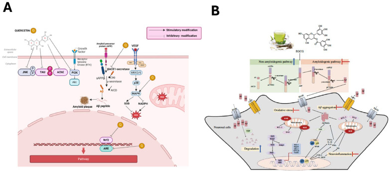
4.2. Biological Pathways of Flavonoids in Dementia-Related Diseases
Flavonoids represent one of the most abundant biologically active plant-derived polyphenols in the human diet [ref. 69]. There is significant evidence from epidemiological dietary pattern studies that flavonoid intake is linked with reduced risk of AD and related dementias (ADRD) and reduced loss of cognitive abilities [ref. 70,ref. 71,ref. 72,ref. 73,ref. 74]. These epidemiological effects have been proposed to be mediated by the neuroprotective effects of flavonoids. Molecular and cellular studies indicate that flavonoid-mediated neuroprotection operates at several levels and involves multiple pathways related to the development of ADRD, the modulation of oxidative stress protection, and signaling pathways that impact the health of neuronal cells.
4.2.1. Inhibition of Aβ Aggregation and Promotion of Clearance
Inhibition of Beta-Site Amyloid Cleaving Enzyme 1 (BACE1)
BACE1 is the rate-limiting enzyme of the amyloidogenic pathway, and its inhibition has long been a therapeutic goal in AD. Multiple flavonoids, including quercetin, luteolin, myricetin, and kaempferol, have been shown to inhibit BACE1 activity directly through competitive or non-competitive binding, reducing Aβ generation. Structural modeling identified the catechol moiety on the B ring and the hydroxyl group at position 3 of the C ring as particularly important determinants of BACE1 inhibitory potency. Luteolin is notable for providing a complementary mechanism: in addition to BACE1 inhibition, it upregulates the insulin-degrading enzyme (IDE), a zinc-dependent metalloprotease responsible for Aβ clearance, thereby reducing amyloid burden through parallel routes [ref. 75].
Disruption of Aβ Aggregation
Several flavonoids interact directly with Aβ monomers and oligomers to destabilize their assembly into toxic fibrils, with EGCG being the most extensively studied compound in this context. Biophysical studies using thioflavin T fluorescence, circular dichroism, and atomic force microscopy have shown that EGCG redirects amyloidogenic polypeptides into unstructured oligomers that are neither toxic nor capable of seeding further aggregation [ref. 76]. This occurs by flavonoids forming hydrogen bonds with Aβ peptides and sterically obstructing the β-sheet conformation required for fibril elongation. Quercetin and rutin (the glycoside form of quercetin) similarly exhibit dose-dependent anti-amyloid and fibril-disaggregating activity in vitro and potent antioxidant activity in APP-overexpressing cell lines [ref. 77]. Taxifolin has shown efficacy in models of cerebral amyloid angiopathy, preventing both cognitive impairment and vascular amyloid deposition [ref. 78]. The catechins, epigallocatechin and epicatechin-3-gallate, block Aβ assembly at sub-micromolar concentrations, adding further structural diversity to the class of flavonoid amyloid-assembly agents [ref. 79].
Enhancement of Autophagy-Mediated Clearance
Beyond direct anti-aggregation effects, certain flavonoids promote the clearance of existing Aβ and tau deposits through the autophagy-lysosomal pathway. Fisetin, a flavonol found in strawberries and apples, activates the transcription factor EB (TFEB), which drives lysosomal biogenesis and autophagic flux, thereby accelerating degradation of phosphorylated tau and Aβ oligomers [ref. 80]. EGCG similarly promotes autophagy through Sirtuin 1 (SIRT1) [ref. 81], an additional mechanism that complements its direct anti-aggregation properties.
4.2.2. Suppression of Tau Hyperphosphorylation and Aggregation
Tau hyperphosphorylation in AD is largely mediated by the dysregulated kinases Glycogen synthase kinase-3β (GSK-3β) and cyclin-dependent kinase 5 (CDK5), and multiple flavonoids affect these targets directly or through upstream signaling effects. Quercetin inhibits GSK-3β activity, reducing tau phosphorylation at Ser396 and Thr231 residues, while also activating the Phosphoinositide 3-kinase (PI3K)/Ak strain transforming (Akt) pathway, which phosphorylates and thereby inactivates GSK-3β through a complementary indirect mechanism [ref. 82]. Kaempferol modulates GSK-3β through similar Akt-dependent mechanisms, and polyherbal formulations combining kaempferol with quercetin show synergistic reductions in tau pathology in rodent models [ref. 83]. Rutin inhibits tau aggregation in cell-free assays and disrupts pre-formed tau fibrils. EGCG prevents tau self-assembly through direct binding that interferes with the intermolecular contacts required for fiber formation [ref. 84,ref. 85]. Fisetin, through its TFEB-mediated activation of autophagy, also stimulates degradation of phosphorylated tau and has reversed cognitive deficits in P301L tau-expressing mice [ref. 80,ref. 86].
4.2.3. Attenuation of Neuroinflammation
NF-κB Pathway Inhibition
NF-κB is the master transcriptional regulator of pro-inflammatory gene expression in the nervous system, controlling production of Tumor Necrosis Factor alpha (TNF-α), interleukin-1β (IL-1β), IL-6, inducible nitric oxide synthase (iNOS), and cyclooxygenase-2 (COX-2). Multiple flavonoids suppress this pathway in AD-relevant models, with direct evidence linking NF-κB inhibition to cognitive improvement. In the 3 × Tg-AD triple-transgenic mouse model that recapitulates both Aβ and tau pathology, luteolin dose-dependently improved spatial learning and memory in the Morris water maze and was accompanied by significant reductions in TNF-α, IL-1β, IL-6, nitric oxide, COX-2, and iNOS protein levels in brain tissue, along with suppression of astrocyte overactivation [ref. 87]. In a complementary Aβ1-42 intracerebroventricular injection model, luteolin directly attenuated phosphorylated NF-κB p65 (Ser536) expression in the frontal cortex and hippocampus, confirming reduced nuclear NF-κB activation in both regions. In vitro studies using a c-Jun NH2-terminal kinase Keap1 (JNK) inhibitor indicate that this effect may be mediated through upstream JNK suppression [ref. 88].
The flavonol quercetin suppresses NF-κB activation in microglial cells via a distinct upstream mechanism based on activating SIRT1, causing reduced acetylation of high mobility group box 1 (HMGB1) and restricting its nucleocytoplasmic shuttling, thereby suppressing downstream toll-like receptor 4 (TLR4)/myeloid differentiation primary response 88 (MyD88)/NF-κB signaling [ref. 89]. In lipopolysaccharides (LPS)-stimulated BV2 microglia, kaempferol inhibits IκB kinase (IKK) complex activity, preventing Inhibitor of nuclear factor kappa B alpha (IκBα) degradation and NF-κB nuclear translocation, with accompanying reductions in TNF-α, IL-1β, nitric oxide, iNOS, and COX-2 [ref. 90]. These microglial pathway studies, while not conducted in ADRD models specifically, support the mechanistic hypothesis of kaempferol and quercetin as NF-κB modulators in the brain inflammatory environment observed in ADRD, and are further supported by parallel findings from hypoxic–ischemic brain injury models in which quercetin’s suppression of TLR4/MyD88/NF-κB signaling in BV2 microglia translated to reduced cerebral infarct volume and rescued cognitive and motor function in neonatal mice [ref. 91].
Microglial Polarization
Microglia exhibit a range of activation states, ranging from the pro-inflammatory M1-like phenotype that is characterized by cytokine and reactive oxygen species (ROS) release to the anti-inflammatory M2-like cells, which secrete neuroprotective factors and promote Aβ phagocytosis. Several flavonoids shift microglial polarization toward the M2-like state, simultaneously reducing neuroinflammatory activation and enhancing amyloid clearance capacity. Naringenin directly promotes M2 polarization in microglia and upregulates Aβ-degrading enzymes, including neprilysin and insulin-degrading enzyme, suggesting a mechanistic link between polarization state and amyloid clearance [ref. 92]. In the amyloid precursor protein (APP)/presenilin1 (PS1) mouse model for AD, naringenin reduces Aβ deposition, suppresses microglial and astrocytic activation, and induces Aβ-degrading enzymes in M2 microglia [ref. 93]. Naringenin was also shown to promote microglia M1/M2 polarization to the M2 anti-inflammatory state; this was dependent on reduced c-Jun NH2-terminal kinase (JNK) inactivation, thus implicating mitogen-activated protein kinase (MAPK) signaling as being important [ref. 94]. Apigenin reduces M1 microglial activation and decreases cluster of differentiation 68 (CD68) expression and ionized calcium-binding adaptor molecule 1 (Iba-1)+ proliferation in cortical neuron-glia co-cultures exposed to Aβ oligomers, LPS, and IL-1β, with increased brain-derived neurotrophic factor (BDNF) expression and neuronal survival [ref. 95]. Similarly, chronic apigenin supplementation reduced hippocampal microgliosis in a chronic neuroinflammatory model relevant to ADRD; however, spatial memory recall impairment was not improved [ref. 96]. An additional mechanism through which flavonoids can promote anti-inflammatory microglial polarization in the AD-relevant context is peroxisome proliferator-activated receptor gamma (PPAR-γ) activation in concert with upregulation of triggering receptor expressed on myeloid cell 2 (TREM2) and reduction in Aβ through increased Aβ42 phagocytosis [ref. 97].
4.2.4. Antioxidant Defense and Mitochondrial Protection
Direct Radical Scavenging
There is strong evidence that flavonoids have the potential to act directly as antioxidant agents. The polyhydroxylated structure of most flavonoids confers potent hydrogen atom transfer and single-electron transfer radical scavenging capacity. The B-ring catechol group (3′,4′-dihydroxylation), the C2=C3 double bond, and the C3 and C5 hydroxyl groups contribute most to this activity. Quercetin and EGCG rank among the most potent natural radical scavengers characterized to date, with measured antioxidant capacities exceeding those of vitamins C and E in several standardized assays [ref. 98]. Flavonoids also chelate the redox-active transition metals Fe2+ and Cu2+ that can catalyze Fenton chemistry and generate highly damaging hydroxyl radicals in proximity to neuronal membranes and deoxyribonucleic acid (DNA) [ref. 99].
In AD brains, iron- and Aβ-catalyzed free-radical attack on neuronal membrane polyunsaturated fatty acids generates the toxic aldehyde 4-hydroxynonenal (HNE) at significantly elevated levels beginning at the earliest disease stages, with HNE-protein adducts impairing synaptic and metabolic function [ref. 100,ref. 101]. Quercetin pretreatment of primary hippocampal neurons in vitro directly attenuates Aβ-induced HNE formation, protein oxidation, and apoptosis in a dose-dependent manner, demonstrating that flavonoid radical scavenging may be able to interrupt this membrane oxidative damage cascade at a biologically relevant point [ref. 102].
Nuclear Factor Erythroid 2-Related Factor 2 (Nrf2)/Antioxidant Response Element (ARE) Pathway Activation
At the transcriptional level, flavonoids activate the Nrf2/ARE pathway, a master regulator of cellular antioxidant defense. Under basal conditions, Nrf2 is retained in the cytoplasm by its inhibitor Keap1, which targets it for ubiquitin-mediated proteasomal degradation. Flavonoids, including EGCG, genistein, quercetin, and apigenin, promote Nrf2 release and nuclear translocation through mechanisms that include Keap1 protein reduction and upstream PI3K/Akt kinase signaling, activating transcription of heme oxygenase -1 (HO-1), NAD(P)H quinone oxidoreductase 1 (NQO1), superoxide dismutase, catalase, glutathione S-transferase, and glutamate-cysteine ligase [ref. 103]. In AD-relevant neuronal models, quercetin activates the kelch-like enoyl-CoA hydratase-associated protein (Keap1)/Nrf2/HO-1 axis in Aβ-challenged PC12 cells and increases cell survival and proliferation [ref. 104]. Genistein activates Nrf2 via the PI3K/Akt/Nrf2/Keap1 signaling cascade in primary hippocampal neurons exposed to Aβ25-35, reducing ROS accumulation and neuronal apoptosis [ref. 105].
Mitochondrial Protection
Flavonoids protect mitochondrial integrity through several converging mechanisms: they prevent mitochondrial membrane depolarization, inhibit cytochrome c release and thereby suppress the intrinsic apoptosis pathway, reduce mitochondrial ROS generation, and enhance ATP synthesis efficiency [ref. 106,ref. 107]. Quercetin has shown well-characterized mitochondrial protective activity in models of dopaminergic neurodegeneration and AD, including inhibition of caspase-3 activation and Bcl-2-associated X protein (Bax) upregulation [ref. 103,ref. 108,ref. 109].
4.2.5. Modulation of Key Signal Transduction Pathways
PI3K/Akt/mTOR Pathway
The PI3K/Akt pathway is a central pro-survival cascade that promotes neuronal survival, inhibits apoptosis, regulates synaptic plasticity, and, through phosphorylation and inactivation of GSK-3β, attenuates tau hyperphosphorylation [ref. 110]. Quercetin and several flavonol-enriched plant extracts activate PI3K/Akt signaling, leading to downstream effects on GSK-3β and mTOR [ref. 82]. Chronic blueberry supplementation in aged rodents increases hippocampal Akt phosphorylation, activates mechanistic target of rapamycin (mTOR), elevates activity-regulated cytoskeletal-associated protein (Arc/Arg3.1), an activity-regulated cytoskeletal protein important for long-term potentiation consolidation, and improves spatial working memory [ref. 111].
MAPK/ERK and CREB Signaling
ERK1/2 activation links growth factor receptor signaling to CREB phosphorylation, which drives transcription of synaptic plasticity genes, including BDNF, Arc, and zif268 [ref. 112]. Flavonoids and their metabolites interact with MEK1/2 to activate the extracellular signal-regulated kinase (ERK)–cAMP response element-binding protein (CREB) axis and promote memory consolidation. (-)Epicatechin at physiologically relevant nanomolar concentrations activates ERK and stimulates CREB phosphorylation through PI3K-dependent mechanisms in primary cortical neurons, concurrently upregulating glutamate ionotropic receptor AMPA type subunit 2 (GluR2) glutamate receptor subunit expression [ref. 113]. Chronic green tea catechin administration activates the protein kinase A (PKA)/CREB pathway and upregulates synaptic plasticity-related proteins, including BDNF, postsynaptic density protein 95 (PSD95), and calcium/calmodulin-dependent protein kinase II (CaMKII), in the hippocampus of senescence-accelerated mouse prone 9 (SAMP8) AD-model mice. This correlated with reductions in Aβ1-42 oligomers and improved spatial memory performance [ref. 114]. Flavanols also protect primary neurons and astrocytes from apoptosis by suppressing the pro-apoptotic JNK pathway and its downstream effectors c-jun and caspase-3, driving the signaling pathway toward net neuronal survival [ref. 113,ref. 115].
GSK-3β Inhibition
GSK-3β occupies a pivotal node in AD molecular pathology, phosphorylating tau at multiple disease-relevant epitopes, promoting Aβ production through APP processing modulation, and suppressing neuronal survival signals [ref. 116]. Quercetin, kaempferol, myricetin, fisetin, and luteolin reduce tau hyperphosphorylation and enhance cell survival signaling by inhibiting GSK-3β activity through direct adenosine triphosphate (ATP)-competitive kinase inhibition and through PI3K/Akt-mediated phosphorylation of GSK-3β [ref. 82,ref. 117,ref. 118].
SIRT1 Activation
SIRT1 is a nicotinamide adenine dinucleotide (NAD)+-dependent deacetylase whose expression is significantly reduced in AD brain tissue, with postmortem studies demonstrating a 29–45% decline in SIRT1 mRNA and protein in the parietal cortex of AD patients relative to age-matched controls, and it is correlated with Braak stage and tau accumulation [ref. 119]. Through deacetylation of NF-κB p65, SIRT1 suppresses transcriptional activation of pro-inflammatory genes, providing a direct mechanistic link between sirtuin activity and neuroinflammation [ref. 120]. SIRT1 also deacetylates and activates peroxisome proliferator-activated receptor gamma coactivator 1-α (PGC-1α), a master transcriptional coactivator of mitochondrial biogenesis, as part of the 5′ adenosine monophosphate-activated protein kinase (AMPK)/SIRT1/PGC-1α axis, which is disrupted in AD. Aβ oligomers reduce SIRT1 and PGC-1α levels in hippocampal neurons, impairing mitochondrial biogenesis [ref. 121]. Additional studies showed that SIRT1 is upregulated in AD-related models and that increasing SIRT1 activity by either SIRT1 overexpression or by resveratrol treatment protects against neurodegeneration, reduces hippocampal neuronal loss, and improves learning deficits in the p25 transgenic mouse model [ref. 122].
Both quercetin and fisetin activate these SIRT1-dependent pathways, though through partially distinct mechanisms. Quercetin activates SIRT1 to attenuate neuroinflammation via SIRT1/NF-κB-related signaling, including SIRT1/iNOS/NF-κB, and has also been linked to SIRT1-dependent autophagy [ref. 123]. Fisetin has been shown to upregulate SIRT1 expression and activate SIRT1-mediated deacetylation in aging brain models, contributing to anti-inflammatory and pro-autophagic effects [ref. 124]. Fisetin also promotes autophagic clearance of phosphorylated tau via TFEB and Nrf2 transcription factor activation through mechanistic target of rapamycin complex 1 (mTORC1) inhibition in cortical neurons, an AD-relevant mechanism that operates in parallel to its SIRT1 effects [ref. 80].
4.2.6. BDNF Signaling and Neurotrophic Support
BDNF/tropomyosin receptor kinase B (TrkB) signaling is a particularly important target for flavonoid-mediated neuroprotection given the well-documented reduction in BDNF expression in AD [ref. 125]. Multiple flavonoid subclasses appear to converge on BDNF/TrkB through overlapping transcriptional and post-translational mechanisms. Upon binding mature BDNF, TrkB activates three principal downstream cascades: MAPK/ERK for differentiation and synaptic plasticity, PI3K/Akt for cell survival, and phospholipase Cγ (PLCγ)/inositol 1,4,5-triphosphate (IP3) for calcium homeostasis and neurotransmission [ref. 126,ref. 127].
Among the compounds with the most direct relevance to this pathway is 7,8-dihydroxyflavone (7,8-DHF), a naturally occurring flavonoid that functions as a potent TrkB agonist. 7,8-DHF rescues cognitive deficits, synaptic loss, and amyloid burden in multiple AD transgenic mouse models [ref. 128,ref. 129,ref. 130]. Beyond direct TrkB agonism, a wide range of flavonoids upregulate BDNF gene expression: chronic blueberry supplementation elevates hippocampal BDNF levels and CREB phosphorylation; quercetin upregulates BDNF in Aβ-challenged neuronal systems; and kaempferol enhances BDNF-TrkB signaling in hippocampal neurons [ref. 131,ref. 132,ref. 133,ref. 134].
4.2.7. Promotion of Adult Neurogenesis
Adult hippocampal neurogenesis involves the generation of new neurons from neural stem cells in the subgranular zone of the dentate gyrus. This process is substantially reduced in AD patients and in transgenic AD models, and this reduction correlates with memory impairment [ref. 135]. Several flavonoids promote neurogenesis through BDNF upregulation, PI3K/Akt/mTOR activation, and stimulation of the wingless-related integration site (Wnt)/β-catenin pathway [ref. 126]. Quercetin promotes hippocampal neurogenesis in AD-model mice partly through inhibition of endoplasmic integrated stress response signaling that normally suppresses this process [ref. 136]. Blueberry-derived anthocyanins and flavanols increase hippocampal neurogenesis in aged rodents, and these histological improvements correlate with enhancements in spatial reference memory [ref. 137,ref. 138].
4.2.8. Inhibition of AChE and Augmentation of Cholinergic Transmission
A major driver of AD is the progressive degeneration of basal forebrain cholinergic neurons, and this degeneration is thought to be responsible for the episodic memory and attentional deficits associated with AD. In AD, cortical AChE activity is abnormally elevated while choline acetyltransferase (ChAT) is depleted, creating a synaptic ACh deficit that correlates closely with cognitive impairment. As the disease advances, butyrylcholinesterase (BChE) has a major hydrolytic role and both enzymes additionally promote Aβ fibril formation through non-catalytic mechanisms. Thus, there is a dual rationale for their inhibition beyond ACh preservation alone [ref. 139,ref. 140]. Several flavonoids function as natural inhibitors of AChE and BChE, increasing synaptic acetylcholine availability through mechanisms that overlap pharmacodynamically with approved AD therapies. Among the catechins, EGCG is a competitive inhibitor of both AChE and BChE, and completely reverses the AChE elevation and associated cognitive deficit in a streptozotocin dementia model [ref. 141,ref. 142]. Flavonoids enhance cholinergic neurotransmission through dual mechanisms: inhibition of ACh degradation and enhancement of ACh synthesis. Quercetin, kaempferol, luteolin, and chrysin inhibit both AChE and BChE activity, slowing synaptic ACh hydrolysis [ref. 140,ref. 142]. Additionally, quercetin has been shown to upregulate ChAT activity in the hippocampus and prefrontal cortex, thereby directly enhancing ACh synthesis [ref. 143].
4.3. Biological Pathways of Alkaloids in Dementia-Related Diseases
Several alkaloid families, including indole, isoquinoline, and bisbenzylisoquinoline scaffolds, have been widely investigated for therapeutic actions and show promising neuroprotective effects relevant to dementia therapy [ref. 144,ref. 145,ref. 146]. These compounds engage multi-target mechanisms across enzymes, receptors, transcription factors, mitochondrial dysfunction, autophagy and mitophagy imbalance, and proteostasis pathways, positioning them as strong candidates for addressing the complex pathology of AD and related dementias [ref. 51,ref. 147].
4.3.1. Neurotransmission, Neurogenesis, and Synaptic Plasticity
A central mechanism of alkaloids in dementia involves cholinergic signaling. Naturally occurring plant-derived alkaloids such as galantamine act as reversible AChE inhibitors and positive allosteric modulators of α7 and α4β2 nicotinic acetylcholine receptors, enhancing cholinergic tone critical for cognition [ref. 148]. Several alkaloids also inhibit monoamine oxidases (MAO-A/B), supporting neurotransmitter balance and reducing oxidative burden. This activity is characteristic of β-carboline alkaloids such as harmine and harman, as well as isoquinoline alkaloids like berberine [ref. 144,ref. 149].
Several isoquinoline, bisbenzylisoquinoneline, and β-carboline alkaloids directly disrupt Aβ fibrillogenesis by binding to Aβ monomers or oligomers, thereby preventing β-sheet formation and destabilizing pre-formed fibrils. Berbamine hydrochloride inhibits the deposition of Aβ plaque by binding to the Aβ fibrils, as demonstrated by molecular docking, chemical kinetics analysis, and atomic force microscopy [ref. 150]. P4B is a derivative of papaverine—a benzylisoquinoline alkaloid—with butyrate, which was reported to prevent Aβ aggregation and attenuate oxidative stress in aged APP/PS1 mice [ref. 151].
In parallel, alkaloids, such as harmine derivatives, allocryptopine, tetrahydropalmatine, tetrahydroberberine, kopoffines, and ‘dendrobium nobile lindl. alkaloid’ (included in Dendrobium nobile Lindl., a traditional Chinese medicinal herb, abbreviated as DNLA), exert significant anti-tau effects. The effects are mediated by (1) suppressing tau phosphorylating kinases such as GSK-3β (harmine derivatives, allocryptopine, tetrahydropalmatine, tetrahydroberberine) [ref. 152,ref. 153] and CDK5 (kopoffines) [ref. 154], and (2) activating pro-survival signaling pathways including PI3K/Akt/GSK-3β (DNLA) [ref. 155]. These effects collectively contribute to reducing tau hyperphosphorylation and neurofibrillary tangle formation [ref. 156]. By inhibiting dual specificity tyrosine phosphorylation regulated kinase 1A (DYRK1A), harmine has demonstrated its potential in reducing tau hyperphosphorylation, limiting aberrant amyloid precursor protein processing, and enhancing neprilysin-mediated clearance of Aβ [ref. 157].
Several studies demonstrated the solid links between alkaloids and adult neurogenesis, including berberine, huperzine A, harmine, galantamine, and vinpocetine. Berberine was suggested to enhance nerve regeneration in hippocampal neurons by modulating the insulin-like growth factor receptor (IGFR)-mediated JNK-Akt signaling pathway [ref. 158] and rescue neuronal integrity by suppressing nucleotide-binding oligomerization domain-like receptor family pyrin domain containing 3 (NLRP3) activation, thereby restoring synaptic plasticity and promoting neurogenesis [ref. 159]. Ma et al. found that huperzine A promotes hippocampal neurogenesis both in vitro and in vivo by activating the MAPK/ERK signaling pathway [ref. 160]. One strong piece of evidence linking the β-carboline alkaloid family to neurogenic pathways is that harmine markedly increases the proliferation of human neural progenitor cells (hNPCs) by inhibiting the dual-specificity tyrosine-phosphorylation-regulated kinase (DYRK1A) [ref. 161]. Recently, Jiang et al. showed that galantamine upregulates the insulin-like growth factor 2 (IGF-2) pathway, leading to the elevation of adult neuronal differentiation and neurite outgrowth in neural stem and progenitor cells (NSPCs) [ref. 162].
4.3.2. Anti-Inflammatory and Immunomodulatory Pathways
Many alkaloids attenuate neuroinflammation through suppression of the NF-κB and MAPK pathways. Bisbenzylisoquinolines such as liensinine, neferine, and isoliensinine reduce cytokine release and inhibit IκBα phosphorylation in microglia [ref. 146,ref. 163]. Isoliensinine also diminishes BV2 microglial inflammation by modulating MAPK/NF-κB signaling, reducing oxidative stress and mitochondrial dysfunction [ref. 164]. Tetrandrine further downregulates NF-κB/MAPK/signal transducer and activator of transcription 3 (STAT3) pathways [ref. 163]. Vinpocetine complements these actions by directly inhibiting IKK, preventing canonical NF-κB activation and promoting neural progenitor survival and differentiation [ref. 165]. The natural β-carboline alkaloid harmine suppresses neuroinflammation by downregulating the TLR4/NF-κB pathway and NLRP3-dependent inflammatory signaling [ref. 157].
4.3.3. Antioxidant, Mitochondrial, and Pro-Survival Processes
Isoquinoline alkaloids like berberine activate pro-survival signaling cascades, including PI3K/Akt, AMPK, CREB, MAPK, and Nrf2, protecting mitochondria and lowering inflammation [ref. 166,ref. 167,ref. 168]. Berberine also elevates the activities of major antioxidant enzymes, including superoxide dismutase, glutathione peroxide, and catalase, thereby strengthening cellular defense against oxidative stress [ref. 169]. Sinomenine, an alkaloid extracted from the Chinese herb qingfenteng, was tested in the APP/PS1 transgenic AD mouse model and Aβ-42 oligopeptide-stimulated differentiated SH-SY5Y neuronal AD cell model. Oxidative stress, Aβ aggregation, and mitochondrial dysfunction were inhibited via the α7 nicotinic acetylcholine receptor (α7nAChR)/Nrf2/Keap1 pathway, indicating the α7 nicotinic acetylcholine receptor as a new target for AD prevention [ref. 170]. Rich alkaloid extracts from Glaucium grandiflorum (GGAE), including allocryptopine, tetrahydropalmatine, and tetrahydroberberine N-oxide, demonstrated their neuroprotective capacity by maintaining intracellular calcium homeostasis and dephosphorylating stress-related proteins such as ERK1/2, JNK, and p38 [ref. 153].
4.3.4. Proteostasis and Autophagy Modulation
Alkaloids regulate proteostasis by inhibiting Aβ aggregation, modulating tau-related kinases, and promoting autophagic clearance. Novel harmine derivatives were reported to exhibit dual inhibitory activity against AChE and Aβ fibril formation at sub-micromolar potencies while maintaining low toxicity, highlighting their multi-target therapeutic potential [ref. 171]. Berberine, a natural alkaloid, has shown an impressive record in the inhibition of Aβ formation in previous studies, making it a potential candidate for the treatment of dementia-related diseases. Berberine counteracts ribosylation-driven Aβ pathology by suppressing the mTOR/p70 Ribosomal S6 Kinase (p70S6K) pathway, leading to improved spatial learning and memory in a widely used transgenic AD mouse model that carries human mutations in the APP and presenilin-1 (PS1) [ref. 172]. It also activates autophagy through the class III PI3K/beclin-1 signaling to facilitate the autophagic removal of tau protein in the triple-transgenic mouse model of AD (3 × Tg AD) [ref. 173]. Immunofluorescence data reported by Sun et al. revealed that berberine treatment in the five familial AD mutation (5 × FAD) mice and Aβ-induced SH-SY5Y cell models resulted in attenuation of Aβ plaque aggregation, suppression of autophagy, and induction of ferroptosis via the JNK-p38MAPK pathway [ref. 174]. GGAE alkaloid extract stimulates the phosphorylation of Akt and GSK-3β and concurrently ameliorates the phosphorylation of Tau, suggesting the association of the Akt/GSK-3β/Tau pathway in the treatment of neuroprotective diseases [ref. 153].
4.4. Biological Pathways of Terpenoids in Dementia-Related Diseases
Terpenes and terpenoids comprise the largest single class of plant secondary metabolites, with over 80,000 structurally characterized compounds built from repeating C5 isoprene units [ref. 175], and this structural diversity is matched by extensive pharmacological diversity. Diverse terpene and terpenoid classes, including small volatile monoterpenes, glycosylated triterpene saponins, and carotenoid polyterpenoids, affect multiple molecular targets implicated in AD and related dementias. Preclinical evidence supports terpene and terpenoid activity across the major pathological hallmarks of ADRD, and a subset of compounds, most notably the ginkgo diterpene trilactones and saffron carotenoids, have demonstrated cognitive benefits in replicated randomized controlled trials [ref. 12,ref. 47,ref. 176,ref. 177,ref. 178,ref. 179].
4.4.1. Inhibition of Aβ Aggregation and Promotion of Clearance
BACE-1 is a key factor in amyloidogenic APP processing, and its inhibition reduces Aβ generation upstream of aggregation. Ginkgolides from Ginkgo biloba reduce BACE-1 activity and limit Aβ production [ref. 180,ref. 181]. Ginsenoside Rg1 from Panax ginseng reduces Aβ generation by suppressing BACE-1 gene expression rather than directly inhibiting the enzyme, a mechanism mediated by PPARγ nuclear translocation [ref. 182], while cryptotanshinone, an abietane diterpenoid from Salvia miltiorrhiza, reduces Aβ production by upregulating α-secretase activity through the PI3K pathway, shifting APP processing toward the non-amyloidogenic route [ref. 183]. Notably, several of the GSK-3β inhibitory effects described below further reduce BACE-1 transcription as a secondary effect, linking the tau and amyloid arms of the pathological cascade through a shared regulatory pathway [ref. 183].
At the level of fibril assembly, ginkgolide B, the diterpene trilactone component of EGb 761, inhibits Aβ fibril formation through direct molecular interactions [ref. 180]. Crocin, the water-soluble carotenoid from Crocus sativus, similarly inhibits Aβ fibrillogenesis in vitro [ref. 184]. Bacosides, the triterpenoid saponins of Bacopa monnieri, act at multiple points: they inhibit both BACE-1 and Aβ fibrillation and additionally reduce cortical Aβ levels in transgenic AD mouse models [ref. 47,ref. 185]. Withanolides from Withania somnifera, when administered orally in APP/PS1 and APPSwInd transgenic mice, significantly increased hepatic low-density lipoprotein receptor-related protein 1 (LRP1) expression and plasma-soluble LPR1. This LRP1 reservoir may serve as a peripheral sink to accelerate Aβ clearance from the brain [ref. 186].
4.4.2. Suppression of Tau Hyperphosphorylation
Tau hyperphosphorylation in AD is driven primarily by the dysregulated kinases GSK-3β and CDK5, and PI3K/Akt-mediated inactivation of GSK-3β (via phosphorylation at Ser9) is a common mechanism across multiple terpenoid classes. Ginsenoside Rg1 decreases tau hyperphosphorylation and reduces GSK-3β expression in AD models via the Wnt/GSK-3β/β-catenin signaling pathway [ref. 187]. Bacosides reduce tau hyperphosphorylation and preserve microtubule integrity in Aβ42-injected rat models via the same GSK-3β interaction [ref. 47]. Cryptotanshinone reduces tau hyperphosphorylation through PI3K/Akt-mediated GSK-3β inactivation, along with improvements in spatial memory in scopolamine-induced amnesia models [ref. 183]. Withanolides from Withania somnifera (ashwagandha) attenuate tau pathology [ref. 47], and asiaticosides from Centella asiatica reduce tau hyperphosphorylation via GSK-3β inhibition and protein phosphatase 2A (PP2A) activation in AD rat models [ref. 188].
Saffron and its carotenoid constituents have demonstrated improvements in cognitive function and biomarkers in clinical trials of mild-to-moderate AD, although there is limited direct evidence that tau effects are the primary mechanism [ref. 177,ref. 178,ref. 189]. Ursolic acid, a pentacyclic triterpene widely distributed in medicinal plants including rosemary, demonstrates tau-directed activity through mechanisms that may involve CDK5 [ref. 183], providing an additional tau-directed mechanism that operates independently of GSK-3β.
4.4.3. Attenuation of Neuroinflammation
Chronic neuroinflammation, driven largely by activated microglia and astrocytes, contributes to synaptic dysfunction, neuronal loss, and progression of AD pathology. Terpenoids have been reported to influence several inflammatory signaling pathways relevant to AD.
NF-κB Pathway Inhibition
Sustained NF-κB-driven microglial activation, with its linked production of TNF-α, IL-1β, and IL-6, amplifies Aβ deposition and accelerates neuronal loss in AD. Cryptotanshinone and tanshinone IIA from Salvia miltiorrhiza are among the most potent natural inhibitors of NF-κB-driven neuroinflammatory cytokine production described in the literature [ref. 183]. Ginsenoside Rg1 suppresses NF-κB signaling in neuroinflammatory models [ref. 190]. Bacosides suppress NF-κB-mediated neuroinflammation [ref. 47] and reduce pro-inflammatory cytokines including IL-6 and TNF-α in microglial models [ref. 47,ref. 191]. Ursolic acid inhibits NF-κB by blocking IκBα degradation and preventing p65 nuclear translocation [ref. 192], while withanolides suppress NF-κB signaling through targeting IKKβ [ref. 47].
Among the volatile terpenoids, 1,8-cineole (the dominant monoterpene of rosemary essential oil) and linalool (the principal constituent of lavender oil) reduce NF-κB-dependent gene expression via upstream kinase inhibition in cell models [ref. 52]. Linalool additionally potentiates gamma-aminobutyric acid A (GABA-A) receptors in electrophysiological assays [ref. 193] and inhibits glutamate binding at N-methyl-D-aspartate (NMDA) receptors in cortical membrane preparations [ref. 194]. Of mechanistic interest within this class are ginkgolide B and bilobalide from EGb 761, which suppress NLRP3 inflammasome assembly and downstream caspase-1 activation, a specific mechanism directly relevant to IL-1β processing in AD brain tissue that is not widely activated by other natural compounds [ref. 180,ref. 195].
CB2 Receptor-Mediated Microglial Modulation
β-Caryophyllene (BCP), a bicyclic sesquiterpene found at high concentrations in black pepper, cloves, and copaiba oil, is unique among terpenes and terpenoids studied for neuroinflammation because it is a selective agonist of the cannabinoid 2 (CB2) receptor [ref. 196], making it the only Generally Recognized as Safe (GRAS)-classified terpene by the U.S. Food and Drug Administration (FDA) with confirmed phytocannabinoid activity. Because CB2 is expressed primarily in microglia, its activation provides a receptor-mediated route to attenuating NF-κB-driven neuroinflammation, reducing pro-inflammatory cytokine release, and promoting microglial M2 polarization that is mechanistically distinct from the direct kinase inhibition described above [ref. 197]. In APP/PS1 transgenic mice, BCP administration reduced Aβ plaque burden, improved spatial memory in the Morris water maze, and reduced cortical inflammatory marker expression including COX-2, TNF-α, and IL-1β. These protective effects were reversed by the CB2 antagonist AM630 and the PPARγ antagonist GW9662, confirming the receptor-mediated and PPARγ pathway mechanism [ref. 198].
4.4.4. Antioxidant Defense and Mitochondrial Protection
Oxidative stress and mitochondrial dysfunction are prominent features of AD and contribute to neuronal injury, impaired bioenergetics, and increased vulnerability to excitotoxic and inflammatory damage. Multiple terpenoid classes have been reported to influence antioxidant and mitochondrial pathways.
Nrf2/ARE Pathway Activation
Nrf2 is the master transcriptional regulator of cellular antioxidant defense, driving expression of HO-1, NQO1, glutathione peroxidase (GPx), SOD, and glutathione biosynthetic enzymes through the antioxidant response element (ARE). Its activity declines with age and is further compromised in the AD brain, creating an opportunity for pharmacological rescue. Withanolides from ashwagandha activate Nrf2 nuclear translocation, upregulating HO-1 and downstream antioxidant enzymes in microglial and cortical models [ref. 47]. Withaferin A, the most pharmacologically characterized withanolide, has similarly been shown to activate Nrf2/HO-1 signaling in neurodegeneration models [ref. 199]. Ginsenosides from Panax ginseng also contribute to antioxidant defense through pathways including Nrf2 activation and reduction in oxidative stress markers in AD models [ref. 47]. Asiaticosides from Centella asiatica induce antioxidant enzyme expression through Nrf2 activation in oxidative stress models [ref. 47]. The antioxidant effects of crocin are notable for having been supported by patient studies. In a randomized double-blind trial of saffron adjunctive to donepezil in mild-to-moderate AD, crocin-containing saffron elevated GPx, mitochondrial superoxide dismutase (SOD), and glutathione reductase activity and reduced total oxidative stress in clinical samples, providing human validation of the antioxidant response element (ARE)-dependent transcriptional program observed preclinically [ref. 200]. A common upstream mechanism across many of these terpenoid classes often involves PI3K/Akt or AMPK pathway activation, leading to Keap1-Nrf2 interaction disruption and Nrf2 nuclear translocation.
Mitochondrial Protection
Bilobalide, the sesquiterpene trilactone component of ginkgo extract EGb 761, is particularly well-characterized with respect to mitochondrial protection. It stabilizes mitochondrial membranes, upregulates NADH dehydrogenase gene expression in mitochondria, and modulates calcium channels to prevent excitotoxic calcium overload [ref. 180,ref. 201,ref. 202]. In cultured chick embryonic neurons and rat hippocampal mixed cultures, bilobalide rescues neurons from apoptosis induced by serum deprivation and by staurosporine and represents one of the most potent anti-apoptotic constituents in the EGb 761 extract [ref. 201]. In focal cerebral ischemia in gerbil models, oral bilobalide at 3–6 mg/kg/day for 7 days before transient global ischemia progressively protected CA1 neurons from death and preserved mitochondrial COX III mRNA levels, with effects comparable to the full EGb 761 extract [ref. 202].
Other terpenoid classes may also support mitochondrial integrity. Bacosides reduce mitochondrial oxidative damage and restore mitochondrial function in AD models [ref. 47]. Crocin has also shown neuroprotective effects in Aβ-exposed models, including reduction in neuronal injury in association with PI3K/Akt signaling [ref. 203].
4.4.5. Modulation of Key Signal Transduction Pathways
PI3K/Akt/mTOR Pathway
The PI3K/Akt pathway is a central pro-survival cascade that promotes neuronal survival, inhibits apoptosis, regulates synaptic plasticity, and attenuates tau hyperphosphorylation through GSK-3β inactivation. Crocin activates PI3K/Akt signaling in Aβ25-35-injected mice, reducing neuroinflammatory cytokine production and hippocampal neuron injury [ref. 203]. Cornel iridoid glycoside (CIG) from Cornus officinalis activates the PI3K/Akt/mTOR pathway as its dominant proposed mechanism of action, improving memory performance in rodent models of both vascular dementia and AD, with corresponding reductions in hippocampal Aβ levels, tau phosphorylation, and neuroinflammatory markers [ref. 204]. Ginsenoside Rg1 engages PI3K/Akt as part of a broader neuroprotective program that extends through BDNF/TrkB signaling to support hippocampal neuroplasticity and long-term potentiation in APP/PS1 transgenic mice [ref. 190,ref. 205].
MAPK/ERK Signaling
The MAPK cascades, including p38, ERK1/2, and JNK arms, are relevant to both pro-inflammatory signaling and to neuroprotective and synaptic plasticity programs, depending on context and cell type. Saffron and its constituents crocin and safranal have demonstrated beneficial effects on cognitive outcomes in clinical trials of mild-to-moderate AD, with preclinical evidence supporting activity across multiple neuroprotective mechanisms [ref. 177,ref. 178,ref. 189]. Ginsenoside Rg1 engages ERK signaling through inhibition of ERK-mediated PPARγ phosphorylation, which in turn upregulates insulin-degrading enzyme (IDE) expression to promote Aβ degradation in a primary hippocampal neuron AD model [ref. 206].
GSK-3β Inhibition
GSK-3β occupies a key node in AD-related signaling because it contributes to tau phosphorylation and can influence amyloidogenic processing. Several terpenoid classes, including ginsenosides, bacosides, cryptotanshinone, and Centella asiatica-derived compounds, have been reported to reduce GSK-3β activity either directly or through upstream signaling pathways such as PI3K/Akt [ref. 47,ref. 183,ref. 187,ref. 188]. Thus, GSK-3β inhibition may provide a mechanistic link between tau-directed and amyloid-related effects, although the extent of this convergence differs among compounds and experimental models.
4.4.6. BDNF Signaling and Neurotrophic Support
Loss of neurotrophic support is a major contributor to synaptic dysfunction and impaired plasticity in AD. BDNF and its receptor TrkB are particularly important for hippocampal long-term potentiation, neuronal survival, and memory formation. Ginsenoside Rg1 upregulates hippocampal BDNF and phospho-CREB in SAMP8 mice alongside dose-dependent reductions in soluble Aβ1-40 [ref. 207], and increases BDNF/p-TrkB protein expression with corresponding restoration of long-term potentiation in APP/PS1 transgenic mice [ref. 205]. Bacosides activate CREB-mediated neuroplasticity and support kinase-driven neuronal repair [ref. 47]. The withanolides function through a qualitatively distinct mechanism: withanolide A from ashwagandha promotes neurite outgrowth and synaptogenesis in cortical neurons through direct protein-level interactions, representing a potentially reconstructive effect rather than simply a neuroprotective one [ref. 208].
Asiaticosides from Centella asiatica consistently promote axonal elongation in culture models, and Centella asiatica extract has demonstrated improvements in neuronal morphology and Aβ plaque load in transgenic AD mouse models [ref. 47]. CIG, the combined morroniside and loganin fraction from Cornus officinalis, promotes nerve growth factor (NGF) and BDNF expression to support cholinergic neuron survival in fimbria-fornix lesion models of AD-like cholinergic deficit [ref. 204], a neurotrophic mechanism that is not activated by most other terpenoid classes and is relevant to the cholinergic neuronal loss central to AD. At the synaptic level, ginkgolides protect against Aβ1-42-induced synaptotoxicity and preserve synaptic protein expression in vitro [ref. 209], while a ginkgolide preparation also reduced Aβ deposition and neuroinflammation in transgenic mouse models [ref. 195].
4.4.7. Promotion of Adult Neurogenesis
Adult hippocampal neurogenesis is substantially reduced in AD patients and transgenic AD models, and this reduction correlates with memory impairment. Compared with amyloid, tau, or inflammatory mechanisms, evidence for terpenoid effects on neurogenesis is less extensive, but several classes converge on signaling pathways relevant to neuronal differentiation, survival, and synaptic maturation.
Ginsenoside Rg1 promotes hippocampal neuroplasticity through BDNF/TrkB signaling, restoring long-term potentiation in APP/PS1 transgenic mice [ref. 205], and upregulating phospho-CREB and BDNF in SAMP8 mice [ref. 207]. Bacosides support neurogenesis through BDNF/CREB-mediated pathways [ref. 47]. Centella asiatica extract has demonstrated improvements in neuronal morphology in transgenic AD mouse models [ref. 47], with neurotrophic effects also documented in healthy elderly human volunteers [ref. 210]. The neurogenesis evidence for terpenoids is generally less extensively characterized at the mechanistic level than for flavonoids, but the convergence of ginsenosides, bacosides, and Centella-derived asiaticosides on BDNF/CREB and PI3K/Akt pathways that regulate neuroprogenitor survival and differentiation provides a plausible biological basis for these effects.
4.4.8. Inhibition of AChE and Augmentation of Cholinergic Transmission
The progressive degeneration of basal forebrain cholinergic neurons is a major driver of the episodic memory and attentional deficits in AD, and inhibition of AChE and BChE to preserve synaptic acetylcholine is the mechanism of all currently approved AD therapies. AChE and BChE inhibitory activity is notably widespread across terpenoid structural classes [ref. 211]. Among the monoterpenes, 1,8-cineole, α-pinene, linalool, safranal, citral, and citronellal have been reported to exhibit AChE and BChE inhibitory activity in vitro, though potency varies considerably across compounds and assay conditions [ref. 52,ref. 212,ref. 213]. The significance of 1,8-cineole’s cholinesterase inhibitory activity has been directly validated under human conditions. Moss and Oliver [ref. 214] measured plasma 1,8-cineole concentrations in healthy volunteers following ambient rosemary essential oil exposure and found a significant positive correlation with both speed and accuracy of cognitive performance. Although suggestive, this does not directly establish cholinesterase activity as the primary mechanism. Safranal also shows AChE inhibitory activity in vitro [ref. 215], providing a cholinergic complement to crocin’s carotenoid-mediated effects from compounds derived from the same botanical source.
Among the triterpene classes, ginsenosides support cholinergic neurotransmission and protect cholinergic neurons from Aβ-induced degeneration [ref. 47]. Ginkgo terpenoids modulate cholinergic neurotransmission indirectly through neurotransmitter release mechanisms rather than direct enzyme inhibition [ref. 180], and ursolic acid demonstrates AChE inhibitory activity in vitro [ref. 183]. It is noteworthy that the lemon balm monoterpene aldehydes citral and citronellal, which are the principal constituents of Melissa officinalis essential oil extracts, were shown in radioligand binding assays to displace [3H]-nicotine from nicotinic ACh receptors and [3H]-scopolamine from muscarinic ACh receptors in human cerebral cortical membranes, a cholinomimetic receptor pharmacology that operates independently of AChE inhibition [ref. 216]. Clinical trials of Melissa officinalis essential oil in dementia have produced mixed results, with one placebo-controlled randomized controlled trial (RCT) reporting reduced agitation [ref. 217] and another finding no significant benefit over placebo or donepezil [ref. 218].
4.5. Biological Pathways of Phenolic Components in Dementia-Related Diseases
Phenolic acids, including caffeic acid, ferulic acid, gallic acid, rosmarinic acid, and protocatechuic acid, exert multi-target neuroprotective effects relevant to AD and related dementias [ref. 219,ref. 220]. Their mechanisms span antioxidant defense, anti-inflammatory signaling, mitochondrial support, amyloid and tau modulation, and synaptic protection [ref. 51,ref. 221].
4.5.1. Antioxidant Defense and Redox Homeostasis
Phenolic acids exhibit potent antioxidant activity due to their hydroxyl groups, making them powerful radical scavengers, which neutralize reactive oxygen species (ROS), including superoxide anions, hydroxyl radicals, and peroxynitrite [ref. 221]. A synthetic caffeic acid phenethyl ester 4-O-glucoside, FA-97, mitigates oxidative stress-induced neuronal apoptosis and alleviates scopolamine-driven cognitive deficits by activating the Nrf2/HO-1 signaling pathway [ref. 222]. Treatment with sesamol, a natural phenolic compound, helps maintain redox homeostasis, safeguarding mitochondrial function, enhancing antioxidant enzyme expression by activating the Nrf2 pathway and promoting its nuclear translocation in H2O2-exposed SH-SY5Y cells [ref. 223]. Administration of monomeric polyphenol berry extract not only reinstates the reduced levels of tyrosine hydroxylase and dopamine transporter but also diminishes the buildup of α-synuclein within the midbrain via the NF-κB/Nrf2/Bax pathway [ref. 224].
4.5.2. Modulation of Neuroinflammation
Phenolic acids inhibit microglial overactivation by suppressing key inflammatory pathways such as NF-κB/MAPK (urolithin) [ref. 225], SIRT1/NF-κB (resveratrol) [ref. 226], PPARγ/NF-κB (phloretin) [ref. 227], ERK/JNK/p38 (gallic acid and resveratrol) [ref. 228], GSK3β/PTEN/PI3K/Akt (trahydrocurcumin) [ref. 229], SIRT3/SOD2 (trilobatin) [ref. 230] and COX-2 [ref. 231]. Rosmarinic acid, in particular, downregulates pro-inflammatory cytokines such as TNF-α and IL-1β, thereby preventing chronic neuroinflammation associated with cognitive decline [ref. 232]. Gallic acid markedly reduces nitric oxide, iNOS, and IL-1β expression in the substantia nigra of lipopolysaccharide-infused rats, further alleviating neuroinflammation [ref. 233]. In D-galactose–treated mice, sesamol not only enhanced the mRNA and protein expression of the antioxidant enzymes HO-1 and NAD(P)H:quinone oxidoreductase 1 (NQO1), but also lowered the serum concentrations of the pro-inflammatory cytokines TNF-α and IL-1β [ref. 223].
4.5.3. Enhancement of Synaptic Plasticity, Neurotransmission, and Neurogenesis
Many phenolic acids block Aβ aggregation by directly binding to Aβ monomers or destabilizing β-sheet-rich fibrils. Caffeic and rosmarinic acids inhibit BACE1, the β-secretase enzyme responsible for initiating Aβ generation, resulting in decreased Aβ plaque formation. Additionally, ferulic acid disrupts Aβ oligomerization and protects synaptic structure from Aβ-induced toxicity [ref. 234]. Phenolic compounds modulate tau pathology through inhibition of GSK-3β and CDK5, the two principal kinases responsible for tau hyperphosphorylation [ref. 219]. The use of different combinations of ferulic acid, curcumin, and phosphatidylserine results in remarkable decreases in Aβ, IL-1 β, and phosphorylated tau in addition to the elevation of brain-derived neurotrophic factor (BDNF) level [ref. 235]. Propofol enhances synaptic signaling by upregulating synaptic proteins such as synaptophysin and postsynaptic density protein 95 (PSD-95), thereby alleviating the cognitive impairment observed in the offspring of pregnant C57BL/6 mice exposed to isoflurane-induced anesthesia [ref. 236].
Many phenolic acids, such as caffeic acid [ref. 58], ferulic acid [ref. 237], rosmarinic acid [ref. 238], and gallic acid [ref. 239], have exhibited significant inhibition of acetylcholinesterase activity. Phenolic-rich extract of Pinus densiflora not only enhances long-term potentiation (LTP) induction but also counteracts the LTP suppression caused by scopolamine (SCOP), a muscarinic receptor antagonist, and reduces AChE activity [ref. 240]. Vanillic acid has been shown to mitigate SCOP-induced deficits in learning and memory by safeguarding hippocampal cholinergic function, reducing oxidative damage, and supporting synaptic plasticity [ref. 241]. After screening thousands of Chinese traditional medicines, forsythoside A has emerged as the most potent inhibitor of Aβ deposition by interfering with the pre-formed fibrils, thereby extending the lifespan of the Caenorhabditis elegans AD transgenic models [ref. 242].
Phenolic compounds promote neurogenesis not only through their antioxidant properties, but also by re-establishing a supportive neurogenic microenvironment. In an amyloid-β1–42-induced Alzheimer’s mouse model, rosmarinic acid treatment significantly restored hippocampal neurogenesis markers such as Ki-67 and doublecortin (DCX) and improved social memory, indicating enhanced neuronal proliferation and differentiation [ref. 243]. In addition, caffeic acid alkyl esters have been shown to stimulate extracellular ERK1/2 and Akt serine threonine phosphorylation in the rat PC12 neuronal cells, thereby promoting neurotrophic effects such as cell survival and differentiation [ref. 244]. Jiang et al. demonstrated that gallic acid stimulates the proliferation and differentiation of NSCs through activation of the MAPK/ERK pathway [ref. 245]. Moreover, Yu Ding et al. found that gallic acid enhances hippocampal neurogenesis and alleviates cognitive deficits in APP/PS1 mice via the GSK3β–Nrf2 signaling axis [ref. 246].
4.5.4. Mitochondrial Protection and Homeostasis
Phenolic acids stabilize mitochondrial membrane potential, preserve ATP synthesis, and inhibit cytochrome-c release [ref. 234]. By regulating mitochondrial ROS balance partly through activation of the AMPKs/PGC-1α/Sirt3 pathway, honokiol counteracts NaF-induced oxidative stress and mitochondrial dysfunction. This leads to the prevention of neuronal and synaptic damage as well as the resulting cognitive impairments [ref. 247]. Polyphenol, when combined with physical activities, can promote mitochondrial biogenesis through the activation of Peroxisome proliferator-activated receptor-gamma coactivator 1-α (PCG-1α) in age-related central nervous system (CNS) disorder [ref. 248]. Anti-apoptotic actions include upregulation of B-cell lymphoma 2 (Bcl-2), p-PI3K, p-Akt, p-GSK-3β, Microtubule-associated protein 2 (MAP2), and synaptophysin (SYN) concurrently with suppression of Bax and caspase-3 were observed in the diabetic mice treated with salidroside [ref. 249].
4.6. Biological Pathways of Phytosterols in Dementia-Related Diseases
Phytosterols are plant-derived sterol alcohols that share the tetracyclic core of cholesterol but carry additional alkyl or unsaturated substituents at C-24, giving rise to the major dietary members: β-sitosterol, stigmasterol, campesterol, and the algal sterols fucosterol and 24(S)-saringosterol [ref. 250,ref. 251]. The relatively minor structural differences observed in phytosterols result in functionally distinct interactions with membrane lipid microdomains, sterol-sensing receptors, and nuclear transcription factors that are directly relevant to neuronal cholesterol homeostasis. Phytosterols cannot be synthesized by mammals and are exclusively diet-derived, with the richest concentrations found in unrefined vegetable oils, nuts, seeds, and legumes [ref. 252]. Unlike cholesterol, which is synthesized within the brain and largely excluded from CNS entry by the blood–brain barrier, dietary and circulating phytosterols have been shown in mouse studies to cross the blood–brain barrier and accumulate in brain parenchyma [ref. 253,ref. 254].
Evidence that phytosterol levels in the brain affect cognitive health comes primarily from epidemiological studies. Mendelian randomization analyses using genome-wide association data from the International Genomics of Alzheimer’s Project and the UK Biobank found that higher circulating stigmasterol levels were associated with a reduced risk of AD, and a similar inverse association was identified for sitosterol, while campesterol and brassicasterol showed no significant effects; however, these associations were attenuated after adjustment for blood lipids [ref. 255]. Other population-based studies in elderly subjects found that baseline sitosterol levels were associated with cognitive impairment, and that longitudinal increases in stigmasterol, campestanol, and sitostanol over five years were associated with cognitive decline. Thus, phytosterols appear to have complex and sterol-specific relationships with circulating phytosterol levels and cognition, with some sex-specific effects observed [ref. 256,ref. 257]. These human studies provide an indication that some phytosterols may have promise for enhancing cognitive health; however, compared to other classes of phytochemicals, there is significantly less mechanistic data available to support this potential use.
4.6.1. Modulation of Amyloidogenic APP Processing
The proteolytic cleavage of APP by BACE1 and γ-secretase is strongly favored within cholesterol-enriched lipid raft microdomains, where APP and presenilin-1 co-localize to drive Aβ production. Because phytosterols differ structurally from cholesterol at C-24, their incorporation into neuronal membranes alters raft composition and shifts APP processing toward the non-amyloidogenic α-secretase route [ref. 252].
Among the common dietary phytosterols, stigmasterol has the most extensively characterized anti-amyloidogenic activity. Studies in APP-overexpressing neuroblastoma cells and in mice fed stigmasterol-enriched diets showed that stigmasterol reduces Aβ generation by directly decreasing BACE1 enzymatic activity, suppressing transcription of all γ-secretase complex subunits, reducing cholesterol and presenilin-1 from detergent-resistant lipid raft fractions, and reducing BACE1 internalization to the endosomal compartments where amyloidogenic cleavage of APP takes place. In in vivo studies, both BACE1 and γ-secretase activities were reduced in brain tissue, and Aβ40 and Aβ42 levels fell correspondingly, without any change in total brain cholesterol. This suggests that the effect arose from membrane compositional changes rather than bulk sterol displacement [ref. 252]. β-Sitosterol similarly promotes non-amyloidogenic APP processing by redistributing APP from lipid rafts to non-raft membrane regions, and treatment of APP/PS1 double transgenic mice improved spatial learning and recognition memory, reduced plaque load, and reversed hippocampal synaptic deficits normally observed in this model [ref. 252,ref. 258]. Complementary sterol profiling of aged APP transgenic mouse brain demonstrated that sterol metabolism is altered relative to non-transgenic controls, consistent with the proposition that amyloidogenic APP processing is coupled to broader dysregulation of brain sterol homeostasis [ref. 259].
Fucosterol, the predominant sterol in brown algae, inhibits BACE1 through non-competitive enzyme kinetics in vitro, with molecular docking analysis indicating interaction with residues of the BACE1 active site [ref. 260]. Pharmacological studies of fucosterol also identified liver X receptor β (LXR-β), TrkB, and TLR2/4 among its predicted targets, suggesting multiple mechanisms of neuroprotection [ref. 261]. In silico docking studies of β-sitosterol and stigmasterol isolated from Polygonum hydropiper likewise showed favorable binding geometries against BACE1 and monoamine oxidase, though these computational findings await experimental confirmation [ref. 262].
4.6.2. Attenuation of Neuroinflammation
AMPK/NF-κB and NLRP3 Pathway Inhibition by Stigmasterol
Chronic microglial activation driven by NF-κB and NLRP3 inflammasome signaling amplifies Aβ deposition and accelerates synaptic injury throughout AD progression. Stigmasterol suppresses these pathways through AMPK activation, an upstream energy-sensing kinase whose activity dampens NF-κB nuclear translocation and NLRP3 inflammasome assembly. In APPswe/PS1dE9 transgenic mice, stigmasterol treatment attenuated cognitive deficits and reduced cortical and hippocampal Aβ42 concentrations. In BV2 microglia stimulated with Aβ42 oligomers, stigmasterol suppressed pro-inflammatory cytokine release and lowered M1 microglial polarization through this AMPK-dependent mechanism, thus reducing caspase-1 activation and IL-1β processing [ref. 263]. The engagement of an upstream metabolic sensing pathway distinguishes stigmasterol’s anti-inflammatory mechanism from the direct kinase inhibitory mechanisms of many flavonoid and terpene compounds.
Anti-Inflammatory Effects of β-Sitosterol
β-Sitosterol modulates neuroinflammatory signaling through several complementary mechanisms. Dietary studies in a murine model showed that a plant sterol-poor diet was associated with elevated pro-inflammatory lipid mediators in the brain, with thromboxane B2 and prostaglandin D2 concentrations inversely correlated with campesterol and β-sitosterol levels across brain regions. This suggests that endogenous phytosterol levels in the brain may reduce neuroinflammatory signaling [ref. 264]. In a cardiovascular disease model, β-sitosterol suppressed MAPK pathway signaling [ref. 265] and NLRP3 inflammasome activation and upregulated antioxidant enzymes through Nrf2 [ref. 266]. Although these findings derive from non-CNS models, the involvement of MAPK and NLRP3, both implicated in AD neuroinflammation, provides a plausible mechanistic basis for analogous effects in the brain.
Gut–Brain Axis Modulation
An emerging potential mechanism through which phytosterols may reduce neuroinflammation involves modulation of the gut–brain axis. Gut microbiota dysbiosis, characterized by reductions in short-chain fatty acid (SCFA)-producing bacterial populations, has been linked to increased Aβ deposition, tau hyperphosphorylation, blood–brain barrier disruption, and heightened neuroinflammation in AD [ref. 265]. Phytosterols have been proposed to partially correct this dysbiosis by promoting SCFA-producing bacteria, resulting in increased secondary bile acids that signal through the Farnesoid X receptor (FXR) and Takeda Gprotein-coupled receptor 5 (TGR5) receptors to suppress neuroinflammatory tone. The preclinical AD model evidence reviewed by Ganamurali et al. [ref. 265] supports phytosterol-driven gut–brain axis effects, although direct mechanistic studies specifically identifying the gut microbiota contribution to phytosterol-mediated CNS outcomes remain limited.
4.6.3. Antioxidant Defense and Membrane Protection
PI3K/GSK-3β Signaling and Membrane Antioxidant Effects
β-Sitosterol incorporated into neuronal plasma membranes protects against oxidative stress and lipid peroxidation through estrogen receptor-dependent signaling. Studies in hippocampal cell lines and primary hippocampal neurons demonstrated that membrane-bound β-sitosterol activates PI3K and recruits it to lipid rafts, leading to downstream phosphorylation and inactivation of GSK-3β. Estrogen receptor antagonism or PI3K inhibition abolished these protective effects, confirming receptor-dependent signal transduction [ref. 267]. The dual effects—the reduction in oxidative membrane damage in concert with GSK-3β inactivation—are particularly relevant to AD, where lipid peroxidation is elevated from early disease stages and GSK-3β drives tau hyperphosphorylation.
Nrf2 Pathway Activation
There is little evidence that phytosterols affect this important pathway in the development of ADRD. Stigmasterol has been shown to upregulate Nrf2 expression and attenuate oxidative stress in a rheumatoid arthritis chondrocyte model [ref. 268], while in the ApoE−/− cardiovascular model, β-sitosterol activated Nrf2 and upregulated antioxidant enzyme activity [ref. 266]. Further studies are required to determine if these results translate into a contribution to neuroprotection in cognitive decline.
4.6.4. LXR Activation and Cholesterol Homeostasis
Liver X receptors (LXRα and LXRβ) are nuclear cholesterol sensors whose activation in the brain upregulates ATP-Binding Cassette Transporter A1 (ABCA1) and ATP-Binding Cassette Transporter G1 (ABCG1) cholesterol transporters, increases apolipoprotein E (ApoE) secretion from astrocytes and microglia, and promotes cholesterol efflux from neurons [ref. 269]. The lipidation state of ApoE governs whether it promotes Aβ clearance or aggregation, and ApoE itself is an LXR target gene. Thus, LXR-mediated cholesterol redistribution provides a potential pathway for reducing amyloid burden.
Several phytosterols, including β-sitosterol, stigmasterol, and fucosterol, have been identified as LXR ligands in cell-based reporter systems, though systematic screening suggests that the major dietary phytosterols at physiological plasma concentrations have limited capacity to activate LXRs [ref. 270]. The algal oxyphytosterol 24(S)-saringosterol from Sargassum fusiforme is, by contrast, a potent selective LXRβ agonist. Dietary supplementation with Sargassum fusiforme or its lipid extract improved short-term memory, substantially reduced hippocampal Aβ plaque load in APPswePS1ΔE9 mice, and activated LXR-responsive genes in brain tissue without inducing hypertriglyceridemia or hepatic steatosis [ref. 270]. A subsequent study using purified 24(S)-saringosterol showed that it prevented cognitive decline and reduced microglial Iba1 expression in the cortex of APPswePS1ΔE9 mice; however, this occurred without a significant effect on Aβ plaque load, suggesting that the full neuroprotective activity of the seaweed extract may require contributions from multiple constituents that work in concert with 24(S)-saringosterol [ref. 271]. The semi-synthetic derivative 22-ketositosterol has also shown LXR agonism and has been reported to slow AD progression in transgenic mouse models [ref. 272].
4.6.5. Inhibition of Acetylcholinesterase and Augmentation of Cholinergic Transmission
Progressive loss of basal forebrain cholinergic neurons and the resulting acetylcholine deficit are core features of AD. Multiple phytosterols have demonstrated cholinesterase inhibitory activity in experimental systems, suggesting mechanistic overlap with existing AD treatments.
Stigmasterol has been evaluated in a scopolamine-induced amnesia model, in which oral administration reversed memory impairment in passive avoidance and Morris water maze tasks. Hippocampal ERK1/2 and CREB phosphorylation were elevated in stigmasterol-treated animals, and the cognitive effects were abolished by NMDA-receptor and estrogen receptor antagonism, indicating that memory enhancement proceeds through NMDA and estrogen receptor-linked signaling rather than simple cholinesterase inhibition alone [ref. 273].
β-Sitosterol isolated from Polygonum hydropiper inhibited both AChE and BChE in vitro. In transgenic mice, cholinesterase inhibitory activity was also observed in the frontal cortex and hippocampus and was correlated with behavioral improvement. Molecular docking further supported the direct binding of β-sitosterol to the active sites of both enzymes [ref. 274]. Fucosterol has also demonstrated moderate AChE inhibitory activity, consistent with network pharmacology predictions identifying AChE as a potential significant binding target [ref. 261].
4.6.6. Modulation of Key Signal Transduction Pathways
PI3K/Akt/GSK-3β Pathway
β-Sitosterol activates PI3K/Akt through an estrogen receptor-dependent membrane-mediated mechanism, resulting in downstream GSK-3β inactivation [ref. 267]. Since GSK-3β phosphorylates tau at multiple disease-relevant epitopes and can upregulate BACE1 transcription, its inactivation provides a potential point of intersection between phytosterol membrane effects and both tau- and amyloid-related pathology. However, unlike several flavonoids and terpenoids, current phytosterol studies do not directly demonstrate reduced tau phosphorylation or inhibition of tau fibril formation. Network pharmacology analysis of fucosterol similarly identified PI3K/Akt signaling as a primary enriched pathway, alongside TNF and MAPK cascades [ref. 261]. More studies are needed to confirm and define the effects of phytosterols on this critical pathway.
ERK/CREB Signaling
The stigmasterol-dependent upregulation of ERK1/2 and CREB phosphorylation in the hippocampus of the scopolamine amnesia model demonstrates potential phytosterol involvement in memory-consolidating signal transduction downstream of NMDA receptor and estrogen receptor activation [ref. 273]. This ERK/CREB cascade overlaps with the PI3K/Akt and BDNF/TrkB signaling described for flavonoids and terpenes in the preceding sections and represents a recurring convergence point across diverse phytochemical classes that activate neuroprotective and synaptic plasticity gene expression. Network pharmacology predictions identified TrkB as a predicted target of fucosterol [ref. 261], suggesting that algal phytosterols may support neurotrophic signaling, although experimental validation in cell or animal models is required.
4.7. Summary of the Phytochemical Compounds in Section 4
Table 5 and Figure 3 summarize the examples of phytochemical compounds described in Section 4. These are only examples among the broad range of phytochemical compounds studied. Other than being included in essential oils, many of them are included in tea (for example, EGCG), and there are many available as supplements as well (for example, quercetin).
Table 5: Summary of the examples of phytochemical compounds shown in the main text of Section 3.
| Groups(Location in the Text) | Inhibition/Suppression of Aβ Aggregation | Inhibition/Suppression of Tau Aggregation | Inhibition/Suppression of Neuro-Inflammation | Anti-Oxidation | Modulation of Key Signal Transduction Pathways | Adult Neurogenesis | Inhibition/Suppression of AChE, BChE |
|---|---|---|---|---|---|---|---|
| Flavonoids(Section 4.2) | EGCG, Epigallocatechin, fisetin, kaempferol, luteolin, myricetin, naringenin quercetin, rutin, taxifolin | EGCG, fisetin kaempferol, luteolin, quercetin, myricetin rutin | apigenin, kaempferol, luteolin, naringenin, quercetin | apigenin, EGCG, genistein, quercetin | cyanidin-3-O-glucoside (-)epicatechin, 7,8-dihydroxyflavone fisetin, kaempferol, luteolin, myricetin, naringenin quercetin | anthocyanins, flavanols, quercetin | chrysin, EGCG, kaempferol, luteolin, quercetin |
| Alkaloids(Section 4.3) | berbamine, berberine, harmine, P4B, sinomenine | allocryptopine, berberine, dendrobium nobile lindl. alkaloid, harmine, kopoffines, tetrahydropalmatine, tetrahydroberberine | allocryptopine, harmine, isoliensinine, liensinine, neferine, tetrandrine, tetrahydropalmatine, tetrahydroberberine N-oxide, vinpocetine | berberine, harman, harmine, sinomenine | Dendrobium nobile lindl. alkaloid, tetrandrine, isoliensinine, berberin, sinomenine, vinpocetine | berberine, huperzine A, harmine, galantamine, vinpocetine | galantamine, harmine derivatives, berberine |
| Terpenoids(Section 4.4) | bacosides, β-caryophyllene crocin, cornel iridoid glycoside, cryptotanshi-none, ginkgolide B, withanolides | asiaticosides, bacosides, cornel iridoid glycoside, cryptotanshi-none, ginsenoside Rg1, ursolic acid, withanolides | bacosides, bilobalide, β-caryophyllene, 1,8-cineole, cornel iridoid glycoside, cryptotanshi-none, ginkgolide B, ginsenoside Rg1, linalool, tanshinone IIA, ursolic acid, withanolides | asiaticosides, bacosides, bilobalide, crocin, ginsenosides, withaferin A | asiaticosides, bacosides, crocin, cornel iridoid glycoside, cryptotanshi-none, ginkgolides, ginsenoside Rg1, safranal, withanolides | asiaticosides, bacosides, ginsenoside Rg1 | 1,8-cineole, citral, citronellal, ginsenosides, linalool, α-pinene safranal, ursolic acid |
| Phenolic components (Section 4.5) | Caffeic acid, ferulic acid, forsythoside A, rosmarinic acids | ferulic acid | Gallic acid, phloretin, protocatechuic acid resveratrol, rosmarinic acid, sesamol, tetrahydrocurcumin, trilobatin | phenethyl ester 4-O-glucoside, sesamol | Caffeic acid phenethyl ester 4-O-glucoside, honokiol, urolithin, resveratrol, phloretin, resveratrol, tetrahydrocurcumin, trilobatin, gallic acid | Propofol, vanillic acid, caffeic acid alkyl esters, rosmarinic acid, gallic acid | Caffeic acid, ferulic acid, rosmarinic acid, gallic acid, vanillic acid |
| Phytosterols(Section 4.6) | fucosterol, β-sitosterol, stigmasterol | campesterol, β-sitosterol, stigmasterol | β-sitosterol, stigmasterol | β-sitosterol, stigmasterol, fucosterol, 24(S)-saringosterol | fucosterol, β-sitosterol, stigmasterol |
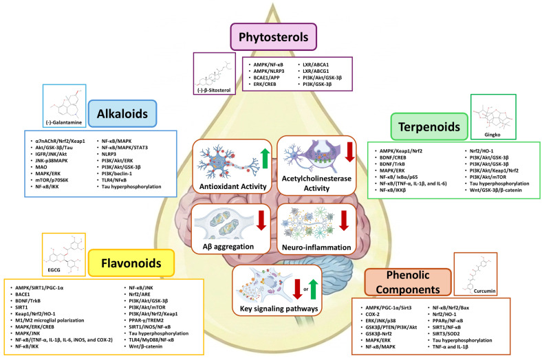
5. Studies on the Effects of Essential Oils on Dementia
5.1. Observations of the Effects of Essential Oils on Nursing Home Patients by Aromatherapists
Reports by aromatherapists who have years of experience conducting, for example, hand massages on healthy clients and patients with dementia are important resources of information on the possible effects of the phytochemicals in essential oils. Table 6 summarizes examples of observations on reactions following hand massage using essential oils. These reports most likely include the effects of hand massage itself and the effects of interaction with the aromatherapist in addition to the effects of phytochemicals in the essential oils (unpublished data by KT and YM), and there could be synergistic influences of each factor as well.
Table 6: Examples of observations of changes after hand massage using essential oils (unpublished data). Age > 80. One male patient and all others were female patients.
| Patterns | Client | Before | After |
|---|---|---|---|
| Pattern 1(mood) | Dementia patient | The patient is usually agitated, upset, or grumpy | About 6 min after the massage started, the patient calmed down with a calm look |
| Pattern 2(mood) | Dementia patient | No description about before the hand massage | Started to smile, and the face lit up |
| Pattern 3(talking) | Dementia patient | Patient did not talk about family at the facility; Patient had negative self-esteem | Started to talk about their family; Started to talk with the aromatherapist; Repeatedly talked about the same content; Started to talk, thank, and smile |
| Pattern 4(falling asleep) | Dementia patient | A patient who talks usually calmly or quietly | Around the time the massage of one hand is over and switching to the other hand (about 12 min for each hand), the patient started to fall asleep |
| Pattern 5(emotional) | Dementia patient | Has emotional instability, which gradually became worse as the symptoms of dementia progressed. Many patients showed an increase in anxiety late in the afternoon. | Suddenly became overcome with emotion and started to cry when the hand massage started, then the mood became uplifted, starting to happily say it feels very nice, returning to her/his room calmly |
| Pattern 6(curiosity) | Healthy 97-year-old senior | Previously worked in a company related to drug development | Showed a strong interest in the name of the essential oil and asked for its name |
5.2. Effects of Essential Oils on Cognitive Functions
Numerous studies have shown the effects of essential oils or their chemical constituents on improving cognitive function or neuropsychiatric symptoms. Woo et al. [ref. 275] showed that cognitive function increased by 226% in healthy seniors over 60 years in age who used a diffuser for two hours in the evening with seven different essential oils rotating daily [ref. 275]. Cognitive function was assessed using the Ray Auditory Verbal Learning test and the Weschler Adult Intelligence Scale (ver.3) (WAIS-III). The remarkable increase in cognitive function test scores suggests that essential oils can be a promising new agent for preventing the decline of cognitive functions in seniors. The study did not compare the effects of the seven essential oils used, nor did it analyze their chemical compositions, which can vary substantially even among oils with the same name. This information would be important to know whether all the essential oils were effective or, if not, which one caused the observed effects. The differences due to the types of essential oils, if any, are also important for determining which phytochemical compounds had significant effects in improving cognitive functions.
In another study, 28 patients with either AD, AD and cerebrovascular lesions, or vascular dementia were exposed to a mixture of lemon and rosemary essential oils in the morning (9:00 to 11 am) and a mixture of lavender and orange essential oils in the evening (7:30 pm to 9 pm) for 4 weeks using a diffuser system [ref. 276,ref. 277]. The effects were evaluated using the Functional Assessment Staging of Alzheimer’s disease (FAST), Hasegawa’s dementia scale (HDS-R), a Japanese version of the Gottfries, Braine, Steen Scale (GBSS-J) on cognitive function (GBSS-J-A), spontaneity (GBSS-J-B), feeling function (GBSS-J-C), other moral condition (GBSS-J-D), and movement function (GBSS-J-E). The study found that there was significant improvement in cognitive function, specifically in abstract function (GBSS-J-A-13), in the patients with mild to moderate AD [ref. 277].
Other than essential oils, there are some studies showing the effects of medicinal plants on improving cognitive function or memory [ref. 278,ref. 279]. In a review study by Kirubakaran [ref. 278], lavender (Lavandura angustifolia) and common sage (Salvia officinalis), which are well-known for their essential oils, were listed as herbal remedies for treatment of AD. In addition, various herbs and spices, which are not usually used as essential oils, were also listed to improve cognitive function: for example, ginkgo (Ginkgo biloba), waterhyssop (Bocopa monnieri), rhodiola (Rhodiola rosea), and fir moss (Huperzia serrata) [ref. 278]. Another study using extract drops of lemon balm (Melissa officinalis) showed improved cognitive function (measured by the Alzheimer’s disease assessment scale (ADAS-cog)) after four months without side effects [ref. 280].
Indian pennywort (or gotu kola, Asiatic pennywort, Ji Xue Cao, Centella asiatica) is well-known as one of the medicinal plants used in Ayurveda [ref. 281] (a traditional medical system of India) [ref. 282] and in traditional Chinese medicine [ref. 283]. In a review, Sun et al. [ref. 283] reported that Centella asiatica improved cognitive functions in studies using mice or rats as animal models. Wattanathorn et al. [ref. 210] conducted a clinical study in which 28 healthy seniors (average age 65) took a capsule that contained either 250, 500, or 750 mg (or a placebo) of extract of Centella asiatica daily for 8 weeks. They found that participants who took Centella asiatica showed faster reaction times and higher accuracy in cognitive function tests after the 8 weeks [ref. 210]. There is also currently an ongoing Phase I clinical study in the U.S. by Soumyanath et al. (accessed on 11 March 2026, https://clinicaltrials.gov/study/NCT05591027) testing the effects of taking Centella asiatica for 6 weeks, indicating growing interest in the effects of Centella asiatica. Pharmacokinetic and pharmacodynamic studies of the group also showed that the aqueous extract of Centella asiatica was safe [ref. 11].
Waterhyssop (Bacopa monnieri) is another herb used in Ayurveda. Essential oil derived from B. monnieri is called bacopa oil or brahmi oil. A clinical trial in Australia tested the effects of waterhyssop in 101 participants (age 40 to 70 with self-reported memory and attention problems; in total, 40 participants who took 300 mg of B. monnieri extract daily and 47 participants who took placebo completed a 12-week period). Although there were no significant differences in the cognitive test performances, the self-reported stress level and fatigue after the cognitive tasks were significantly lower [ref. 284].
Another plant well-known as one of the medicinal plants used in Ayurveda [ref. 281] and traditional Chinese medicine [ref. 283] is the false daisy (other names of the same plant: bhangra, bhringaraj, or Han Lian Cao, Eclipta prostrata, or E. alba). We are not aware of any clinical trials on its effects on dementia yet, although there are some studies using rats as animal models showing that it improved learning and memory [ref. 285].
5.3. Effects of Essential Oils on Neuropsychiatric Symptoms
There have been studies on utilizing essential oils for treating neuropsychiatric symptoms [ref. 217,ref. 280,ref. 286,ref. 287], in which lemon balm (Melissa officinalis) [ref. 217,ref. 280] and lavender (Lavandula angustifolia) [ref. 287] were used, showing positive effects on suppressing agitation. In these studies, Ballard et al. [ref. 217] used lemon balm essential oil by mixing it into a base lotion (sunflower was used as the placebo for the control group) and applying it on the face and arms of the patients twice a day for four weeks [ref. 217]. Agitation (evaluated using the Cohen-Mansfield Agitation Inventory (CMAI)) and Quality of Life Indices (measured by Dementia Care Mapping) both showed improvement in the group treated with lemon balm essential oil. In the study by Lin et al. [ref. 287], diffusers were used. Two drops of pure lavender (Lavandula angustifolia) certified by the Aromatherapy Organization Council (sunflower was used in the control group) were put on cosmetic cotton and placed in diffusers, and two diffusers were placed on each side of the pillows of the patient at night for at least 1 h [ref. 287]. Effects were evaluated after three weeks using the Chinese version of the Neuropsychiatric Inventory (CNPI) and CMAI, and significant improvements in both CNPI and CMAI were observed. The treatment’s effects were especially significant for agitation, irritability/lability, aberrant motor behaviors, and night-time behaviors [ref. 287]. There are some studies using lavender that did not show measurable effects [ref. 288], which could be due to differences in methods or the essential oil chemical profiles, and further tests will be required.
Other than bergamot and lavender, numerous candidate essential oils have been tested using animal models and in human studies, not necessarily related to dementia but more focused on depression and anxiety [ref. 289]. Sweet orange (Citrus sinensis), yuzu (Citrus junos), lemon (Citrus limon), chamomile (Matricaria chamomilla), rosemary (Salvia Rosmarinus), sage (Salvia officinalis), Spanish sage (Salvia lavandulaefolia), Japanese sweet flag (Acorus gramineus), perilla (Perilla frutescens), coriander (Coriandrum sativum), asarum (Asarum caudatum), rose (Rosa disambiguation), and ylang-ylang (Cananga odorata) are listed as having effects on increasing serotonin secretion and locomotor activity in animal model studies and antidepressant effects and anxiolytic effects in human studies [ref. 289]. These studies suggest that there are many candidate essential oils that may have effects on dementia but are still in need of testing.
As briefly mentioned in Section 3.2.3, drugs for neuropsychiatric symptoms such as Brexpiprazole can increase the risk of stroke, resulting in a higher chance of death [ref. 41,ref. 42,ref. 43]. The development of methods that have fewer risks is important, and the possibility of fewer life-threatening adverse events using essential oils suggests that they may offer a promising alternative to these drugs, which have an increased risk of death (see Section 6.2 for studies on improving the stability of and reducing the adverse events caused by some essential oils).
5.4. Effects of Essential Oils on Factors Associated with Dementia
There are many studies testing the effects of each phytochemical compound on Aβ, AChE, etc. Table 7 shows examples of the essential oils or the plants that showed effects on factors associated with dementia, either in clinical studies or animal model studies. This suggests there is a high possibility of identifying the phytochemical compounds, and plants rich in them, which can become future drugs for dementia. It is also possible to see from the table that the same plant/essential oils have multiple effects. This is not surprising considering that each disease mechanism (such as Aβ, tau, AChE, etc.) is not an independent factor, and considering that each plant has hundreds of chemical constituents and that multiple constituents often possess bioactive properties. The large number of plants in these examples indicates the difficulties in going through an extensive number of plants to identify the best candidate(s) for further study. To facilitate this process, we propose a network machine learning strategy (see Section 6.1), in which the mechanisms of action of phytochemical compounds can be compared to the drugs for dementia to identify the plants that contain many of them.
Table 7: Examples of the essential oils or the plants and their major constituents with effects on factors associated with dementia.
| Effects | Essential Oil | Chemical Constituents Proposed to Have Effects on Dementia | References |
|---|---|---|---|
| Anti-Aβ | Ashwagandha * (Withania somnifera) | Withanolides #4.4 | [ref. 47] |
| Asian ginseng * (Panax ginseng) | Ginsenoside #4.4 | [ref. 47,ref. 290] | |
| Aleppo pine * (Pinus halepensis) | β-Caryophyllene #4.4, α-pinene #4.4 and myrcene | [ref. 291,ref. 292] | |
| Cinnamon (Cinnamomum zeylanicum) | Linalool #4.4, (E)-cinnamaldehyde and (E)-cinnamyl acetate, eugenol [ref. 293]; italicene [ref. 294] | [ref. 293,ref. 294] | |
| Coriander (Coriandrum sativum) | Linalool #4.4 | [ref. 295] | |
| Ginkgo * (Ginkgo biloba) | Ginkgolides #4.4 (A,B,C,J), bilobalide #4.4 | [ref. 47] | |
| Indian pennywort (or gotu kola, Asian pennywort, Ji Xue Cao, Centella asiatica) | Asiaticoside #4.4, madecassoside, asiatic acid, madecassic acid; isochlorogenic acid A, 1,5-dicaffeoylquinic acid | [ref. 47,ref. 283,ref. 296,ref. 297] | |
| Lavender (Lavandula angustifolia) | Linalool #4.4, linalyl acetate, β-caryophyllene **** #4.4 | [ref. 295] | |
| Lemon balm (Melissa officinalis) | 2,2-Dimethoxybutane, 3′,4′,5,7-tetrahydroxyflavone, 3′-O-β-D-glucuronopyranoside, γ-O-β-D-glucopyranoside and 2,3,19,23-tetrahydroxy-12-ursen-28-oic acid-23-sulfate, 28-O-β-D-glucopyranosyl ester, sajerinic acid [ref. 298]; solely through rosmarinic acid #4.5 [ref. 47] | [ref. 47,ref. 298] | |
| Rosemary * (Rosmarinus officinalis) | Oleanolic acid | [ref. 294] | |
| Saffron * (Crocus sativus) | Crocin #4.4, safranal #4.4 | [ref. 47] | |
| Sage ** (Salvia officinalis) | Pinocembrin [ref. 294]; rosmarinic acid #4.5, carnosic acid | [ref. 47,ref. 294] | |
| Turmeric (Curcuma longa) | Curcumin #4.5, demethoxycurcumin, bisdemethoxycurcumin | [ref. 47] | |
| Waterhyssop * (Bacopa monnieri) | Bacosides A #4.4, bacosides B #4.4 | [ref. 47] | |
| Sweet wormwood (Artemisia annua) | Artemisinin | [ref. 299,ref. 300] | |
| Anti-tau | Cinnamon (Cinnamomum zeylanicum) | Chamigrene, italicene | [ref. 294] |
| Rosemary (Rosmarinus officinalis) | Luteolin #4.2 | [ref. 294] | |
| Sage ** (Salvia officinalis) | Myricetin #4.2 | [ref. 294] | |
| Saffron * (Crocus sativus) | Crocin #4.4, safranal #4.4 | [ref. 47] | |
| Sweet wormwood (Artemisia annua) | Artemisinin | [ref. 299,ref. 300] | |
| Anti-α-Synuclein | Citron * (Citrus medica) | Linalool #4.4 | [ref. 293] |
| Sage ** (Salvia officinalis) | 1,8-Cineole (eucalyptol) #4.4 | [ref. 293] | |
| Thyme (Thymus vulgaris) | [ref. 292] | ||
| Anti-AChE | Ashwagandha * (Withania somnifera) | Withanolides #4.4 | [ref. 47] |
| Cinnamon (Cinnamomum zeylanicum) | Linalool #4.4, (E)-cinnamaldehyde and (E)-cinnamyl acetate, eugenol | [ref. 293] | |
| Clove (Syzygium aromaticum) | [ref. 301] | ||
| False daisy * (Eclipta prostrata) | Wedelolactone | [ref. 302,ref. 303] | |
| Garlic (Allium sativum) * | Allicin [ref. 304] | [ref. 304,ref. 305] | |
| Greater galangal * (Alpinia galanga) | α-Humulene [ref. 306] | [ref. 306,ref. 307,ref. 308] | |
| Lemon balm (Melissa officinalis) | 2,2-Dimethoxybutane, 3′,4′,5,7-tetrahydroxyflavone, 3′-O-β-D-glucuronopyranoside, γ-O-β-D-glucopyranoside and 2,3,19,23-tetrahydroxy-12-ursen-28-oic acid-23-sulfate, 28-O-β-D-glucopyranosyl ester, sajerinic acid [ref. 298] | [ref. 47,ref. 309] | |
| Myrtle (Myrtus cummunis) | [ref. 310] | ||
| Oregano (Origanum vulgare) | Thymol, carvacrol | [ref. 292] | |
| Black pine (Pinus nigra) | [ref. 311] | ||
| Pistacia khinjuk * | [ref. 305] | ||
| Rosemary (Rosmarinus officinalis) | 1,8-Cineole (eucalyptol) #4.4 | [ref. 292,ref. 294] | |
| Tetraclinis articulata * | [ref. 312] | ||
| True cinnamon tree (Cinnamomum verum) | (E)-Cinnamaldehyde [ref. 310] | [ref. 310] | |
| Anti-butyrylcholinesterase | False daisy *(Eclipta prostrata) | Wedelolactone | [ref. 302,ref. 303] |
| Myrtle (Myrtus cummunis) | [ref. 310] | ||
| Pinus nigra | [ref. 311] | ||
| True cinnamon tree (Cinnamomum verum) | (E)-Cinnamaldehyde [ref. 310] | [ref. 310] | |
| Anti-beta-Secretase 1 | Lemon balm (Melissa officinalis) | Triethyl citrate, 3′,4′,5,7-tetrahydroxyflavone, 3′-O-β-D-glucuronopyranoside, γ-O-β-D-glucopyranoside and 2,3,19,23-tetrahydroxy-12-ursen-28-oic acid-23-sulfate, 28-O-β-D-glucopyranosyl ester, sajerinic acid | [ref. 298] |
| True cinnamon tree (Cinnamomum verum) | (E)-Cinnamaldehyde [ref. 310] | [ref. 310] | |
| Turmeric (Curcuma longa) | α-Turmerone, β-turmerone and turmerone | [ref. 313] | |
| Anti-neuroinflammation | Bergamot *** (Citrus bergamia) | Limonene, linalyl acetate ****, Linalool **** #4.4 | [ref. 292] |
| Clove (Syzygium aromaticum) | Eugenol | [ref. 292] | |
| Ginger (Zingiber officinale) | 10-gingerol, 6-shogaol | [ref. 292] | |
| Lavender (Lavandula angustifolia) | Linalool #4.4, linalyl acetate | [ref. 292] | |
| Peppermint (Mentha piperita) | Menthol, 1,8-Cineole **** | [ref. 292] | |
| Satureja khuzistanica * | Carvacrol | [ref. 292] | |
| Tea tree (Melaleuca alternifolia) | Terpinene-4-ol, 1,8-Cineole **** #4.4 | [ref. 292] | |
| Thyme (Thymus vulgaris) | [ref. 292] | ||
| Sweet wormwood (Artemisia annua) | Artemisinin | [ref. 299,ref. 300] |
*: Not commonly used as essential oils but on the market as supplements; **: Contains thujone, which is safe for culinary use but needs caution when used in higher doses as tea, medicines, supplements, and essential oils. See Section 6.2; ***: Contains bergapten, which is a furocoumarin. See Section 6.2; ****: Chemical constituents usually listed to be contained in the essential oils of these plants by the Japan Aromacoordinator Association (JAA) and found to have effects on dementia but not specifically referred to in the paper cited; #: Chemical compounds with description in Section 4 with the section number. For example, #4.4 indicates it is described in Section 4.4.
5.5. Phytochemical Compounds in the Essential Oils Can Reach the Brain Through the Nose-to-Brain Pathway and the Transdermal Pathway
5.5.1. Nose-to-Brain Phytochemical Pathway and the Pathway in Sensing Odors
Utilization of the nose to deliver a drug to the brain has been receiving increasing interest as it enables bypassing the blood–brain barrier (BBB), delivers the drug to the CNS quickly, and is noninvasive [ref. 314,ref. 315,ref. 316,ref. 317,ref. 318]. The route through the nose includes the olfactory pathway (Figure 4A), the trigeminal pathways (Figure 4B), and the respiratory pathway (Figure 4(C1,C2)) [ref. 319], among which the nose-to-brain pathway utilizes the olfactory pathway as a gate to the brain. There are recent studies trying to establish this method as a method to deliver drugs to patients with dementia as an alternative to IV injections or pills [ref. 320,ref. 321,ref. 322]. This indicates that the inhalation of an essential oil either from a diffuser or when it is used in a hand massage can also deliver the phytochemicals contained in it from the nose to the brain, i.e., nose-to-brain phytochemical delivery. Thus, the phytochemicals that have bioactive properties that are beneficial in treating dementia can reach the brain, bypassing the BBB, in a fast and noninvasive way.
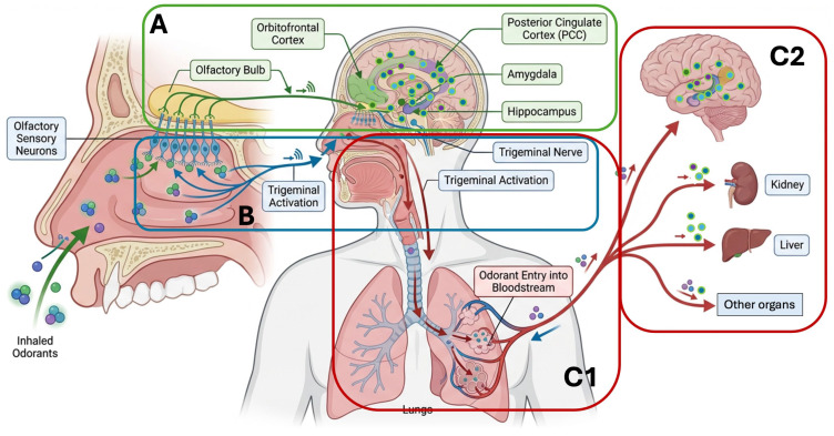
A key question is where in the brain these phytochemicals reach via nose-to-brain drug delivery and how they do so. A confusing fact is that, as they have smells, we tend to misunderstand that the mechanisms of our olfactory sense, which we use in detecting, recognizing, and memorizing smells by the activation of olfactory receptors on olfactory sensory neurons (Figure 4A locations written in green boxes), are involved. The route of olfaction is based on the activation of olfactory receptors, which does not require the transport of the chemical compounds that activate the olfactory receptors. The activated signals reach the olfactory bulbs, and from there, the orbitofrontal cortex (olfactory cortex) for recognition, the amygdala to affect hormone secretion, and the hippocampus for memorization.
How phytochemical compounds are delivered from the nose to the brain needs to be considered separately from how the smells are detected. This is critically important because we need the phytochemicals to be ‘delivered’ to the areas where there is neuroinflammation, aggregation of Aβ, and accumulation of other enzymes and proteins related to dementia, causing negative effects on neuronal health. The details on how phytochemicals become transported through the nose-to-brain pathway are not well known. However, in a recent study by Shen et al. [ref. 317], the authors used mice as an animal model system and administered commensal bacteria labeled with fluorescein isothiocyanate (FITC) to the nasal cavity. It was known that FITC would be released gradually from the bacteria, with the peak release within 48 h. This allowed them to trace the travel of FITC after the commensal bacteria were administered intranasally. They found that FITC traveled through the olfactory nerve bundles in the lamina propria under the olfactory epithelium, from there to the olfactory bulb, and eventually to broad regions in the brain [ref. 317] (Figure 5). In another study using mice as an animal model, Oguro et al. [ref. 323] exposed mice to linalool (4ppm as a mist) for 2 h and measured the levels of linalool in the brain and in the other organs of the mice using GC-MS. Linalool was found in the olfactory bulb, cerebral cortex, cerebellum, lung, and blood [ref. 323], showing that phytochemical compounds can reach various locations inside the brain within two hours by inhalation.
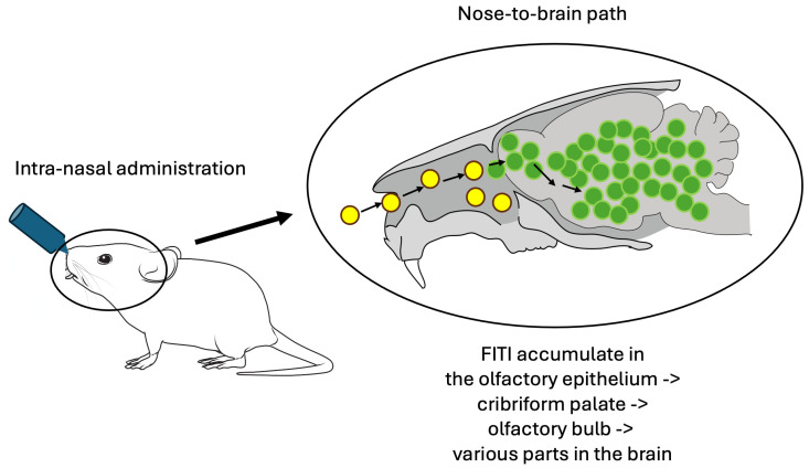
5.5.2. Transdermal Pathway
When essential oils are applied by hand massage, multiple routes are involved (Figure 6A). As they are highly volatile, we can smell the odor of the essential oils when they are applied to the skin, which indicates that we inhale the phytochemical compounds contained in the oils. This allows the routes through the nose as already described above (see Section 5.5.1). Here we will focus on the transdermal pathway.
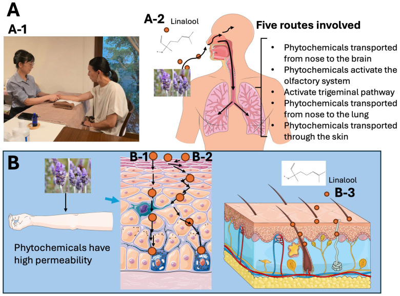
Essential oils are well-known to permeate skin easily, and they are even used as one of the methods of permeation enhancers in transdermal drug delivery [ref. 324,ref. 325,ref. 326,ref. 327]. There are three routes involved in the transdermal pathway (Figure 6B): intracellular (Figure 6(B-1)), intercellular (Figure 6(B-2)), and through the hair follicle and sweat gland ducts (Figure 6(B-3)). Importantly, when essential oils are used in hand massage, they are diluted using carrier oils [ref. 328,ref. 329] to avoid contact dermatitis [ref. 330,ref. 331] and evaporation, as well as to maintain/manage the concentration. Oils from jojoba (Simmondsia chinensis), almond (Prunus amygdalus), seeds of grape (Vitis vinifera), coconut (Cocos nucifera), apricot (Prunus armeniaca), and calendula (Calendula officinalis) are often used as carriers. The dilution rates recommended by the Japan Aromacoordinator Association (JAA) are 2 to 3%, which depends on the types of essential oil and also on the types of carriers [ref. 329]. Diluting essential oils may trigger questions about concentration and effects. In a study by Komori et al. [ref. 328], a mixture of three different essential oils diluted with jojoba oil was used. The dilution rates of the essential oils to jojoba oil were 1:150 to 1:300 (0.33% to 0.67%): lemon (Citrus limon) (0.67%), tuberose (Polianthes tuberosa) (0.33%), and labdanam (Cistus ladanifer) (0.33%). They asked the participants to go through the Uchida-Kraepelin Performance test for 30 min, and then the participants were provided with either a hand massage with jojoba oil without essential oil, a full hand massage with essential oil, a simpler hand massage with essential oil, or no treatment as a control. Though the dilution rates they used were much lower than those recommended by the JAA, they still found significantly reduced stress levels in the participants that received a hand massage with essential oils in both the full massage and simpler massage groups compared to the no-treatment group and hand massage with only-carrier-oil group, which came out in the middle, between the no-treatment and essential oil groups [ref. 328].
6. Conclusions and Future Perspective
As we have summarized so far, there is a high potential for utilizing essential oils or plant-derived supplements and medicines for patients with dementia. One of the biggest obstacles in the case of essential oils could be the ‘image’ of essential oils that many people still have, i.e., that they are non-scientific (often using the word ‘holistic’ in that meaning) and that they are only for relaxing and enjoying the scents. Unfortunately, we receive these comments often, and we hear surprise that there is actually solid scientific research on the basic chemistry of phytochemical compounds. One of the goals of our review is to enhance the understanding of phytochemical compounds and their effects.
The more we know about phytochemical compounds and their bioactive properties, the more we notice there are several questions that need to be answered: for example, (1) which one should be used for what condition, (2) can we or should we improve the delivery by using a chemical formulation, (3) should we use them as a single chemical compound or as a combination or as a whole extract, and (4) what is the best delivery method? It must be emphasized that the phytochemical approaches and aromatherapy strategies discussed in this review are intended to be evaluated as complementary interventions alongside, not as replacements for, established pharmacotherapy. Patients receiving FDA-approved treatments for dementia should not discontinue or modify their medications based on preclinical or early-phase clinical evidence for plant-derived compounds without consultation with their healthcare providers.
6.1. Finding the Most Promising Plant for the Treatment or Prevention of Dementia: Network Machine Learning
The extensive number of medicinal plants and their extracts, with an even more extensive number of phytochemical compounds, is overwhelming. Which one(s) should be used is a difficult question to answer quickly, and we see papers on the effects of essential oils without the rationale on how/why the oils were selected. It would be extraordinarily helpful if we could select them based on scientific evidence on the phytochemical constituents.
In 2019, Veselkov et al. [ref. 332] conducted an extensive machine learning analysis of 7692 bioactive phytochemical compounds that possess the same mechanisms of action as the existing, clinically approved anti-cancer therapies [ref. 332]. Then they conducted an analysis of foods that contain these phytochemical compounds (naming them ‘Hyperfoods’ from their cancer-beating nature) [ref. 332]. Using the same methodology, they also determined the ‘Hyperfoods’ for COVID-19 [ref. 333]. In 2023, using the same methodology but focusing on olive oil, they determined the phytochemical compounds included in olive oil that have a high possibility of suppressing the onset and progression of AD [ref. 334]. Table 8 shows the top 10 phytochemical compounds included in olive oil identified in their study to have beneficial effects on the suppression or treatment of AD [ref. 334]. These approaches have a high possibility of providing rationales for selection from the broad number of plants if the analyses are expanded to include a large number of phytochemical compounds, as the authors did for their studies on cancer and COVID-19 [ref. 332,ref. 333].
Table 8: Phytochemical compounds with a high correlation to drugs for AD (from Rita et al. [ref. 334]).
| Phytochemical Compound | Correlation % |
|---|---|
| Quercetin | 78 |
| Genistein | 75.5 |
| Luteolin | 73.9 |
| Palmitoleate | 69.5 |
| Stearic acid | 67.5 |
| Apigenin | 67.4 |
| Epicatechin | 66.4 |
| Kaempferol | 65.1 |
| Squalene | 63.7 |
| Daidzein | 62.1 |
6.2. Enhancing Stability and Reducing Toxicity
There are multiple methods for extracting essential oils, and depending on the methods, there are large differences in their chemical constituents [ref. 335,ref. 336,ref. 337,ref. 338,ref. 339]. Traditional methods include hydro-distillation, steam distillation, solvent extraction, pressured liquid extraction, and cold pressing extraction [ref. 335]. There are many recently developed methods which use, for example, enzyme-assisted extraction, fractional distillation, microwave-assisted hydro-distillation, solvent-free microwave extraction, and so on [ref. 335]. The differences in the chemical constituents depending on the distillation methods also affect the bioactive properties [ref. 335,ref. 337,ref. 338]. Mohanty et al. [ref. 338] used three different Curcuma plant species and compared the chemical constituents and the bioactive properties of their essential oils when hydro-distillation and solvent-free microwave extraction were used as extraction methods [ref. 338]. They found that, although the major chemical constituents were qualitatively the same, solvent-free microwave extraction produced more essential oil volume, with significantly different quantities, and the essential oils made using this method showed better results in their cytotoxicity [ref. 338]. These studies suggest that new technologies for making essential oils are more efficient than traditional methods.
New technologies also allow totally different strategies, for example, to stabilize unstable chemical constituents and eliminate furocoumarins, which can cause skin reactions called phytophotodermatitis, after topical application and exposure to UV [ref. 340,ref. 341]. Furocoumarins, or furanocoumarins, are tricyclic aromatic compounds found especially in citrus (Rutaceae) such as citrus fruits (bergamot, lime, lemons, etc.) and Apiaceae family plants such as carrots, celery, parsley, etc. [ref. 341,ref. 342]. Although they can cause phytophotodermatitis, they also have ‘photo-protective effects’ on terpenes, suppressing their degradation [ref. 343], which indicates that simply excluding furocoumarins does not provide a complete solution, because, if they are chemically excluded, it is necessary to protect the remaining chemical constituents from degradation.
As an attempt to solve this problem, Scuteri et al. [ref. 344] developed bergamot without bergapten [ref. 344,ref. 345], the major furocoumarin included in bergamot (bergapten-free bergamot, BEO-BF), which maintained the pharmacological properties of bergamot. In addition, in order to enhance the stability of the remaining chemical constituents, they used solid lipid nanoparticles (SLN), chemically encapsulating BEO-BF (NanoBEO) [ref. 344]. This chemically formulated bergamot without bergapten showed significant anti-nociceptive and anti-allodynic effects in their study using mice as an animal model [ref. 344]. Scuteri et al. [ref. 43] conducted a clinical trial with patients with dementia, applying NanoBEO on their arms once daily for four weeks, and determined that NanoBEO significantly reduced the frequency and level of agitation in the patients [ref. 43]. As such, new chemical strategies are being incorporated into or replacing the traditional methods, leading to a decrease in adverse events from the chemical constituents and improving the outcomes.
Another chemical compound with known toxicity at high doses is α-thujone. α-Thujone is found in plants such as common sage (Salvia officinalis) and wormwood (Artemisia absinthium) (Spanish sage, Salvia lavandulifolia, and sweet wormwood, Artemisia annua, contain low or non-detectable amounts of thujone). Although the concentration of α-thujone in common sage and wormwood is safe for culinary use, it is suggested that higher doses included in essential oils may cause serious health problems. Studies have shown that α-thujone causes GABAergic inhibition, with its strongest effects on GABAA receptor αaβ2δ receptors [ref. 346], causing convulsion. The notorious health problems called absinthism in the past were determined to be not due to thujone but rather caused by alcohol intoxication from wormwood spirit absinthe, as the spirit did not contain a high enough dose of thujone to cause problems [ref. 347,ref. 348]. A study testing the toxicity of the essential oils of common sage (S. officinalis), wormwood (A. absinthium), northern white cedar (Thuja occidentalis), and tansy (Tanacetum vulgare) has shown that toxicity was not solely due to thujones in the oils [ref. 349]. These studies indicate that further studies are necessary. There are studies showing that wormwood essential oils improved the cognitive functions of rats with AD (50 mg/kg) [ref. 350]. There are also studies showing that thujone has antioxidant and neuroprotective effects, as well as other effects unrelated to dementia [ref. 351]. With cautions on the concentrations included in the oils, there are possibilities of utilizing it for AD and other types of dementia.
Enhancing stability is also important, particularly for the purpose of delaying oxidation. For example, the oxidized product of BCP, which is caryophyllene oxide, can be an allergen of moderate strength, with potential to cause skin sensitization [ref. 352]. Studies on enhancing the stability of phytochemical compounds while maintaining their pharmacological properties will be important.
6.3. The Entourage Effects (Synergistic Effects by Multiple Chemical Compounds)
The entourage effect is best known in studies on the cannabinoid system [ref. 353]. Ben-Shabat et al. [ref. 354] were the first to call the synergistic effects of multiple chemical compounds ‘the entourage effects’ in their studies on the cannabinoid system. Their study was not on phytochemical compounds but on an endocannabinoid called 2-arachidonoyl-glycerol (2-AG), which showed enhanced binding to cannabinoid receptors 1 and 2 (CB1 and CB2) by endogenous monoglycerides 2-linoleoyl-glycerol (2-LG) and 2-palmitoyl-glycerol (2-PG) [ref. 354]. Entourage effects are found in many phytochemical compounds [ref. 355,ref. 356,ref. 357]. For example, a combined use of CBD and BCP produced synergistic analgesic effects [ref. 358]. Another well-known example is the combinatory use of curcumin and piperine, improving its bioavailability [ref. 359]. Scuteri et al. [ref. 360] cited Ribeiro [ref. 361] as stating that “the strongest effect of EOs is due to the whole phytocomplex made up of various plant components that need to be present in a precise ratio to exert the so-called entourage effect” (EOs: essential oils) [ref. 360,ref. 361]. There are multiple factors considered to be involved in producing the synergistic effects, i.e., the entourage effects: (1) enhanced bioavailability by generating a phytocomplex, like in the case of curcumin and piperine [ref. 359], (2) additive effects of interfering with different inflammatory pathways, and (3) additive effects of interfering with the same inflammatory pathways [ref. 356,ref. 362]. Importantly, these synergistic/entourage effects enable reducing the concentration of each phytochemical compound to produce the targeted effects, which can lead to reducing the adverse events, if any. It is important to identify the combinations that work most efficiently and to adjust the concentrations or doses or frequencies depending on the conditions, in addition to the individual effects of each phytochemical compound. For example, factors such as whether it is for prevention or treatment, the specific symptoms of each person, the age, the sex, and other parameters such as the genotype of cytochrome P450 (CYP) [ref. 363], which is involved in metabolizing various chemical compounds, should be considered.
6.4. Limitations of the Evidence
The reader should be aware that the evidence base for most phytochemicals discussed in this review remains largely preclinical. The majority of mechanistic studies cited were conducted in cell culture systems or rodent models, and the translational validity of such models for complex neurodegenerative diseases like AD is inherently limited. Of the compounds reviewed, those with the most direct and robust clinical evidence in human dementia populations are: EGb 761 (Ginkgo biloba extract), with replicated RCT evidence in mild-to-moderate AD and MCI [ref. 12,ref. 176]; saffron (Crocus sativus carotenoids), with two completed RCTs in mild-to-moderate AD [ref. 177,ref. 178]; curcumin, with clinical trial data in healthy older adults and subjective cognitive decline [ref. 9]; and rosmarinic acid, with a completed MCI trial showing improvement on CDR-SB [ref. 10]. For all other compounds discussed, clinical translation has not yet been established, and the review’s mechanistic descriptions should be interpreted as hypothesis-generating rather than as evidence of clinical efficacy. Furthermore, many of the clinical studies cited are limited by small sample sizes, short durations, heterogeneous patient populations, and the absence of validated biomarker endpoints. Larger, better-powered trials with standardized formulations and mechanistically relevant endpoints are needed across all compound classes described in this review.
6.5. Possibility of Utilizing Plant-Origin Natural Products in Both Prevention and Treatment for ‘Health Span’ in the Longer Life Span
Studies have shown that human life expectancy increased by about 30 years during the 1990s [ref. 1], leading to an increase in the number of patients with dementia. How to extend health span or health longevity (the life with good health) is a critical issue in our lives. The importance of exercise and nutrition is often discussed in the literature [ref. 364,ref. 365]. In addition, phytochemical compounds have a high potential to be used as ‘drugs’ for dementia. A broad range of studies are still needed to learn how to improve the stability of the chemicals, enhance bioavailability, and rationalize the best compounds for specific indications (precision phytochemicals). We can also develop supplements and drugs in addition to using essential oils, and it is important to conduct network machine learning analyses to determine the ‘Hyperplants’ for this goal as well. Medicinal plants have been used since ancient times, but we are still in the early stage of fully employing them based on modern scientific techniques and the evidence from integrated ‘omics’ methodologies with advanced data analyses.
Despite the abundance of preclinical evidence reviewed here, only a small fraction of the phytochemicals with demonstrated activity in cellular and animal models of AD have been evaluated in rigorous human clinical trials. Several interrelated barriers account for this discrepancy. First, many polyphenols, terpenoids, and alkaloids display poor oral bioavailability, undergoing rapid first-pass hepatic metabolism and producing circulating metabolites whose neuroprotective properties may differ substantially from the parent compound. Second, CNS penetration is limited for many hydrophilic or high-molecular-weight phytochemicals, and P-glycoprotein efflux at the blood–brain barrier (BBB) further restricts brain accumulation of some compounds. Third, the chemical heterogeneity of plant-derived preparations, whose composition varies by cultivar, geographic origin, extraction method, and processing conditions, makes dose standardization and reproducibility across trials a significant challenge. Fourth, the typically slow and multifactorial progression of AD demands long-duration, large-cohort trials with validated cognitive and biomarker endpoints that are costly and organizationally complex to maintain for the duration of the trial. Addressing these barriers through improved formulation strategies (as discussed in Section 6.2), rigorous product characterization, and the use of validated surrogate biomarkers in early-phase trials will be essential to translating the preclinical promise of phytochemicals into clinically meaningful therapies.
References
- S.J. Olshansky, B.J. Willcox, L. Demetrius, H. Beltrán-Sánchez. Implausibility of Radical Life Extension in Humans in the Twenty-First Century. Nat. Aging, 2024. [DOI | PubMed]
- F. Prinelli, A. Mastropietro, S. Bernini, T. Camboni, C. Consolandi, S. Conti, A. Costa, M. Cotta Ramusino, N. Jesuthasan, O. Pansarasa. Impacts of an Antioxidant-Rich Diet and Lifestyle Factors on Gut Microbiota Diversity and Brain Health: An Exploratory Analysis from the NutBrain Study. Clin. Nutr., 2026. [DOI | PubMed]
- Z. Li, Z. Zhang, B. Yu. Unlocking the Therapeutic Potential of Natural Products for Alzheimer’s Disease. J. Med. Chem., 2025. [DOI | PubMed]
- N.D. Anderson. State of the Science on Mild Cognitive Impairment (MCI). CNS Spectr., 2019. [DOI | PubMed]
- G. Fernández-Rubio, P. Vuust, M.L. Kringelbach, L. Bonetti. The Neurophysiology of Healthy and Pathological Aging: A Comprehensive Systematic Review. Brain Struct. Funct., 2025. [DOI | PubMed]
- R.C. Petersen, G.E. Smith, S.C. Waring, R.J. Ivnik, E.G. Tangalos, E. Kokmen. Mild Cognitive Impairment: Clinical Characterization and Outcome. Arch. Neurol., 1999. [DOI | PubMed]
- E. Hessen. Mild Cognitive Impairment and Neuropsychological Examination. Front. Psychol., 2025. [DOI | PubMed]
- R.A. Dunne, D. Aarsland, J.T. O’Brien, C. Ballard, S. Banerjee, N.C. Fox, J.D. Isaacs, B.R. Underwood, R.J. Perry, D. Chan. Mild Cognitive Impairment: The Manchester Consensus. Age Ageing, 2021. [DOI | PubMed]
- G.W. Small, P. Siddarth, Z. Li, K.J. Miller, L. Ercoli, N.D. Emerson, J. Martinez, K.-P. Wong, J. Liu, D.A. Merrill. Memory and Brain Amyloid and Tau Effects of a Bioavailable Form of Curcumin in Non-Demented Adults: A Double-Blind, Placebo-Controlled 18-Month Trial. Am. J. Geriatr. Psychiatry, 2018. [DOI | PubMed]
- M. Mahboubi. Melissa officinalis and Rosmarinic Acid in Management of Memory Functions and Alzheimer Disease. Asian Pac. J. Trop. Biomed., 2019. [DOI]
- K.M. Wright, M. Bollen, J. David, A.B. Speers, M.S. Brandes, N.E. Gray, A. Alcázar Magaña, C. McClure, J.F. Stevens, C.S. Maier. Pharmacokinetics and Pharmacodynamics of Key Components of a Standardized Centella Asiatica Product in Cognitively Impaired Older Adults: A Phase 1, Double-Blind, Randomized Clinical Trial. Antioxidants, 2022. [DOI | PubMed]
- G.L.d.O. Pagotto, L.M.O.d. Santos, N. Osman, C.B. Lamas, L.F. Laurindo, K.T. Pomini, L.M. Guissoni, E.P.d Lima, R.d.A. Goulart, V.M.C.S. Catharin. Ginkgo Biloba: A Leaf of Hope in the Fight against Alzheimer’s Dementia: Clinical Trial Systematic Review. Antioxidants, 2024. [DOI | PubMed]
- J. Hugo, M. Ganguli. Dementia and Cognitive Impairment: Epidemiology, Diagnosis, and Treatment. Clin. Geriatr. Med., 2014. [DOI | PubMed]
- D.S. Knopman, R.C. Petersen. Mild Cognitive Impairment and Mild Dementia: A Clinical Perspective. Mayo Clin. Proc., 2014. [DOI | PubMed]
- M. Grundman. Mild Cognitive Impairment Can Be Distinguished From Alzheimer Disease and Normal Aging for Clinical Trials. Arch. Neurol., 2004. [DOI | PubMed]
- M.M. Rahman, C. Lendel. Extracellular Protein Components of Amyloid Plaques and Their Roles in Alzheimer’s Disease Pathology. Mol. Neurodegener., 2021. [DOI | PubMed]
- M.H. Janeiro, C.G. Ardanaz, N. Sola-Sevilla, J. Dong, M. Cortés-Erice, M. Solas, E. Puerta, M.J. Ramírez. Biomarkers in Alzheimer’s Disease. Adv. Lab. Med., 2021. [DOI | PubMed]
- M.R. Shaikh, A. Jeyabose, R.V. Arjunan. Deep Learning for Alzheimer’s Disease: Advances in Classification, Segmentation, Subtyping, and Explainability. BioMed Eng. OnLine, 2025. [DOI | PubMed]
- 2025 Alzheimer’s Disease Facts and Figures. Alzheimer’s Dement., 2025. [DOI]
- R. Ossenkoppele, R. van der Kant, O. Hansson. Tau Biomarkers in Alzheimer’s Disease: Towards Implementation in Clinical Practice and Trials. Lancet Neurol., 2022. [DOI | PubMed]
- P. Calabresi, A. Mechelli, G. Natale, L. Volpicelli-Daley, G. Di Lazzaro, V. Ghiglieri. Alpha-Synuclein in Parkinson’s Disease and Other Synucleinopathies: From Overt Neurodegeneration Back to Early Synaptic Dysfunction. Cell Death Dis., 2023. [DOI | PubMed]
- J. Burré, M. Sharma, T.C. Südhof. Cell Biology and Pathophysiology of α-Synuclein. Cold Spring Harb. Perspect. Med., 2018. [DOI | PubMed]
- R. Harada, N. Okamura, S. Furumoto, K. Yanai. Imaging Protein Misfolding in the Brain Using β-Sheet Ligands. Front. Neurosci., 2018. [DOI | PubMed]
- S. Gunes, Y. Aizawa, T. Sugashi, M. Sugimoto, P.P. Rodrigues. Biomarkers for Alzheimer’s Disease in the Current State: A Narrative Review. Int. J. Mol. Sci., 2022. [DOI | PubMed]
- M. Shoji, T. Kawarabayashi, T. Nakamura, T. Sugawara, K. Ishizawa, M. Amari, R. Takahashi, H. Kasahara, N. Koganezawa, A. Higa. Drebrin Is a Novel Biomarker of Cognitive Deterioration in Alzheimer’s Disease. J. Alzheimer’s Dis., 2026. [DOI | PubMed]
- A.M. Brickman, J.J. Manly, L.S. Honig, D. Sanchez, D. Reyes-Dumeyer, R.A. Lantigua, P.J. Lao, Y. Stern, J.P. Vonsattel, A.F. Teich. Plasma P-Tau181, p-Tau217, and Other Blood-Based Alzheimer’s Disease Biomarkers in a Multi-Ethnic, Community Study. Alzheimer’s Dement., 2021. [DOI | PubMed]
- A. Inamdar, P. Bugadannavar, M. Palled, S. Umarani, P. Salve, B. Gurupadayya, P. Patil, H. Sharma. Biological Determinants of Blood-Based Biomarker Levels in Alzheimer’s Disease: Role of Nutrition, Inflammation, and Metabolic Factors. Front. Aging Neurosci., 2025. [DOI | PubMed]
- H.E. Dark, M.R. Duggan, K.A. Walker. Plasma Biomarkers for Alzheimer’s and Related Dementias: A Review and Outlook for Clinical Neuropsychology. Arch. Clin. Neuropsychol., 2024. [DOI | PubMed]
- C.E. Teunissen, I.M.W. Verberk, E.H. Thijssen, L. Vermunt, O. Hansson, H. Zetterberg, W.M. van der Flier, M.M. Mielke, M. Del Campo. Blood-Based Biomarkers for Alzheimer’s Disease: Towards Clinical Implementation. Lancet Neurol., 2022. [DOI | PubMed]
- S. Rani, S.B. Dhar, A. Khajuria, D. Gupta, P.K. Jaiswal, N. Singla, M. Kaur, G. Singh, R.P. Barnwal. Advanced Overview of Biomarkers and Techniques for Early Diagnosis of Alzheimer’s Disease. Cell. Mol. Neurobiol., 2023. [DOI | PubMed]
- P. Gomes, F. Tzouanou, K. Skolariki, A. Vamvaka-Iakovou, C. Noguera-Ortiz, K. Tsirtsaki, C.L. Waites, P. Vlamos, N. Sousa, B. Costa-Silva. Extracellular Vesicles and Alzheimer’s Disease in the Novel Era of Precision Medicine: Implications for Disease Progression, Diagnosis and Treatment. Exp. Neurol., 2022. [DOI | PubMed]
- A.R.R. Da Conceicao, J. Marinatto, L.S. Pinheiro, T. Rody, F.G. De Felice. The Multifaceted Role of Extracellular Vesicles in Alzheimer’s Disease. J. Neurochem., 2025. [DOI | PubMed]
- Z.-R. Chen, J.-B. Huang, S.-L. Yang, F.-F. Hong. Role of Cholinergic Signaling in Alzheimer’s Disease. Molecules, 2022. [DOI | PubMed]
- R.B. Rocha, F.F. Bomtempo, G.B. Nager, G.I. Cenci, J.P.M. Telles. Dual Orexin Receptor Antagonists for the Treatment of Insomnia: Systematic Review and Network Meta-Analysis. Arq. Neuropsiquiatr., 2023. [DOI | PubMed]
- A.K. Dubey, S.S. Handu, P.K. Mediratta. Suvorexant: The First Orexin Receptor Antagonist to Treat Insomnia. J. Pharmacol. Pharmacother., 2015. [DOI | PubMed]
- C. Liguori. Orexin and Alzheimer’s Disease. Curr. Top. Behav. Neurosci., 2017. [DOI | PubMed]
- Y.H. Um, H.K. Lim. Orexin and Alzheimer’s Disease: A New Perspective. Psychiatry Investig., 2020. [DOI | PubMed]
- M. Markovic, A. Gallipani, K.H. Patel, M. Maroney. Brexpiprazole. Ann. Pharmacother., 2017. [DOI | PubMed]
- Y. Nakamura, J. Adachi, N. Hirota, K. Iba, K. Shimizu, M. Nakai, K. Takahashi, N. Mori. Brexpiprazole Treatment for Agitation in Alzheimer’s Dementia: A Randomized Study. Alzheimer’s Dement., 2024. [DOI | PubMed]
- D. Lee, M. Slomkowski, N. Hefting, D. Chen, K.G. Larsen, E. Kohegyi, M. Hobart, J.L. Cummings, G.T. Grossberg. Brexpiprazole for the Treatment of Agitation in Alzheimer Dementia: A Randomized Clinical Trial. JAMA Neurol., 2023. [DOI | PubMed]
- J. Zissimopoulos, J. Thunell, M. Jacobson, S. Haye, B. Tysinger, P. Ferido, G. Joyce. Real-World Evidence of Brexpiprazole Use and 6-Month Mortality, Hospitalization, and Emergency Department Visits Among Persons With Dementia. Neurology, 2025. [DOI | PubMed]
- K. Johnell, G. Jonasdottir Bergman, J. Fastbom, B. Danielsson, N. Borg, P. Salmi. Psychotropic Drugs and the Risk of Fall Injuries, Hospitalisations and Mortality among Older Adults. Int. J. Geriatr. Psychiatry, 2017. [DOI | PubMed]
- D. Scuteri, D. Pierobon, M. Pagliaro, K. Hamamura, T. Hayashi, L. Pignolo, P. Nicotera, G. Bagetta, M.T. Corasaniti. Clinical and Market Analysis of NanoBEO: A Public-Worth, Innovative Therapy for Behavioral and Psychological Symptoms of Dementia (BPSD)-Emerging Evidence and Its Implications for a Health Technology Assessment (HTA) and Decision-Making in National Health Systems. Pharmaceutics, 2024. [DOI | PubMed]
- J.M. Jakubowski, A.A. Orr, D.A. Le, P. Tamamis. Interactions between Curcumin Derivatives and Amyloid-β Fibrils: Insights from Molecular Dynamics Simulations. J. Chem. Inf. Model., 2020. [DOI | PubMed]
- B. Khurshid, A.U. Rehman, S. Muhammad, A. Wadood, J. Anwar. Toward the Noninvasive Diagnosis of Alzheimer’s Disease: Molecular Basis for the Specificity of Curcumin for Fibrillar Amyloid-β. ACS Omega, 2022. [DOI | PubMed]
- M.T. Bayo Jimenez, L. Rivas-García, C. Sánchez-González, G. Grosso, V. Lipari, L. Vera-Ramírez, M. Battino, F. Giampieri, J.L. Quiles, T.Y. Forbes-Hernández. Natural Products in Alzheimer’s Disease: A Systematic Review of Clinical Trials and Underlying Molecular Mechanisms. Int. J. Mol. Sci., 2025. [DOI | PubMed]
- S. Pavlov, S.K. Prajapati, D. Yadav, A. Marcano-Rodriguez, H. Yadav, S. Jain. Advances in Bioactive Compounds from Plants and Their Applications in Alzheimer’s Disease. Biomolecules, 2025. [DOI | PubMed]
- G. Fu, M. Pan, Q. Sun, G. Yue, T. Lei. Natural Product-Based Strategies Targeting Inflammation in Alzheimer’s Disease: Mechanisms, Drug Delivery, and Clinical Trials. J. Pharm. Anal., 2025. [DOI]
- S.T. Abdelkhalek, J.-H. Shi, M.-X. Jin, S.S. Abdelgayed, M.-Q. Wang. Classification of Phytochemicals in Plants with Herbal Value. Herbal Medicine Phytochemistry, 2024
- S. Koyama, A. Purk, M. Kaur, H.A. Soini, M.V. Novotny, K. Davis, C.C. Kao, H. Matsunami, A. Mescher. Beta-Caryophyllene Enhances Wound Healing through Multiple Routes. PLoS ONE, 2019. [DOI | PubMed]
- E.P. de Lima, L.F. Laurindo, V.C.S. Catharin, R. Direito, M. Tanaka, I. Jasmin Santos German, C.B. Lamas, E.L. Guiguer, A.C. Araújo, A.M.R. Fiorini. Polyphenols, Alkaloids, and Terpenoids Against Neurodegeneration: Evaluating the Neuroprotective Effects of Phytocompounds Through a Comprehensive Review of the Current Evidence. Metabolites, 2025. [DOI | PubMed]
- M. Qneibi, S. Bdir, C. Maayeh, M. Bdair, D. Sandouka, D. Basit, M. Hallak. A Comprehensive Review of Essential Oils and Their Pharmacological Activities in Neurological Disorders: Exploring Neuroprotective Potential. Neurochem. Res., 2024. [DOI | PubMed]
- B.-H. Yoon, V.-L. Truong, W.-S. Jeong. Phytosterols: Extraction Methods, Analytical Techniques, and Biological Activity. Molecules, 2025. [DOI | PubMed]
- C. Liu, J. Huang, J. Qiu, H. Jiang, S. Liang, Y. Su, J. Lin, J. Zheng. Quercitrin Improves Cardiac Remodeling Following Myocardial Infarction by Regulating Macrophage Polarization and Metabolic Reprogramming. Phytomedicine, 2024. [DOI | PubMed]
- D. Tungmunnithum, A. Thongboonyou, A. Pholboon, A. Yangsabai. Flavonoids and Other Phenolic Compounds from Medicinal Plants for Pharmaceutical and Medical Aspects: An Overview. Medicines, 2018. [DOI | PubMed]
- W. Yi, J. Fischer, C.C. Akoh. Study of Anticancer Activities of Muscadine Grape Phenolics in Vitro. J. Agric. Food Chem., 2005. [DOI | PubMed]
- B. Rohida. The Classification of Plant Bioactive Compounds, Their Structure and Applications in Daily Life. Plant Secondary Metabolites—Occurrence, Structure and Role, 2025
- G. Oboh, O.M. Agunloye, A.J. Akinyemi, A.O. Ademiluyi, S.A. Adefegha. Comparative Study on the Inhibitory Effect of Caffeic and Chlorogenic Acids on Key Enzymes Linked to Alzheimer’s Disease and Some pro-Oxidant Induced Oxidative Stress in Rats’ Brain-in Vitro. Neurochem. Res., 2013. [DOI | PubMed]
- M.F. Roberts. Alkaloids: Biochemistry, Ecology, and Medicinal Applications, 1998
- F. Starý, H. Storchová. The Natural Guide to Medicinal Herbs and Plants, 1991
- M.A. Saiqali, S.S. Aziz, A.V. Reddy. Current Status on Phytochemicals Classification, Structure-Activity Relationship, Stereochemistry and AI-Driven Applications: A Systematic Review. Pharmacogn. Rev., 2025. [DOI]
- M.E. Bergman, B. Davis, M.A. Phillips. Medically Useful Plant Terpenoids: Biosynthesis, Occurrence, and Mechanism of Action. Molecules, 2019. [DOI | PubMed]
- A.J. Uttu, M.S. Sallau, H. Ibrahim, O.R.A. Iyun. Isolation, Characterization, and Docking Studies of Campesterol and β-Sitosterol from Strychnos Innocua (Delile) Root Bark. J. Taibah Univ. Med. Sci., 2023. [DOI | PubMed]
- R.L. von Holtz, C.S. Fink, A.B. Awad. Beta-Sitosterol Activates the Sphingomyelin Cycle and Induces Apoptosis in LNCaP Human Prostate Cancer Cells. Nutr. Cancer, 1998. [DOI | PubMed]
- Z. Khan, N. Nath, A. Rauf, T.B. Emran, S. Mitra, F. Islam, D. Chandran, J. Barua, M.U. Khandaker, A.M. Idris. Multifunctional Roles and Pharmacological Potential of β-Sitosterol: Emerging Evidence toward Clinical Applications. Chem. Biol. Interact., 2022. [DOI | PubMed]
- O. Gabay, C. Sanchez, C. Salvat, F. Chevy, M. Breton, G. Nourissat, C. Wolf, C. Jacques, F. Berenbaum. Stigmasterol: A Phytosterol with Potential Anti-Osteoarthritic Properties. Osteoarthr. Cartil., 2010. [DOI | PubMed]
- O.-D. Frenț, L. Stefan, C.M. Morgovan, N. Duteanu, I.L. Dejeu, E. Marian, L. Vicaș, F. Manole. A Systematic Review: Quercetin—Secondary Metabolite of the Flavonol Class, with Multiple Health Benefits and Low Bioavailability. Int. J. Mol. Sci., 2024. [DOI | PubMed]
- K. Youn, C.-T. Ho, M. Jun. Multifaceted Neuroprotective Effects of (-)-Epigallocatechin-3-Gallate (EGCG) in Alzheimer’s Disease: An Overview of Pre-Clinical Studies Focused on β-Amyloid Peptide. Food Sci. Hum. Wellness, 2022. [DOI]
- A. Scalbert, G. Williamson. Dietary Intake and Bioavailability of Polyphenols. J. Nutr., 2000. [DOI | PubMed]
- L. Letenneur, C. Proust-Lima, A. Le Gouge, J.F. Dartigues, P. Barberger-Gateau. Flavonoid Intake and Cognitive Decline over a 10-Year Period. Am. J. Epidemiol., 2007. [DOI | PubMed]
- J. Zheng, B. Yang, T. Huang, Y. Yu, J. Yang, D. Li. Green Tea and Black Tea Consumption and Prostate Cancer Risk: An Exploratory Meta-Analysis of Observational Studies. Nutr. Cancer, 2011. [DOI | PubMed]
- E. Shishtar, G.T. Rogers, J.B. Blumberg, R. Au, P.F. Jacques. Long-Term Dietary Flavonoid Intake and Risk of Alzheimer Disease and Related Dementias in the Framingham Offspring Cohort. Am. J. Clin. Nutr., 2020. [DOI | PubMed]
- T. Minocha, H. Birla, A.A. Obaid, V. Rai, P. Sushma, C. Shivamallu, M. Moustafa, M. Al-Shehri, A. Al-Emam, M.A. Tikhonova. Flavonoids as Promising Neuroprotectants and Their Therapeutic Potential against Alzheimer’s Disease. Oxid. Med. Cell. Longev., 2022. [DOI | PubMed]
- T.M. Holland, P. Agarwal, Y. Wang, K. Dhana, S.E. Leurgans, K. Shea, S.L. Booth, K.B. Rajan, J.A. Schneider, L.L. Barnes. Association of Dietary Intake of Flavonols With Changes in Global Cognition and Several Cognitive Abilities. Neurology, 2023. [DOI | PubMed]
- J. Kruszka, J. Martyński, K. Szewczyk-Golec, A. Woźniak, J. Nuszkiewicz. The Role of Selected Flavonoids in Modulating Neuroinflammation in Alzheimer’s Disease: Mechanisms and Therapeutic Potential. Brain Sci., 2025. [DOI | PubMed]
- D.E. Ehrnhoefer, J. Bieschke, A. Boeddrich, M. Herbst, L. Masino, R. Lurz, S. Engemann, A. Pastore, E.E. Wanker. EGCG Redirects Amyloidogenic Polypeptides into Unstructured, off-Pathway Oligomers. Nat. Struct. Mol. Biol., 2008. [DOI | PubMed]
- K. Jiménez-Aliaga, P. Bermejo-Bescós, J. Benedí, S. Martín-Aragón. Quercetin and Rutin Exhibit Antiamyloidogenic and Fibril-Disaggregating Effects in Vitro and Potent Antioxidant Activity in APPswe Cells. Life Sci., 2011. [DOI | PubMed]
- T. Inoue, S. Saito, M. Tanaka, H. Yamakage, T. Kusakabe, A. Shimatsu, M. Ihara, N. Satoh-Asahara. Pleiotropic Neuroprotective Effects of Taxifolin in Cerebral Amyloid Angiopathy. Proc. Natl. Acad. Sci. USA, 2019. [DOI | PubMed]
- T. Chen, Y. Yang, S. Zhu, Y. Lu, L. Zhu, Y. Wang, X. Wang. Inhibition of Aβ Aggregates in Alzheimer’s Disease by Epigallocatechin and Epicatechin-3-Gallate from Green Tea. Bioorg. Chem., 2020. [DOI | PubMed]
- S. Kim, K.J. Choi, S.-J. Cho, S.-M. Yun, J.-P. Jeon, Y.H. Koh, J. Song, G.V.W. Johnson, C. Jo. Fisetin Stimulates Autophagic Degradation of Phosphorylated Tau via the Activation of TFEB and Nrf2 Transcription Factors. Sci. Rep., 2016. [DOI | PubMed]
- J.-H. Lee, J.-H. Moon, S.-W. Kim, J.-K. Jeong, U.M. Nazim, Y.-J. Lee, J.-W. Seol, S.-Y. Park. EGCG-Mediated Autophagy Flux Has a Neuroprotection Effect via a Class III Histone Deacetylase in Primary Neuron Cells. Oncotarget, 2015. [DOI | PubMed]
- W. Jiang, T. Luo, S. Li, Y. Zhou, X.-Y. Shen, F. He, J. Xu, H.-Q. Wang. Quercetin Protects against Okadaic Acid-Induced Injury via MAPK and PI3K/Akt/GSK3β Signaling Pathways in HT22 Hippocampal Neurons. PLoS ONE, 2016. [DOI | PubMed]
- C. Alexander, A. Parsaee, M. Vasefi. Polyherbal and Multimodal Treatments: Kaempferol- and Quercetin-Rich Herbs Alleviate Symptoms of Alzheimer’s Disease. Biology, 2023. [DOI | PubMed]
- X.-Y. Sun, L.-J. Li, Q.-X. Dong, J. Zhu, Y.-R. Huang, S.-J. Hou, X.-L. Yu, R.-T. Liu. Rutin Prevents Tau Pathology and Neuroinflammation in a Mouse Model of Alzheimer’s Disease. J. Neuroinflamm., 2021. [DOI]
- S.K. Sonawane, H. Chidambaram, D. Boral, N.V. Gorantla, A.A. Balmik, A. Dangi, S. Ramasamy, U.K. Marelli, S. Chinnathambi. EGCG Impedes Human Tau Aggregation and Interacts with Tau. Sci. Rep., 2020. [DOI | PubMed]
- A. Currais, M. Prior, R. Dargusch, A. Armando, J. Ehren, D. Schubert, O. Quehenberger, P. Maher. Modulation of P25 and Inflammatory Pathways by Fisetin Maintains Cognitive Function in Alzheimer’s Disease Transgenic Mice. Aging Cell, 2014. [DOI | PubMed]
- J.-J. Kou, J.-Z. Shi, Y.-Y. He, J.-J. Hao, H.-Y. Zhang, D.-M. Luo, J.-K. Song, Y. Yan, X.-M. Xie, G.-H. Du. Luteolin Alleviates Cognitive Impairment in Alzheimer’s Disease Mouse Model via Inhibiting Endoplasmic Reticulum Stress-Dependent Neuroinflammation. Acta Pharmacol. Sin., 2022. [DOI | PubMed]
- S. Ahmad, M.H. Jo, M. Ikram, A. Khan, M.O. Kim. Deciphering the Potential Neuroprotective Effects of Luteolin against Aβ1-42-Induced Alzheimer’s Disease. Int. J. Mol. Sci., 2021. [DOI | PubMed]
- Z. Chen, F. Ruan, D. Wu, X. Yu, Y. Jiang, W. Bao, H. Wen, J. Hu, H. Bi, L. Chen. Quercetin Alleviates Neonatal Hypoxic-Ischaemic Brain Injury by Rebalancing Microglial M1/M2 Polarization through Silent Information Regulator 1/High Mobility Group Box-1 Signalling. Inflammopharmacology, 2025. [DOI | PubMed]
- S.E. Park, K. Sapkota, S. Kim, H. Kim, S.J. Kim. Kaempferol Acts through Mitogen-Activated Protein Kinases and Protein Kinase B/AKT to Elicit Protection in a Model of Neuroinflammation in BV2 Microglial Cells. Br. J. Pharmacol., 2011. [DOI | PubMed]
- K. Le, Z. Song, J. Deng, X. Peng, J. Zhang, L. Wang, L. Zhou, H. Bi, Z. Liao, Z. Feng. Quercetin Alleviates Neonatal Hypoxic-Ischemic Brain Injury by Inhibiting Microglia-Derived Oxidative Stress and TLR4-Mediated Inflammation. Inflamm. Res., 2020. [DOI | PubMed]
- Z. Yang, T. Kuboyama, C. Tohda. Naringenin Promotes Microglial M2 Polarization and Aβ Degradation Enzyme Expression. Phytother. Res., 2019. [DOI | PubMed]
- Y. Zhu, X. Guo, S. Li, Y. Wu, F. Zhu, C. Qin, Q. Zhang, Y. Yang. Naringenin Ameliorates Amyloid-β Pathology and Neuroinflammation in Alzheimer’s Disease. Commun. Biol., 2024. [DOI | PubMed]
- B. Zhang, Y.-Z. Wei, G.-Q. Wang, D.-D. Li, J.-S. Shi, F. Zhang. Targeting MAPK Pathways by Naringenin Modulates Microglia M1/M2 Polarization in Lipopolysaccharide-Stimulated Cultures. Front. Cell. Neurosci., 2018. [DOI | PubMed]
- N.S. Dourado, C.D.S. Souza, M.M.A. de Almeida, A. Bispo da Silva, B.L. Dos Santos, V.D.A. Silva, A.M. De Assis, J.S. da Silva, D.O. Souza, M.d.F.D. Costa. Neuroimmunomodulatory and Neuroprotective Effects of the Flavonoid Apigenin in in Vitro Models of Neuroinflammation Associated With Alzheimer’s Disease. Front. Aging Neurosci., 2020. [DOI | PubMed]
- R. Chesworth, R. Gamage, F. Ullah, S. Sonego, C. Millington, A. Fernandez, H. Liang, T. Karl, G. Münch, G. Niedermayer. Spatial Memory and Microglia Activation in a Mouse Model of Chronic Neuroinflammation and the Anti-Inflammatory Effects of Apigenin. Front. Neurosci., 2021. [DOI | PubMed]
- Sanjay, J.-H. Shin, M. Park, H.-J. Lee. Cyanidin-3-O-Glucoside Regulates the M1/M2 Polarization of Microglia via PPARγ and Aβ42 Phagocytosis Through TREM2 in an Alzheimer’s Disease Model. Mol. Neurobiol., 2022. [DOI | PubMed]
- C.A. Rice-Evans, N.J. Miller, P.G. Bolwell, P.M. Bramley, J.B. Pridham. The Relative Antioxidant Activities of Plant-Derived Polyphenolic Flavonoids. Free Radic. Res., 1995. [DOI | PubMed]
- S.A. van Acker, D.J. van den Berg, M.N. Tromp, D.H. Griffioen, W.P. van Bennekom, W.J. van der Vijgh, A. Bast. Structural Aspects of Antioxidant Activity of Flavonoids. Free Radic. Biol. Med., 1996. [DOI | PubMed]
- W.R. Markesbery, M.A. Lovell. Four-Hydroxynonenal, a Product of Lipid Peroxidation, Is Increased in the Brain in Alzheimer’s Disease. Neurobiol. Aging, 1998. [DOI | PubMed]
- D.A. Butterfield, C.M. Lauderback. Lipid Peroxidation and Protein Oxidation in Alzheimer’s Disease Brain: Potential Causes and Consequences Involving Amyloid Beta-Peptide-Associated Free Radical Oxidative Stress. Free Radic. Biol. Med., 2002. [DOI | PubMed]
- M.A. Ansari, H.M. Abdul, G. Joshi, W.O. Opii, D.A. Butterfield. Protective Effect of Quercetin in Primary Neurons against Abeta(1-42): Relevance to Alzheimer’s Disease. J. Nutr. Biochem., 2009. [DOI | PubMed]
- M. Cheng, C. Yuan, Y. Ju, Y. Liu, B. Shi, Y. Yang, S. Jin, X. He, L. Zhang, D. Min. Quercetin Attenuates Oxidative Stress and Apoptosis in Brain Tissue of APP/PS1 Double Transgenic AD Mice by Regulating Keap1/Nrf2/HO-1 Pathway to Improve Cognitive Impairment. Behav. Neurol., 2024. [DOI | PubMed]
- X. Yu, Y. Li, X. Mu. Effect of Quercetin on PC12 Alzheimer’s Disease Cell Model Induced by Aβ 25-35 and Its Mechanism Based on Sirtuin1/Nrf2/HO-1 Pathway. Biomed. Res. Int., 2020. [DOI | PubMed]
- J. Guo, G. Yang, Y. He, H. Xu, H. Fan, J. An, L. Zhang, R. Zhang, G. Cao, D. Hao. Involvement of α7nAChR in the Protective Effects of Genistein Against β-Amyloid-Induced Oxidative Stress in Neurons via a PI3K/Akt/Nrf2 Pathway-Related Mechanism. Cell. Mol. Neurobiol., 2021. [DOI | PubMed]
- Y. Guan, L. Li, R. Yang, Y. Lu, J. Tang. Targeting Mitochondria with Natural Polyphenols for Treating Neurodegenerative Diseases: A Comprehensive Scoping Review from Oxidative Stress Perspective. J. Transl. Med., 2025. [DOI | PubMed]
- A. Kicinska, W. Jarmuszkiewicz. Flavonoids and Mitochondria: Activation of Cytoprotective Pathways?. Molecules, 2020. [DOI | PubMed]
- S. Pakrashi, J. Chakraborty, J. Bandyopadhyay. Quercetin Alleviates 6-OHDA-Caused Apoptosis in SH-SY5Y Cells. Toxicol. Res., 2024. [DOI]
- D.-M. Wang, S.-Q. Li, W.-L. Wu, X.-Y. Zhu, Y. Wang, H.-Y. Yuan. Effects of Long-Term Treatment with Quercetin on Cognition and Mitochondrial Function in a Mouse Model of Alzheimer’s Disease. Neurochem. Res., 2014. [DOI | PubMed]
- E. Razani, A. Pourbagheri-Sigaroodi, A. Safaroghli-Azar, A. Zoghi, M. Shanaki-Bavarsad, D. Bashash. The PI3K/Akt Signaling Axis in Alzheimer’s Disease: A Valuable Target to Stimulate or Suppress?. Cell Stress Chaperones, 2021. [DOI | PubMed]
- C.M. Williams, M.A. El Mohsen, D. Vauzour, C. Rendeiro, L.T. Butler, J.A. Ellis, M. Whiteman, J.P.E. Spencer. Blueberry-Induced Changes in Spatial Working Memory Correlate with Changes in Hippocampal CREB Phosphorylation and Brain-Derived Neurotrophic Factor (BDNF) Levels. Free Radic. Biol. Med., 2008. [DOI | PubMed]
- J.P.E. Spencer. The Impact of Flavonoids on Memory: Physiological and Molecular Considerations. Chem. Soc. Rev., 2009. [DOI | PubMed]
- H. Schroeter, P. Bahia, J.P.E. Spencer, O. Sheppard, M. Rattray, E. Cadenas, C. Rice-Evans, R.J. Williams. (-)Epicatechin Stimulates ERK-Dependent Cyclic AMP Response Element Activity and up-Regulates GluR2 in Cortical Neurons. J. Neurochem., 2007. [DOI | PubMed]
- Q. Li, H.F. Zhao, Z.F. Zhang, Z.G. Liu, X.R. Pei, J.B. Wang, Y. Li. Long-Term Green Tea Catechin Administration Prevents Spatial Learning and Memory Impairment in Senescence-Accelerated Mouse Prone-8 Mice by Decreasing Abeta1-42 Oligomers and Upregulating Synaptic Plasticity-Related Proteins in the Hippocampus. Neuroscience, 2009. [DOI | PubMed]
- X. Lan, X. Han, Q. Li, J. Wang. (-)-Epicatechin, a Natural Flavonoid Compound, Protects Astrocytes Against Hemoglobin Toxicity via Nrf2 and AP-1 Signaling Pathways. Mol. Neurobiol., 2017. [DOI | PubMed]
- C.L. Sayas, J. Ávila. GSK-3 and Tau: A Key Duet in Alzheimer’s Disease. Cells, 2021. [DOI | PubMed]
- J.L. Johnson, S.G. Rupasinghe, F. Stefani, M.A. Schuler, E.G. de Mejia. Citrus Flavonoids Luteolin, Apigenin, and Quercetin Inhibit Glycogen Synthase Kinase-3β Enzymatic Activity by Lowering the Interaction Energy within the Binding Cavity. J. Med. Food, 2011. [DOI | PubMed]
- Y. Jung, S.Y. Shin, Y.H. Lee, Y. Lim. Flavones with Inhibitory Effects on Glycogen Synthase Kinase 3β. Appl. Biol. Chem., 2017. [DOI]
- C. Julien, C. Tremblay, V. Emond, M. Lebbadi, N. Salem, D.A. Bennett, F. Calon. Sirtuin 1 Reduction Parallels the Accumulation of Tau in Alzheimer Disease. J. Neuropathol. Exp. Neurol., 2009. [DOI | PubMed]
- F. Yeung, J.E. Hoberg, C.S. Ramsey, M.D. Keller, D.R. Jones, R.A. Frye, M.W. Mayo. Modulation of NF-kappaB-Dependent Transcription and Cell Survival by the SIRT1 Deacetylase. EMBO J., 2004. [DOI | PubMed]
- J.D. Panes, P.A. Godoy, T. Silva-Grecchi, M.T. Celis, O. Ramirez-Molina, J. Gavilan, C. Muñoz-Montecino, P.A. Castro, G. Moraga-Cid, G.E. Yévenes. Changes in PGC-1α/SIRT1 Signaling Impact on Mitochondrial Homeostasis in Amyloid-Beta Peptide Toxicity Model. Front. Pharmacol., 2020. [DOI | PubMed]
- D. Kim, M.D. Nguyen, M.M. Dobbin, A. Fischer, F. Sananbenesi, J.T. Rodgers, I. Delalle, J.A. Baur, G. Sui, S.M. Armour. SIRT1 Deacetylase Protects against Neurodegeneration in Models for Alzheimer’s Disease and Amyotrophic Lateral Sclerosis. EMBO J., 2007. [DOI | PubMed]
- Z. Cui, X. Zhao, F.K. Amevor, X. Du, Y. Wang, D. Li, G. Shu, Y. Tian, X. Zhao. Therapeutic Application of Quercetin in Aging-Related Diseases: SIRT1 as a Potential Mechanism. Front. Immunol., 2022. [DOI | PubMed]
- S. Singh, A.K. Singh, G. Garg, S.I. Rizvi. Fisetin as a Caloric Restriction Mimetic Protects Rat Brain against Aging Induced Oxidative Stress, Apoptosis and Neurodegeneration. Life Sci., 2018. [DOI | PubMed]
- S.M. Alqahtani, H.M. Al-Kuraishy, A.I. Al Gareeb, A.K. Albuhadily, A. Alexiou, M. Papadakis, L.R. Hemeda, S.A. Faheem, G. El-Saber Batiha. Unlocking Alzheimer’s Disease: The Role of BDNF Signaling in Neuropathology and Treatment. Neuromolecular Med., 2025. [DOI | PubMed]
- A.A. Singh, S. Katiyar, M. Song. Phytochemicals Targeting BDNF Signaling for Treating Neurological Disorders. Brain Sci., 2025. [DOI | PubMed]
- T. Numakawa, H. Odaka. Brain-Derived Neurotrophic Factor Signaling in the Pathophysiology of Alzheimer’s Disease: Beneficial Effects of Flavonoids for Neuroprotection. Int. J. Mol. Sci., 2021. [DOI | PubMed]
- L. Devi, M. Ohno. 7,8-Dihydroxyflavone, a Small-Molecule TrkB Agonist, Reverses Memory Deficits and BACE1 Elevation in a Mouse Model of Alzheimer’s Disease. Neuropsychopharmacology, 2012. [DOI | PubMed]
- Z. Zhang, X. Liu, J.P. Schroeder, C.-B. Chan, M. Song, S.P. Yu, D. Weinshenker, K. Ye. 7,8-Dihydroxyflavone Prevents Synaptic Loss and Memory Deficits in a Mouse Model of Alzheimer’s Disease. Neuropsychopharmacology, 2014. [DOI | PubMed]
- N.A. Castello, M.H. Nguyen, J.D. Tran, D. Cheng, K.N. Green, F.M. LaFerla. 7,8-Dihydroxyflavone, a Small Molecule TrkB Agonist, Improves Spatial Memory and Increases Thin Spine Density in a Mouse Model of Alzheimer Disease-like Neuronal Loss. PLoS ONE, 2014. [DOI | PubMed]
- C. Rendeiro, D. Vauzour, R.J. Kean, L.T. Butler, M. Rattray, J.P.E. Spencer, C.M. Williams. Blueberry Supplementation Induces Spatial Memory Improvements and Region-Specific Regulation of Hippocampal BDNF mRNA Expression in Young Rats. Psychopharmacology, 2012. [DOI | PubMed]
- C. Rendeiro, D. Vauzour, M. Rattray, P. Waffo-Téguo, J.M. Mérillon, L.T. Butler, C.M. Williams, J.P.E. Spencer. Dietary Levels of Pure Flavonoids Improve Spatial Memory Performance and Increase Hippocampal Brain-Derived Neurotrophic Factor. PLoS ONE, 2013. [DOI | PubMed]
- D. Das, S. Biswal, K.K. Barhwal, O.P. Chaurasia, S.K. Hota. Kaempferol Inhibits Extra-Synaptic NMDAR-Mediated Downregulation of TRkβ in Rat Hippocampus During Hypoxia. Neuroscience, 2018. [DOI | PubMed]
- Z.-X. Ma, R.-Y. Zhang, W.-J. Rui, Z.-Q. Wang, X. Feng. Quercetin Alleviates Chronic Unpredictable Mild Stress-Induced Depressive-like Behaviors by Promoting Adult Hippocampal Neurogenesis via FoxG1/CREB/BDNF Signaling Pathway. Behav. Brain Res., 2021. [DOI | PubMed]
- E. Salta, O. Lazarov, C.P. Fitzsimons, R. Tanzi, P.J. Lucassen, S.H. Choi. Adult Hippocampal Neurogenesis in Alzheimer’s Disease: A Roadmap to Clinical Relevance. Cell Stem Cell, 2023. [DOI | PubMed]
- T. Nakagawa, K. Ohta. Quercetin Regulates the Integrated Stress Response to Improve Memory. Int. J. Mol. Sci., 2019. [DOI | PubMed]
- G. Casadesus, B. Shukitt-Hale, H.M. Stellwagen, X. Zhu, H.-G. Lee, M.A. Smith, J.A. Joseph. Modulation of Hippocampal Plasticity and Cognitive Behavior by Short-Term Blueberry Supplementation in Aged Rats. Nutr. Neurosci., 2004. [DOI | PubMed]
- B. Shukitt-Hale, D.F. Bielinski, F.C. Lau, L.M. Willis, A.N. Carey, J.A. Joseph. The Beneficial Effects of Berries on Cognition, Motor Behaviour and Neuronal Function in Ageing. Br. J. Nutr., 2015. [DOI | PubMed]
- N. Cichon, W. Grabowska, L. Gorniak, M. Stela, P. Harmata, M. Ceremuga, M. Bijak. Mechanistic and Therapeutic Insights into Flavonoid-Based Inhibition of Acetylcholinesterase: Implications for Neurodegenerative Diseases. Nutrients, 2024. [DOI | PubMed]
- A. Calderaro, G.T. Patanè, E. Tellone, D. Barreca, S. Ficarra, F. Misiti, G. Laganà. The Neuroprotective Potentiality of Flavonoids on Alzheimer’s Disease. Int. J. Mol. Sci., 2022. [DOI | PubMed]
- R. Biasibetti, A.C. Tramontina, A.P. Costa, M.F. Dutra, A. Quincozes-Santos, P. Nardin, C.L. Bernardi, K.M. Wartchow, P.S. Lunardi, C.-A. Gonçalves. Green Tea (-)Epigallocatechin-3-Gallate Reverses Oxidative Stress and Reduces Acetylcholinesterase Activity in a Streptozotocin-Induced Model of Dementia. Behav. Brain Res., 2013. [DOI | PubMed]
- E.J. Okello, J. Mather. Comparative Kinetics of Acetyl- and Butyryl-Cholinesterase Inhibition by Green Tea Catechins|Relevance to the Symptomatic Treatment of Alzheimer’s Disease. Nutrients, 2020. [DOI | PubMed]
- J.N. Olayinka, A. Eduviere, O. Adeoluwa, E. Akinluyi, A. Obisesan, O. Akawa, A. Adebanjo. Quercetin Mitigates Scopolamine-Induced Memory Dysfunction: Impact on Oxidative Stress and Cholinergic Mechanisms. Metab. Brain Dis., 2022. [DOI | PubMed]
- Z. Cheng, C. Kang, S. Che, J. Su, Q. Sun, T. Ge, Y. Guo, J. Lv, Z. Sun, W. Yang. Berberine: A Promising Treatment for Neurodegenerative Diseases. Front. Pharmacol., 2022. [DOI | PubMed]
- J. Li, Y. Wu, S. Dong, Y. Yu, Y. Wu, B. Xiang, Q. Li. Research Progress on Neuroprotective Effects of Isoquinoline Alkaloids. Molecules, 2023. [DOI | PubMed]
- X.-L. Meng, L.-C. Zheng, J. Liu, C.-C. Gao, M.-C. Qiu, Y.-Y. Liu, J. Lu, D. Wang, C.-L. Chen. Inhibitory Effects of Three Bisbenzylisoquinoline Alkaloids on Lipopolysaccharide-Induced Microglial Activation. RSC Adv., 2017. [DOI]
- B.K. Utpal, M. Al Amin, M. Zehravi, S.H. Sweilam, U.V.N.V. Arjun, Y.B. Madhuri, J.K. Gupta, L. Yaidikar, T. Tummala, R. Suseela. Alkaloids as Neuroprotectors: Targeting Signaling Pathways in Neurodegenerative Diseases. Mol. Cell. Biochem., 2025. [DOI | PubMed]
- M. Samochocki, A. Höffle, A. Fehrenbacher, R. Jostock, J. Ludwig, C. Christner, M. Radina, M. Zerlin, C. Ullmer, E.F.R. Pereira. Galantamine Is an Allosterically Potentiating Ligand of Neuronal Nicotinic but Not of Muscarinic Acetylcholine Receptors. J. Pharmacol. Exp. Ther., 2003. [DOI | PubMed]
- I. Berlowitz, K. Egger, P. Cumming. Monoamine Oxidase Inhibition by Plant-Derived β-Carbolines; Implications for the Psychopharmacology of Tobacco and Ayahuasca. Front. Pharmacol., 2022. [DOI | PubMed]
- L. Dong, H.-Z. Xie, L. Jia, L. Hong, G. Li. Inhibition of Amyloid β Aggregation and Cytotoxicity by Berbamine Hydrochloride. Chemistry, 2023. [DOI | PubMed]
- A. Kumar, M. Ramesh, J. Bhoi, T. Govindaraju. Papaverine-Derived Dual-Active Modulator Ameliorates Alzheimer’s Disease Pathology in Aged APP/PSEN1 Transgenic Mice. J. Med. Chem., 2026. [DOI | PubMed]
- W. Liu, L. Wu, X. Zeng, H. Chen, X. Ning, X. Sun, W. Xuan, Y. Hao, H. Jia, Z. Li. Discovery of Novel Harmine Derivatives as Potent, Selective, and Brain Permeable GSK3β Inhibitors with Effective In Vivo Anti-AD Activity. Eur. J. Med. Chem., 2026. [DOI | PubMed]
- S. Nigdelioglu Dolanbay, B. Aslim. Allocryptopine, Tetrahydropalmatine, and Tetrahydroberberine N-Oxide Alkaloids Alleviate Cellular Stress by Modulating Calcium Homeostasis and the MAPK and Akt/GSK-3β/Tau Signaling Pathways. Front. Pharmacol., 2025. [DOI | PubMed]
- C. Chen, J.-W. Liu, L.-L. Guo, F. Xiong, X.-Q. Ran, Y.-R. Guo, Y.-G. Yao, X.-J. Hao, R.-C. Luo, Y. Zhang. Monoterpenoid Indole Alkaloid Dimers from Kopsia Arborea Inhibit Cyclin-Dependent Kinase 5 and Tau Phosphorylation. Phytochemistry, 2022. [DOI | PubMed]
- J. Huang, N. Huang, Y. Qiu, J. Shi. Dendrobium Nobile Lindl. Alkaloid Decreases Tau Hyperphosphorylation via Regulating PI3K/Akt/GSK-3β Pathway in Vitro and in Vivo. J. Ethnopharmacol., 2024. [DOI | PubMed]
- M. Chen, G. Tang, Y. Zhang, Z.-J. Qian. Exploring the Protective Effects of Two Alkaloids 1 and 2 from Aspergillus Terreus C23-3 on Neuronal Cells by Combining Bioinformatics Prediction and Experimental Verification. ACS Chem. Neurosci., 2025. [DOI | PubMed]
- X. Shen, X. Dong, J. Nao. Harmine and Its Derivatives: A Promising Multi-Target Therapeutic Avenue for Alzheimer’s Disease. Neuroscience, 2026. [DOI | PubMed]
- H. Zhang, Y. Sun, H. He, H. Li, Q. Xue, Z. Liu, G. Xu, L. Dong. Berberine Promotes Nerve Regeneration through IGFR-mediated JNK-AKT Signal Pathway. Mol. Med. Rep., 2018. [DOI | PubMed]
- Z. Qin, D.-D. Shi, W. Li, D. Cheng, Y.-D. Zhang, S. Zhang, B. Tsoi, J. Zhao, Z. Wang, Z.-J. Zhang. Berberine Ameliorates Depression-like Behaviors in Mice via Inhibiting NLRP3 Inflammasome-Mediated Neuroinflammation and Preventing Neuroplasticity Disruption. J. Neuroinflamm., 2023. [DOI | PubMed]
- T. Ma, K. Gong, Y. Yan, L. Zhang, P. Tang, X. Zhang, Y. Gong. Huperzine A Promotes Hippocampal Neurogenesis in Vitro and in Vivo. Brain Res., 2013. [DOI | PubMed]
- V. Dakic, R.D.M. Maciel, H. Drummond, J.M. Nascimento, P. Trindade, S.K. Rehen. Harmine Stimulates Proliferation of Human Neural Progenitors. PeerJ, 2016. [DOI | PubMed]
- X. Jiang, L. Wu, R. Zhou, M. Quan, X. Xiang. Galanthamine Promotes Neuronal Differentiation and Neurite Outgrowth of Neural Progenitor/Stem Cells by up-Regulating IGF-2. Biochem. Cell Biol., 2025. [DOI]
- W. Xu, S. Chen, X. Wang, J. Kusano, R. Yamamoto, H. Matsuda, Y. Hara, Y. Fujii, S. Hayashi, S. Tanaka. NF-κB Regulation by Bisbenzylisoquinoline Alkaloids in Human T Cells: A Structure–Activity Relationship Study. Med. Chem. Res., 2020. [DOI]
- M. Yuan, J. Hu, L. Gao, W. Long, S. Wang, X. Tan, J. Hu. Isoliensinine Confers Neuroprotection and Alleviates LPS-Induced Neuroinflammation in Microglia by Regulating the MAPK/NF-κB Signaling. Front. Pharmacol., 2025. [DOI | PubMed]
- K.-I. Jeon, X. Xu, T. Aizawa, J.H. Lim, H. Jono, D.-S. Kwon, J.-I. Abe, B.C. Berk, J.-D. Li, C. Yan. Vinpocetine Inhibits NF-kappaB-Dependent Inflammation via an IKK-Dependent but PDE-Independent Mechanism. Proc. Natl. Acad. Sci. USA, 2010. [DOI | PubMed]
- Y. Hu, P. Zhang, X. Wang. Berberine Exerts Neuroprotective Effects in Alzheimer’s Disease by Switching Microglia M1/M2 Polarization Through PI3K-AKT Signaling. Physiol. Res., 2025. [DOI | PubMed]
- E. Tian, G. Sharma, C. Dai. Neuroprotective Properties of Berberine: Molecular Mechanisms and Clinical Implications. Antioxidants, 2023. [DOI | PubMed]
- J. Zhu, D. Cao, C. Guo, M. Liu, Y. Tao, J. Zhou, F. Wang, Y. Zhao, J. Wei, Y. Zhang. Berberine Facilitates Angiogenesis Against Ischemic Stroke Through Modulating Microglial Polarization via AMPK Signaling. Cell. Mol. Neurobiol., 2019. [DOI | PubMed]
- A.M. García-Muñoz, D. Victoria-Montesinos, P. Ballester, B. Cerdá, P. Zafrilla. A Descriptive Review of the Antioxidant Effects and Mechanisms of Action of Berberine and Silymarin. Molecules, 2024. [DOI | PubMed]
- H. Ni, Y. Xiong, M. Liu, J. Liu, M. Liu, L. Zhang, Y. Zhao, L. Yi, R. Zhou, Q. He. Sinomenine Inhibits Oxidative Stress to Attenuate Alzheimer’s Disease Pathology via A7 Nicotinic Acetylcholine Receptor. Phytomedicine, 2026. [DOI | PubMed]
- H. Du, J. Song, F. Ma, H. Gao, X. Zhao, R. Mao, X. He, Y. Yan. Novel Harmine Derivatives as Potent Acetylcholinesterase and Amyloid Beta Aggregation Dual Inhibitors for Management of Alzheimer’s Disease. J. Enzym. Inhib. Med. Chem., 2023. [DOI]
- Y.-Y. Wang, Q. Yan, Z.-T. Huang, Q. Zou, J. Li, M.-H. Yuan, L.-Q. Wu, Z.-Y. Cai. Ameliorating Ribosylation-Induced Amyloid-β Pathology by Berberine via Inhibiting mTOR/p70S6K Signaling. J. Alzheimer’s Dis., 2021. [DOI | PubMed]
- Y. Chen, Y. Chen, Y. Liang, H. Chen, X. Ji, M. Huang. Berberine Mitigates Cognitive Decline in an Alzheimer’s Disease Mouse Model by Targeting Both Tau Hyperphosphorylation and Autophagic Clearance. Biomed. Pharmacother., 2020. [DOI | PubMed]
- C. Sun, X. Gao, S. Sha, S. Wang, Y. Shan, L. Li, C. Xing, H. Guan, H. Du. Berberine Alleviates Alzheimer’s Disease by Activating Autophagy and Inhibiting Ferroptosis through the JNK-p38MAPK Signaling Pathway. Int. Immunopharmacol., 2025. [DOI | PubMed]
- D.W. Christianson. Structural and Chemical Biology of Terpenoid Cyclases. Chem. Rev., 2017. [DOI | PubMed]
- P.L. Le Bars, M.M. Katz, N. Berman, T.M. Itil, A.M. Freedman, A.F. Schatzberg. A Placebo-Controlled, Double-Blind, Randomized Trial of an Extract of Ginkgo Biloba for Dementia. JAMA, 1997. [DOI | PubMed]
- S. Akhondzadeh, M.S. Sabet, M.H. Harirchian, M. Togha, H. Cheraghmakani, S. Razeghi, S.S. Hejazi, M.H. Yousefi, R. Alimardani, A. Jamshidi. Saffron in the Treatment of Patients with Mild to Moderate Alzheimer’s Disease: A 16-Week, Randomized and Placebo-Controlled Trial. J. Clin. Pharm. Ther., 2010. [DOI | PubMed]
- M. Farokhnia, M. Shafiee Sabet, N. Iranpour, A. Gougol, H. Yekehtaz, R. Alimardani, F. Farsad, M. Kamalipour, S. Akhondzadeh. Comparing the Efficacy and Safety of Crocus Sativus L. with Memantine in Patients with Moderate to Severe Alzheimer’s Disease: A Double-Blind Randomized Clinical Trial. Hum. Psychopharmacol., 2014. [DOI | PubMed]
- E. Lima, J. Medeiros. Terpenes as Potential Anti-Alzheimer’s Disease Agents. Appl. Sci., 2024. [DOI]
- A. Nowak, K. Koider, J. Zielonka-Brzezicka, J. Wróbel, M. Bosiacki, M. Fabiańska, M. Wróbel, J. Sołek-Pastuszka, A. Sołek-Pastuszka. The Use of Ginkgo Biloba L. as a Neuroprotective Agent in the Alzheimer’s Disease. Front. Pharmacol., 2021. [DOI | PubMed]
- L. Xie, Q. Zhu, J. Lu. Can We Use Ginkgo Biloba Extract to Treat Alzheimer’s Disease? Lessons from Preclinical and Clinical Studies. Cells, 2022. [DOI | PubMed]
- L.-M. Chen, Z.-Y. Lin, Y.-G. Zhu, N. Lin, J. Zhang, X.-D. Pan, X.-C. Chen. Ginsenoside Rg1 Attenuates β-Amyloid Generation via Suppressing PPARγ-Regulated BACE1 Activity in N2a-APP695 Cells. Eur. J. Pharmacol., 2012. [DOI | PubMed]
- K.-Y. Yoo, S.-Y. Park. Terpenoids as Potential Anti-Alzheimer’s Disease Therapeutics. Molecules, 2012. [DOI | PubMed]
- M.A. Papandreou, C.D. Kanakis, M.G. Polissiou, S. Efthimiopoulos, P. Cordopatis, M. Margarity, F.N. Lamari. Inhibitory Activity on Amyloid-Beta Aggregation and Antioxidant Properties of Crocus Sativus Stigmas Extract and Its Crocin Constituents. J. Agric. Food Chem., 2006. [DOI | PubMed]
- L.A. Holcomb, M. Dhanasekaran, A.R. Hitt, K.A. Young, M. Riggs, B.V. Manyam. Bacopa Monniera Extract Reduces Amyloid Levels in PSAPP Mice. J. Alzheimer’s Dis., 2006. [DOI]
- N. Sehgal, A. Gupta, R.K. Valli, S.D. Joshi, J.T. Mills, E. Hamel, P. Khanna, S.C. Jain, S.S. Thakur, V. Ravindranath. Withania Somnifera Reverses Alzheimer’s Disease Pathology by Enhancing Low-Density Lipoprotein Receptor-Related Protein in Liver. Proc. Natl. Acad. Sci. USA, 2012. [DOI | PubMed]
- Y. Yang, L. Wang, C. Zhang, Y. Guo, J. Li, C. Wu, J. Jiao, H. Zheng. Ginsenoside Rg1 Improves Alzheimer’s Disease by Regulating Oxidative Stress, Apoptosis, and Neuroinflammation through Wnt/GSK-3β/β-Catenin Signaling Pathway. Chem. Biol. Drug Des., 2022. [DOI | PubMed]
- S.M. Chiroma, M.T.H. Baharuldin, C.N. Mat Taib, Z. Amom, S. Jagadeesan, M. Ilham Adenan, O. Mahdi, M.A.M. Moklas. Centella Asiatica Protects D-Galactose/AlCl3 Mediated Alzheimer’s Disease-Like Rats via PP2A/GSK-3β Signaling Pathway in Their Hippocampus. Int. J. Mol. Sci., 2019. [DOI | PubMed]
- Y. Sasaki, K. Kanazawa, K. Shimizu, Y. Shoyama. Pharmacological Activity of Saffron and Crocin on Dementia: A Narrative Review. Longhua Chin. Med., 2022. [DOI]
- J.-J. Wu, Y. Yang, Y. Wan, J. Xia, J.-F. Xu, L. Zhang, D. Liu, L. Chen, F. Tang, H. Ao. New Insights into the Role and Mechanisms of Ginsenoside Rg1 in the Management of Alzheimer’s Disease. Biomed. Pharmacother., 2022. [DOI | PubMed]
- M.D. Nemetchek, A.A. Stierle, D.B. Stierle, D.I. Lurie. The Ayurvedic Plant Bacopa Monnieri Inhibits Inflammatory Pathways in the Brain. J. Ethnopharmacol., 2017. [DOI | PubMed]
- J.-H. Yoon, K. Youn, C.-T. Ho, M.V. Karwe, W.-S. Jeong, M. Jun. P-Coumaric Acid and Ursolic Acid from Corni Fructus Attenuated β-Amyloid(25-35)-Induced Toxicity through Regulation of the NF-κB Signaling Pathway in PC12 Cells. J. Agric. Food Chem., 2014. [DOI | PubMed]
- S. Milanos, S.A. Elsharif, D. Janzen, A. Buettner, C. Villmann. Metabolic Products of Linalool and Modulation of GABAA Receptors. Front. Chem., 2017. [DOI | PubMed]
- L.F. Brum, E. Elisabetsky, D. Souza. Effects of Linalool on [(3)H]MK801 and [(3)H] Muscimol Binding in Mouse Cortical Membranes. Phytother. Res., 2001. [DOI | PubMed]
- G.-Z. Liu, T.-T. Niu, Q. Yu, B.-L. Xu, X.-Q. Li, B.-Y. Yuan, G.-B. Yuan, T.-T. Yang, H.-Q. Li, Y. Sun. Ginkgolide Attenuates Memory Impairment and Neuroinflammation by Suppressing the NLRP3/Caspase-1 Pathway in Alzheimer’s Disease. Aging, 2023. [DOI | PubMed]
- J. Gertsch, M. Leonti, S. Raduner, I. Racz, J.-Z. Chen, X.-Q. Xie, K.-H. Altmann, M. Karsak, A. Zimmer. Beta-Caryophyllene Is a Dietary Cannabinoid. Proc. Natl. Acad. Sci. USA, 2008. [DOI | PubMed]
- C. Ricardi, S. Barachini, G. Consoli, D. Marazziti, B. Polini, G. Chiellini. Beta-Caryophyllene, a Cannabinoid Receptor Type 2 Selective Agonist, in Emotional and Cognitive Disorders. Int. J. Mol. Sci., 2024. [DOI | PubMed]
- Y. Cheng, Z. Dong, S. Liu. β-Caryophyllene Ameliorates the Alzheimer-like Phenotype in APP/PS1 Mice through CB2 Receptor Activation and the PPARγ Pathway. Pharmacology, 2014. [DOI | PubMed]
- E. Logie, W. Vanden Berghe. Tackling Chronic Inflammation with Withanolide Phytochemicals-A Withaferin a Perspective. Antioxidants, 2020. [DOI | PubMed]
- L. Rasi Marzabadi, S. Fazljou, M. Araj-Khodaei, S. Sadigh-Eteghad, A. Naseri, M. Talebi. Saffron Reduces Some Inflammation and Oxidative Stress Markers in Donepezil-Treated Mild-to-Moderate Alzheimer’s Disease Patients: A Randomized Double-Blind Placebo-Control Trial. J. Herbal. Med., 2022. [DOI]
- B. Ahlemeyer, A. Möwes, J. Krieglstein. Inhibition of Serum Deprivation- and Staurosporine-Induced Neuronal Apoptosis by Ginkgo Biloba Extract and Some of Its Constituents. Eur. J. Pharmacol., 1999. [DOI | PubMed]
- K. Chandrasekaran, Z. Mehrabian, B. Spinnewyn, K. Drieu, G. Fiskum. Neuroprotective Effects of Bilobalide, a Component of the Ginkgo Biloba Extract (EGb 761), in Gerbil Global Brain Ischemia. Brain Res., 2001. [DOI | PubMed]
- W. Su, Y. Wang, S. Shao, X. Ye. Crocin Ameliorates Neuroinflammation and Cognitive Impairment in Mice with Alzheimer’s Disease by Activating PI3K/AKT Pathway. Brain Behav., 2024. [DOI | PubMed]
- B. Dinda, M. Dinda, G. Kulsi, A. Chakraborty, S. Dinda. Therapeutic Potentials of Plant Iridoids in Alzheimer’s and Parkinson’s Diseases: A Review. Eur. J. Med. Chem., 2019. [DOI | PubMed]
- F. Li, X. Wu, J. Li, Q. Niu. Ginsenoside Rg1 Ameliorates Hippocampal Long-Term Potentiation and Memory in an Alzheimer’s Disease Model. Mol. Med. Rep., 2016. [DOI | PubMed]
- Q. Quan, X. Ma, M. Li, X. Li, H. Yuan. Ginsenoside Rg1 Promotes Β-amyloid Peptide Degradation through Inhibition of the ERK/PPARγ Phosphorylation Pathway in an Alzheimer’s Disease Neuronal Model. Exp. Ther. Med., 2024. [DOI | PubMed]
- Y.-Q. Shi, T.-W. Huang, L.-M. Chen, X.-D. Pan, J. Zhang, Y.-G. Zhu, X.-C. Chen. Ginsenoside Rg1 Attenuates Amyloid-Beta Content, Regulates PKA/CREB Activity, and Improves Cognitive Performance in SAMP8 Mice. J. Alzheimer’s Dis., 2010. [DOI | PubMed]
- T. Kuboyama, C. Tohda, K. Komatsu. Neuritic Regeneration and Synaptic Reconstruction Induced by Withanolide A. Br. J. Pharmacol., 2005. [DOI | PubMed]
- C. Bate, M. Tayebi, A. Williams. Ginkgolides Protect against Amyloid-Beta1-42-Mediated Synapse Damage in Vitro. Mol. Neurodegener., 2008. [DOI | PubMed]
- J. Wattanathorn, L. Mator, S. Muchimapura, T. Tongun, O. Pasuriwong, N. Piyawatkul, K. Yimtae, B. Sripanidkulchai, J. Singkhoraard. Positive Modulation of Cognition and Mood in the Healthy Elderly Volunteer Following the Administration of Centella Asiatica. J. Ethnopharmacol., 2008. [DOI | PubMed]
- S. Lai Shi Min, S.Y. Liew, N.J.Y. Chear, B.H. Goh, W.-N. Tan, K.Y. Khaw. Plant Terpenoids as the Promising Source of Cholinesterase Inhibitors for Anti-AD Therapy. Biology, 2022. [DOI | PubMed]
- K. Wojtunik-Kulesza, M. Rudkowska, K. Kasprzak-Drozd, A. Oniszczuk, K. Borowicz-Reutt. Activity of Selected Group of Monoterpenes in Alzheimer’s Disease Symptoms in Experimental Model Studies-A Non-Systematic Review. Int. J. Mol. Sci., 2021. [DOI | PubMed]
- W. Chen, K. Chin, K. Tang, S. Agatonovic-Kustrin, K. Yeong. Neuroprotective, Neurite Enhancing, and Cholinesterase Inhibitory Effects of Lamiaceae Family Essential Oils in Alzheimer’s Disease Model. J. Herb. Med., 2023. [DOI]
- M. Moss, L. Oliver. Plasma 1,8-Cineole Correlates with Cognitive Performance Following Exposure to Rosemary Essential Oil Aroma. Ther. Adv. Psychopharmacol., 2012. [DOI | PubMed]
- G.D. Geromichalos, F.N. Lamari, M.A. Papandreou, D.T. Trafalis, M. Margarity, A. Papageorgiou, Z. Sinakos. Saffron as a Source of Novel Acetylcholinesterase Inhibitors: Molecular Docking and in Vitro Enzymatic Studies. J. Agric. Food Chem., 2012. [DOI | PubMed]
- G. Wake, J. Court, A. Pickering, R. Lewis, R. Wilkins, E. Perry. CNS Acetylcholine Receptor Activity in European Medicinal Plants Traditionally Used to Improve Failing Memory. J. Ethnopharmacol., 2000. [DOI | PubMed]
- C.G. Ballard, J.T. O’Brien, K. Reichelt, E.K. Perry. Aromatherapy as a Safe and Effective Treatment for the Management of Agitation in Severe Dementia: The Results of a Double-Blind, Placebo-Controlled Trial with Melissa. J. Clin. Psychiatry, 2002. [DOI | PubMed]
- A. Burns, E. Perry, C. Holmes, P. Francis, J. Morris, M.-J.R. Howes, P. Chazot, G. Lees, C. Ballard. A Double-Blind Placebo-Controlled Randomized Trial of Melissa Officinalis Oil and Donepezil for the Treatment of Agitation in Alzheimer’s Disease. Dement. Geriatr. Cogn. Disord., 2011. [DOI | PubMed]
- M. Tavan, P. Hanachi, M. De La Luz Cádiz-Gurrea, A. Segura Carretero, M.H. Mirjalili. Natural Phenolic Compounds with Neuroprotective Effects. Neurochem. Res., 2024. [DOI | PubMed]
- J. Xie, S. Xiong, Y. Li, B. Xia, M. Li, Z. Zhang, Z. Shi, Q. Peng, C. Li, L. Lin. Phenolic Acids from Medicinal and Edible Homologous Plants: A Potential Anti-Inflammatory Agent for Inflammatory Diseases. Front. Immunol., 2024. [DOI | PubMed]
- J. Godos, G. Carota, G. Caruso, A. Micek, E. Frias-Toral, F. Giampieri, J. Brito-Ballester, C.L. Rodríguez Velasco, J.L. Quiles, M. Battino. Molecular Mechanisms Underlying the Neuroprotective Effects of Polyphenols: Implications for Cognitive Function. EXCLI J., 2025. [DOI | PubMed]
- T. Wan, Z. Wang, Y. Luo, Y. Zhang, W. He, Y. Mei, J. Xue, M. Li, H. Pan, W. Li. FA-97, a New Synthetic Caffeic Acid Phenethyl Ester Derivative, Protects against Oxidative Stress-Mediated Neuronal Cell Apoptosis and Scopolamine-Induced Cognitive Impairment by Activating Nrf2/HO-1 Signaling. Oxid. Med. Cell. Longev., 2019. [DOI | PubMed]
- B. Ren, T. Yuan, Z. Diao, C. Zhang, Z. Liu, X. Liu. Protective Effects of Sesamol on Systemic Oxidative Stress-Induced Cognitive Impairments via Regulation of Nrf2/Keap1 Pathway. Food Funct., 2018. [DOI | PubMed]
- S. Modafferi, F. Molinari, L. Interdonato, R. Fusco, D. Impellizzeri, R. Siracusa, R. D’Amico, A.S. Abdelhameed, U. Wenzel, U. Jacobs. Change in Nfkb/Nrf2/Bax Levels by High Monomeric Polyphenols Berries Extract (HMPBE) in Acute and Chronic Secondary Brain Damage. Cell. Physiol. Biochem., 2024. [DOI | PubMed]
- P. Chen, L. Wu, J. Lei, F. Chen, L. Feng, G. Liu, B. Zhou. The Ellagitannin Metabolite Urolithin C Attenuated Cognitive Impairment by Inhibiting Neuroinflammation via Downregulation of MAPK/NF-kB Signaling Pathways in Aging Mice. Int. Immunopharmacol., 2024. [DOI | PubMed]
- X. le Tang, X. Wang, G. Fang, Y.L. Zhao, J. Yan, Z. Zhou, R. Sun, A.L. Luo, S.Y. Li. Resveratrol Ameliorates Sevoflurane-Induced Cognitive Impairment by Activating the SIRT1/NF-κB Pathway in Neonatal Mice. J. Nutr. Biochem., 2021. [DOI | PubMed]
- W. Chen, M. Liu, Z. Li, Z. Luo, J. Wu. Phloretin Alleviates Sleep Deprivation-Induced Cognitive Impairment by Reducing Inflammation through PPARγ/NF-κB Signaling Pathway. Exp. Neurol., 2024. [DOI | PubMed]
- X. Zhang, S. Huang, H. Xu, Y. Hu, L. Gao. Research Progress on Plant-Derived Natural Compounds Regulating the MAPK Signaling Pathway for the Prevention and Therapy of Alzheimer’s Disease. Front. Pharmacol., 2025. [DOI | PubMed]
- J. Zhang, Y. Gu, W. Sun, L. Yu, T. Li. Tetrahydrocurcumin Protects Against GSK3β/PTEN/PI3K/Akt-Mediated Neuroinflammatory Responses and Microglial Polarization Following Traumatic Brain Injury. Mol. Neurobiol., 2024. [DOI | PubMed]
- J.-M. Gao, X. Zhang, G.-T. Shu, N.-N. Chen, J.-Y. Zhang, F. Xu, F. Li, Y.-G. Liu, Y. Wei, Y.-Q. He. Trilobatin Rescues Cognitive Impairment of Alzheimer’s Disease by Targeting HMGB1 through Mediating SIRT3/SOD2 Signaling Pathway. Acta Pharmacol. Sin., 2022. [DOI | PubMed]
- M. Ayaz, A. Sadiq, M. Junaid, F. Ullah, F. Subhan, J. Ahmed. Neuroprotective and Anti-Aging Potentials of Essential Oils from Aromatic and Medicinal Plants. Front. Aging Neurosci., 2017. [DOI | PubMed]
- J.M.M.A. Fasolo, A.F.K. Vizuete, E.P. Rico, R.B.S. Rambo, N.S.B. Toson, E. Santos, D.L. de Oliveira, C.A.S. Gonçalves, E.E.S. Schapoval, A.T. Heriques. Anti-Inflammatory Effect of Rosmarinic Acid Isolated from Blechnum Brasiliense in Adult Zebrafish Brain. Comp. Biochem. Physiol. C Toxicol. Pharmacol., 2021. [DOI | PubMed]
- Y.-L. Liu, C.-C. Hsu, H.-J. Huang, C.-J. Chang, S.-H. Sun, A.M.-Y. Lin. Gallic Acid Attenuated LPS-Induced Neuroinflammation: Protein Aggregation and Necroptosis. Mol. Neurobiol., 2020. [DOI | PubMed]
- T. Maksimović, C. Gădău, G. Antal, M. Čoban, O. Eșanu, E. Atyim, A. Mioc, C. Șoica. Polyphenol-Based Therapeutic Strategies for Mitochondrial Dysfunction in Aging. Biomolecules, 2025. [DOI | PubMed]
- M. Okuda, Y. Fujita, H. Sugimoto. The Additive Effects of Low Dose Intake of Ferulic Acid, Phosphatidylserine and Curcumin, Not Alone, Improve Cognitive Function in APPswe/PS1dE9 Transgenic Mice. Biol. Pharm. Bull., 2019. [DOI | PubMed]
- Y. Nie, S. Li, T. Yan, Y. Ma, C. Ni, H. Wang, H. Zheng. Propofol Attenuates Isoflurane-Induced Neurotoxicity and Cognitive Impairment in Fetal and Offspring Mice. Anesth. Analg., 2020. [DOI | PubMed]
- V. Mugundhan, A. Arthanari, P.R. Parthasarathy. Protective Effect of Ferulic Acid on Acetylcholinesterase and Amyloid Beta Peptide Plaque Formation in Alzheimer’s Disease: An In Vitro Study. Cureus, 2024. [DOI | PubMed]
- İ. Gülçin, A. Scozzafava, C.T. Supuran, Z. Koksal, F. Turkan, S. Çetinkaya, Z. Bingöl, Z. Huyut, S.H. Alwasel. Rosmarinic Acid Inhibits Some Metabolic Enzymes Including Glutathione S-Transferase, Lactoperoxidase, Acetylcholinesterase, Butyrylcholinesterase and Carbonic Anhydrase Isoenzymes. J. Enzym. Inhib. Med. Chem., 2016. [DOI]
- Y.-S. Cho, S.-K. Kim, C.-B. Ahn, J.-Y. Je. Inhibition of Acetylcholinesterase by Gallic Acid-Grafted-Chitosans. Carbohydr. Polym., 2011. [DOI]
- K.J. Kim, E.-S. Hwang, M.-J. Kim, C.-S. Rha, M.C. Song, S. Maeng, J.-H. Park, D.-O. Kim. Effects of Phenolic-Rich Pinus Densiflora Extract on Learning, Memory, and Hippocampal Long-Term Potentiation in Scopolamine-Induced Amnesic Rats. Antioxidants, 2022. [DOI | PubMed]
- G.-Y. Choi, I.-S. Lee, E. Moon, H. Choi, A.R. Je, J.-H. Park, H.-S. Kweon. Ameliorative Effect of Vanillic Acid against Scopolamine-Induced Learning and Memory Impairment in Rat via Attenuation of Oxidative Stress and Dysfunctional Synaptic Plasticity. Biomed. Pharmacother., 2024. [DOI | PubMed]
- S.-C. Bai, Y.-C. Wang, W.-L. Ye, S.-Y. Wu, L. Jia, L. Hong, H.-Z. Xie, G. Li. Structure-Based Discovery of Forsythoside A as an Effective Inhibitor to Reduce the Neurotoxicity of Amyloid β. Commun. Chem., 2025. [DOI | PubMed]
- F.J. Mirza, S. Zahid. Ursolic Acid and Rosmarinic Acid Ameliorate Alterations in Hippocampal Neurogenesis and Social Memory Induced by Amyloid Beta in Mouse Model of Alzheimer’s Disease. Front. Pharmacol., 2022. [DOI | PubMed]
- R. Hosseini, F. Moosavi, T. Silva, H. Rajaian, S.Y. Hosseini, S. Bina, L. Saso, R. Miri, F. Borges, O. Firuzi. Modulation of ERK1/2 and Akt Pathways Involved in the Neurotrophic Action of Caffeic Acid Alkyl Esters. Molecules, 2018. [DOI | PubMed]
- J. Jiang, J. Hai, W. Liu, Y. Luo, K. Chen, Y. Xin, J. Pan, Y. Hu, Q. Gao, F. Xiao. Gallic Acid Induces Neural Stem Cell Differentiation into Neurons and Proliferation through the MAPK/ERK Pathway. J. Agric. Food Chem., 2021. [DOI | PubMed]
- Y. Ding, J. He, F. Kong, D. Sun, W. Chen, B. Luo, J. Wu, S. Zhang, P. Zhan, C. Peng. Gallic Acid Alleviates Cognitive Impairment by Promoting Neurogenesis via the GSK3β-Nrf2 Signaling Pathway in an APP/PS1 Mouse Model. J. Alzheimer’s Dis. Rep., 2024. [DOI | PubMed]
- D. Wang, L. Cao, X. Zhou, G. Wang, Y. Ma, X. Hao, H. Fan. Mitigation of Honokiol on Fluoride-Induced Mitochondrial Oxidative Stress, Mitochondrial Dysfunction, and Cognitive Deficits through Activating AMPK/PGC-1α/Sirt3. J. Hazard. Mater., 2022. [DOI | PubMed]
- J. Tu. Polyphenols and Exercise in Mitochondrial Biogenesis: Focus on Age-Related CNS Disorders. Mol. Neurobiol., 2025. [DOI | PubMed]
- X.-H. Wang, Z.-F. Zuo, L. Meng, Q. Yang, P. Lv, L.-P. Zhao, X.-B. Wang, Y.-F. Wang, Y. Huang, C. Fu. Neuroprotective Effect of Salidroside on Hippocampal Neurons in Diabetic Mice via PI3K/Akt/GSK-3β Signaling Pathway. Psychopharmacology, 2023. [DOI | PubMed]
- F. Jie, X. Yang, L. Wu, M. Wang, B. Lu. Linking Phytosterols and Oxyphytosterols from Food to Brain Health: Origins, Effects, and Underlying Mechanisms. Crit. Rev. Food Sci. Nutr., 2022. [DOI | PubMed]
- S. Rong, R. Xu, W. Li. Phytosterols and Dementia. Plant Foods Hum. Nutr., 2016. [DOI | PubMed]
- V.K. Burg, H.S. Grimm, T.L. Rothhaar, S. Grösgen, B. Hundsdörfer, V.J. Haupenthal, V.C. Zimmer, J. Mett, O. Weingärtner, U. Laufs. Plant Sterols the Better Cholesterol in Alzheimer’s Disease? A Mechanistical Study. J. Neurosci., 2013. [DOI | PubMed]
- T. Vanmierlo, O. Weingärtner, S. van der Pol, C. Husche, A. Kerksiek, S. Friedrichs, E. Sijbrands, H. Steinbusch, M. Grimm, T. Hartmann. Dietary Intake of Plant Sterols Stably Increases Plant Sterol Levels in the Murine Brain. J. Lipid Res., 2012. [DOI | PubMed]
- P.J. Jansen, D. Lütjohann, K. Abildayeva, T. Vanmierlo, T. Plösch, J. Plat, K. von Bergmann, A.K. Groen, F.C.S. Ramaekers, F. Kuipers. Dietary Plant Sterols Accumulate in the Brain. Biochim. Biophys. Acta, 2006. [DOI | PubMed]
- X. Guo, J. Yu, R. Wang, N. Peng, R. Li. Deciphering the Effect of Phytosterols on Alzheimer’s Disease and Parkinson’s Disease: The Mediating Role of Lipid Profiles. Alzheimer’s Res. Ther., 2024. [DOI | PubMed]
- C. Clark, M. Gholam, L. Zullo, A. Kerksiek, E. Castelao, A. von Gunten, M. Preisig, D. Lütjohann, J. Popp. Plant Sterols and Cholesterol Metabolism Are Associated with Five-Year Cognitive Decline in the Elderly Population. iScience, 2023. [DOI | PubMed]
- M. Spinedi, C. Clark, L. Zullo, A. Kerksiek, G. Pistis, E. Castelao, A. von Gunten, M. Preisig, D. Lütjohann, J. Popp. Cholesterol-Metabolism, Plant Sterols, and Long-Term Cognitive Decline in Older People—Effects of Sex and APOEe4. iScience, 2024. [DOI | PubMed]
- J.-Y. Ye, L. Li, Q.-M. Hao, Y. Qin, C.-S. Ma. β-Sitosterol Treatment Attenuates Cognitive Deficits and Prevents Amyloid Plaque Deposition in Amyloid Protein Precursor/Presenilin 1 Mice. Korean J. Physiol. Pharmacol., 2020. [DOI | PubMed]
- D. Lütjohann, A. Brzezinka, E. Barth, D. Abramowski, M. Staufenbiel, K. von Bergmann, K. Beyreuther, G. Multhaup, T.A. Bayer. Profile of Cholesterol-Related Sterols in Aged Amyloid Precursor Protein Transgenic Mouse Brain. J. Lipid Res., 2002. [DOI | PubMed]
- H.A. Jung, M.Y. Ali, R.J. Choi, H.O. Jeong, H.Y. Chung, J.S. Choi. Kinetics and Molecular Docking Studies of Fucosterol and Fucoxanthin, BACE1 Inhibitors from Brown Algae Undaria Pinnatifida and Ecklonia Stolonifera. Food Chem. Toxicol., 2016. [DOI | PubMed]
- M.A. Hannan, R. Dash, A.A.M. Sohag, I.S. Moon. Deciphering Molecular Mechanism of the Neuropharmacological Action of Fucosterol through Integrated System Pharmacology and In Silico Analysis. Mar. Drugs, 2019. [DOI]
- M. Ayaz, A. Wadood, A. Sadiq, F. Ullah, O. Anichkina, M. Ghufran. In-Silico Evaluations of the Isolated Phytosterols from Polygonum hydropiper L against BACE1 and MAO Drug Targets. J. Biomol. Struct. Dyn., 2022. [DOI | PubMed]
- F. Jie, X. Yang, B. Yang, Y. Liu, L. Wu, B. Lu. Stigmasterol Attenuates Inflammatory Response of Microglia via NF-κB and NLRP3 Signaling by AMPK Activation. Biomed. Pharmacother., 2022. [DOI | PubMed]
- M. Reinicke, J. Leyh, S. Zimmermann, S. Chey, I.B. Brkovic, C. Wassermann, J. Landmann, D. Lütjohann, B. Isermann, I. Bechmann. Plant Sterol-Poor Diet Is Associated with Pro-Inflammatory Lipid Mediators in the Murine Brain. Int. J. Mol. Sci., 2021. [DOI | PubMed]
- N. Ganamurali, V. S B, S. Sabarathinam. Phytosterols as Modulators of Gut-Brain Axis and Neuroinflammation in Alzheimer’s Disease: A Novel Therapeutic Avenue in Aging Research. J. Steroid Biochem. Mol. Biol., 2025. [DOI | PubMed]
- W. Wu, W. Liu, N. Wu, C. Qu, W. Chu, J. Jin. β-Sitosterol Alleviated HFD-Induced Atherosclerosis by Regulating the MAPK/Nrf2/NLRP3 Pathway in ApoE-/- Mice. PLoS ONE, 2025. [DOI | PubMed]
- C. Shi, F. Wu, X.C. Zhu, J. Xu. Incorporation of Beta-Sitosterol into the Membrane Increases Resistance to Oxidative Stress and Lipid Peroxidation via Estrogen Receptor-Mediated PI3K/GSK3beta Signaling. Biochim. Biophys. Acta, 2013. [DOI | PubMed]
- L. Ding, H. Lin, Z. Ma, Y. He, S. Ding, K. Zhang, J. Zhang, W. Li, L. Xiao. Stigmasterol Mitigates Rheumatoid Arthritis Progression by Decreasing Nrf2/NLRP3-Mediated Pyroptosis in Chondrocyte. Mol. Immunol., 2025. [DOI | PubMed]
- N.F. Fitz, K.N. Nam, R. Koldamova, I. Lefterov. Therapeutic Targeting of Nuclear Receptors, Liver X and Retinoid X Receptors, for Alzheimer’s Disease. Br. J. Pharmacol., 2019. [DOI | PubMed]
- J. Bogie, C. Hoeks, M. Schepers, A. Tiane, A. Cuypers, F. Leijten, Y. Chintapakorn, T. Suttiyut, S. Pornpakakul, D. Struik. Dietary Sargassum Fusiforme Improves Memory and Reduces Amyloid Plaque Load in an Alzheimer’s Disease Mouse Model. Sci. Rep., 2019. [DOI | PubMed]
- N. Martens, M. Schepers, N. Zhan, F. Leijten, G. Voortman, A. Tiane, B. Rombaut, J. Poisquet, N. van de Sande, A. Kerksiek. 24(S)-Saringosterol Prevents Cognitive Decline in a Mouse Model for Alzheimer’s Disease. Mar. Drugs, 2021. [DOI | PubMed]
- N. Martens, N. Zhan, S.C. Yam, M. Palumbo, L. Pontini, F.P.J. Leijten, L. van Vark-van der Zee, G. Voortman, S. Friedrichs, A. Gerding. Role for the Liver X Receptor Agonist 22-Ketositosterol in Preventing Disease Progression in an Alzheimer’s Disease Mouse Model. Br. J. Pharmacol., 2025. [DOI | PubMed]
- S.J. Park, D.H. Kim, J.M. Jung, J.M. Kim, M. Cai, X. Liu, J.G. Hong, C.H. Lee, K.R. Lee, J.H. Ryu. The Ameliorating Effects of Stigmasterol on Scopolamine-Induced Memory Impairments in Mice. Eur. J. Pharmacol., 2012. [DOI | PubMed]
- M. Ayaz, M. Junaid, F. Ullah, F. Subhan, A. Sadiq, G. Ali, M. Ovais, M. Shahid, A. Ahmad, A. Wadood. Anti-Alzheimer’s Studies on β-Sitosterol Isolated from Polygonum hydropiper L.. Front. Pharmacol., 2017. [DOI | PubMed]
- C.C. Woo, B. Miranda, M. Sathishkumar, F. Dehkordi-Vakil, M.A. Yassa, M. Leon. Overnight Olfactory Enrichment Using an Odorant Diffuser Improves Memory and Modifies the Uncinate Fasciculus in Older Adults. Front. Neurosci., 2023. [DOI | PubMed]
- S. Durgapal, P. Kumar, A. Loshali, M. Mukhija, B. Singh, A. Singh, S. Uniyal, B.C. Joshi. Unravelling the Potential of Essential Oils for the Management of Dementia: Current Insights. Discov. Appl. Sci., 2025. [DOI]
- D. Jimbo, Y. Kimura, M. Taniguchi, M. Inoue, K. Urakami. Effect of Aromatherapy on Patients with Alzheimer’s Disease. Psychogeriatrics, 2009. [DOI | PubMed]
- D. Kirubakaran. Herbal Remedies for Alzheimer’s Disease: Neuroprotective Mechanisms and Cognitive Enhancement Potential. Digit. Chin. Med., 2025. [DOI]
- V.K. Ganesan, R. Rajyalakshmi, K. Dabburu, D.A.S. Shanmugam, A.D. Balaraman. A Review on Centella Asiatica (L.) Nutrient and Phytochemical Composition and Its Potential Neuroprotective Effect in Alzheimer’s Disease. Arch. Gerontol. Geriatr. Plus, 2026. [DOI]
- S. Akhondzadeh, M. Noroozian, M. Mohammadi, S. Ohadinia, A.H. Jamshidi, M. Khani. Melissa Officinalis Extract in the Treatment of Patients with Mild to Moderate Alzheimer’s Disease: A Double Blind, Randomised, Placebo Controlled Trial. J. Neurol. Neurosurg. Psychiatry, 2003. [DOI | PubMed]
- S.P. Balasubramani, P. Venkatasubramanian, S.K. Kukkupuni, B. Patwardhan. Plant-Based Rasayana Drugs from Ayurveda. Chin. J. Integr. Med., 2011. [DOI | PubMed]
- H.B. Newton. Overview of Ayurveda and Ashwagandha: Bioactive Phytochemicals and Potential Applications to Gliomas. Molecules, 2025. [DOI | PubMed]
- B. Sun, L. Wu, Y. Wu, C. Zhang, L. Qin, M. Hayashi, M. Kudo, M. Gao, T. Liu. Therapeutic Potential of Centella Asiatica and Its Triterpenes: A Review. Front. Pharmacol., 2020. [DOI | PubMed]
- A.L. Lopresti, S.J. Smith. The Effects of a Bacopa Monnieri Extract (Bacumen®) on Cognition, Stress, and Fatigue in Healthy Adults: A Randomized, Double-Blind, Placebo-Controlled Trial. Clin. Drug Investig., 2025. [DOI]
- M.N. Abubakkar, A. Biswas, A. Rathinam, K. Surya, J. Murugesan, A. Sherlin, K.S. Jayachandran, G. Prashanth, M. Anusuyadevi. Herbomineral Form of Eclipta Alba (HMEA) Improves Age-Related Dementia by Increasing Memory Performance in Animal Models: An in Vivo Approach to Investigate the Multitherapeutic Potential of Antioxidant-Based Ayurvedic Drugs. Int. J. Nutr. Pharmacol. Neurol. Dis., 2023. [DOI]
- C.G. Ballard, S. Gauthier, J.L. Cummings, H. Brodaty, G.T. Grossberg, P. Robert, C.G. Lyketsos. Management of Agitation and Aggression Associated with Alzheimer Disease. Nat. Rev. Neurol., 2009. [DOI | PubMed]
- P.W. Lin, W. Chan, B.F. Ng, L.C. Lam. Efficacy of Aromatherapy (Lavandula Angustifolia) as an Intervention for Agitated Behaviours in Chinese Older Persons with Dementia: A Cross-over Randomized Trial. Int. J. Geriatr. Psychiatry, 2007. [DOI | PubMed]
- D.W. O’Connor, B. Eppingstall, J. Taffe, E.S. van der Ploeg. A Randomized, Controlled Cross-over Trial of Dermally-Applied Lavender (Lavandula Angustifolia) Oil as a Treatment of Agitated Behaviour in Dementia. BMC Complement. Altern. Med., 2013. [DOI | PubMed]
- T.K.H. Fung, B.W.M. Lau, S.P.C. Ngai, H.W.H. Tsang. Therapeutic Effect and Mechanisms of Essential Oils in Mood Disorders: Interaction between the Nervous and Respiratory Systems. Int. J. Mol. Sci., 2021. [DOI | PubMed]
- K. Nagori, K.T. Nakhate, K. Yadav, Ajazuddin, M. Pradhan. Unlocking the Therapeutic Potential of Medicinal Plants for Alzheimer’s Disease: Preclinical to Clinical Trial Insights. Future Pharmacol., 2023. [DOI]
- P.A. Postu, F.Z. Sadiki, M. El Idrissi, O. Cioanca, A. Trifan, M. Hancianu, L. Hritcu. Pinus Halepensis Essential Oil Attenuates the Toxic Alzheimer’s Amyloid Beta (1-42)-Induced Memory Impairment and Oxidative Stress in the Rat Hippocampus. Biomed. Pharmacother., 2019. [DOI | PubMed]
- R. Avola, A.G. Furnari, A.C.E. Graziano, A. Russo, V. Cardile. Management of the Brain: Essential Oils as Promising Neuroinflammation Modulator in Neurodegenerative Diseases. Antioxidants, 2024. [DOI | PubMed]
- E. Stylianopoulou, A. Daviti, V. Giourou, E. Gerasimidi, A. Nikolaou, Y. Kourkoutas, M.E. Grigoriou, K.E. Paleologou, G. Skavdis. Assessment of the Anti-Amyloidogenic Properties of Essential Oils and Their Constituents in Cells Using a Whole-Cell Recombinant Biosensor. Brain Sci., 2023. [DOI | PubMed]
- I.E. Mohamed, E.E. Osman, A. Saeed, L.C. Ming, K.W. Goh, P. Razi, A.D.I. Abdullah, M. Dahab. Plant Extracts as Emerging Modulators of Neuroinflammation and Immune Receptors in Alzheimer’s Pathogenesis. Heliyon, 2024. [DOI | PubMed]
- L. Caputo, I. Piccialli, R. Ciccone, P. de Caprariis, A. Massa, V. De Feo, A. Pannaccione. Lavender and Coriander Essential Oils and Their Main Component Linalool Exert a Protective Effect against Amyloid-β Neurotoxicity. Phytother. Res., 2021. [DOI | PubMed]
- Z.M. Hein, P.K. Gopalakrishna, A.K. Kanuri, W. Thomas, F. Hussan, V.R. Naik, N. Shantakumari, M.D. Che Ramli, M.A. Mohd Moklas, C.M.N. Che Mohd Nassir. Centella Asiatica: Advances in Extraction Technologies, Phytochemistry, and Therapeutic Applications. Life, 2025. [DOI | PubMed]
- N.E. Gray, J. Morré, J. Kelley, C.S. Maier, J.F. Stevens, J.F. Quinn, A. Soumyanath. Caffeoylquinic Acids in Centella Asiatica Protect against Amyloid-β Toxicity. J. Alzheimer’s Dis., 2014. [DOI | PubMed]
- A. Kolouei, M. Barati, M. Abbas-Mohammadi. Investigating the Binding Potential of the Melissa Officinalis Oil against Alzheimer’s Targets by Molecular Docking and in Vitro Evaluations. Sci. Rep., 2025. [DOI | PubMed]
- E. Kiss, S. Kins, K. Gorgas, K.H. Venczel Szakács, J. Kirsch, J. Kuhse. Another Use for a Proven Drug: Experimental Evidence for the Potential of Artemisinin and Its Derivatives to Treat Alzheimer’s Disease. Int. J. Mol. Sci., 2024. [DOI | PubMed]
- W. Zhou, B. Lei, C. Yang, M. Silva, X. Xing, H. Yu, J. Lu, W. Zheng. Artemisia Annua Extract Improves the Cognitive Deficits and Reverses the Pathological Changes of Alzheimer’s Disease via Regulating YAP Signaling. Int. J. Mol. Sci., 2023. [DOI | PubMed]
- A. Benny, J. Thomas. Essential Oils as Treatment Strategy for Alzheimer’s Disease: Current and Future Perspectives. Planta Med., 2019. [DOI | PubMed]
- T.K.P. Phan, S.-L. Wang, Q.V. Nguyen, T.Q. Phan, T.T. Nguyen, T.T.T. Tran, A.D. Nguyen, V.B. Nguyen, M.D. Doan. Assessment of the Chemical Profile and Potential Medical Effects of a Flavonoid-Rich Extract of Eclipta prostrata L. Collected in the Central Highlands of Vietnam. Pharmaceuticals, 2023. [DOI | PubMed]
- D.X. Du, N.H.D. Khang, N.H. Tri, P.C. Nam, N.M. Thong. Exploring the Multitarget Activity of Wedelolactone against Alzheimer’s Disease: Insights from In Silico Study. ACS Omega, 2023. [DOI | PubMed]
- S. Kumar. Dual Inhibition of Acetylcholinesterase and Butyrylcholinesterase Enzymes by Allicin. Indian J. Pharmacol., 2015. [DOI | PubMed]
- P. Ghajarbeygi, A. Hajhoseini, M.-S. Hosseini, A. Sharifan. An In Vitro and In Vivo Cholinesterase Inhibitory Activity of Pistacia Khinjuk and Allium Sativum Essential Oils. J. Pharmacopunct., 2019. [DOI]
- X. Tang, C. Xu, Y. Yagiz, A. Simonne, M.R. Marshall. Phytochemical Profiles, and Antimicrobial and Antioxidant Activities of Greater Galangal [Alpinia Galanga (Linn.) Swartz.] Flowers. Food Chem., 2018. [DOI | PubMed]
- W. Chaiyana, S. Sriyab, S. Okonogi. Enhancement of Cholinesterase Inhibition of Alpinia Galanga (L.) Willd. Essential Oil by Microemulsions. Molecules, 2022. [DOI | PubMed]
- B. Zhou, M.-J. Guo, X.-M. Zhao, X.-L. Li, S.-H. Liu, X.-C. Shen, N.-L. Zhang. Terpenoids from Alpinia Galanga and Their Acetylcholinesterase Inhibitory Activity. Nat. Product. Res., 2025. [DOI]
- I. Brinza, R.S. Boiangiu, E. Bagci, E. Todirascu-Ciornea, L. Hritcu, G. Dumitru. Melissa Officinalis Essential Oil Modulates Oxidative Balance, Cholinergic Activity, and Cognitive Performance in a Scopolamine-Induced Zebrafish Model: Implications for Neuroprotective Strategies in Cognitive Disorders. Sci. Rep., 2025. [DOI | PubMed]
- M. Saeedi, A. Iraji, Y. Vahedi-Mazdabadi, A. Alizadeh, N. Edraki, O. Firuzi, M. Eftekhari, T. Akbarzadeh. Cinnamomum verum J. Presl. Bark Essential Oil: In Vitro Investigation of Anti-Cholinesterase, Anti-BACE1, and Neuroprotective Activity. BMC Complement. Med. Ther., 2022. [DOI | PubMed]
- M. Bonesi, F. Menichini, R. Tundis, M.R. Loizzo, F. Conforti, N.G. Passalacqua, G.A. Statti, F. Menichini. Acetylcholinesterase and Butyrylcholinesterase Inhibitory Activity of Pinus Species Essential Oils and Their Constituents. J. Enzym. Inhib. Med. Chem., 2010. [DOI | PubMed]
- F.Z. Sadiki, M.E. Idrissi, O. Cioanca, A. Trifan, M. Hancianu, L. Hritcu, P.A. Postu. Tetraclinis Articulata Essential Oil Mitigates Cognitive Deficits and Brain Oxidative Stress in an Alzheimer’s Disease Amyloidosis Model. Phytomedicine, 2019. [DOI | PubMed]
- S. Matsumura, K. Murata, N. Zaima, Y. Yoshioka, M. Morimoto, H. Kugo, A. Yamamoto, T. Moriyama, H. Matsuda. Inhibitory Activities of Essential Oil Obtained from Turmeric and Its Constituents against β-Secretase. Nat. Prod. Commun., 2016. [DOI | PubMed]
- I. Drath, F. Richter, M. Feja. Nose-to-Brain Drug Delivery: From Bench to Bedside. Transl. Neurodegener., 2025. [DOI | PubMed]
- T.-T.-L. Nguyen, V.-A. Duong. Advancements in Nanocarrier Systems for Nose-to-Brain Drug Delivery. Pharmaceuticals, 2025. [DOI | PubMed]
- A. Bonaccorso, A. Ortis, T. Musumeci, C. Carbone, M. Hussain, V. Di Salvatore, S. Battiato, F. Pappalardo, R. Pignatello. Nose-to-Brain Drug Delivery and Physico-Chemical Properties of Nanosystems: Analysis and Correlation Studies of Data from Scientific Literature. Int. J. Nanomed., 2024. [DOI]
- H. Shen, N. Aggarwal, B. Cui, G.W. Foo, Y. He, S.K. Srivastava, S. Li, M.Z.X. Seah, K.S. Wun, H. Ling. Engineered Commensals for Targeted Nose-to-Brain Drug Delivery. Cell, 2025. [DOI | PubMed]
- Y. Qiu, S. Huang, L. Peng, L. Yang, G. Zhang, T. Liu, F. Yan, X. Peng. The Nasal–Brain Drug Delivery Route: Mechanisms and Applications to Central Nervous System Diseases. MedComm, 2025. [DOI | PubMed]
- L.T.-T. Nguyen, V.-A. Duong. Nose-to-Brain Drug Delivery. Encyclopedia, 2025. [DOI]
- R. Taléns-Visconti, J.V. De Julián-Ortiz, O. Vila-Busó, O. Diez-Sales, A. Nácher. Intranasal Drug Administration in Alzheimer-Type Dementia: Towards Clinical Applications. Pharmaceutics, 2023. [DOI | PubMed]
- A. Alexander, S. Saraf. Nose-to-Brain Drug Delivery Approach: A Key to Easily Accessing the Brain for the Treatment of Alzheimer’s Disease. Neural Regen. Res., 2018. [DOI | PubMed]
- S. Cunha, B. Forbes, J.M. Sousa Lobo, A.C. Silva. Improving Drug Delivery for Alzheimer’s Disease Through Nose-to-Brain Delivery Using Nanoemulsions, Nanostructured Lipid Carriers (NLC) and in Situ Hydrogels. Int. J. Nanomed., 2021. [DOI]
- A. Oguro, T. Fujiyama, H. Sato, T. Yoshida, M. Miyara, Y. Kotake. Linalool Inhalation in Mice Leads to Brain Accumulation and Metabolite-Driven Neuronal Activation. Biochem. Biophys. Res. Commun., 2025. [DOI | PubMed]
- J. Chen, Q.-D. Jiang, Y.-M. Wu, P. Liu, J.-H. Yao, Q. Lu, H. Zhang, J.-A. Duan. Potential of Essential Oils as Penetration Enhancers for Transdermal Administration of Ibuprofen to Treat Dysmenorrhoea. Molecules, 2015. [DOI | PubMed]
- Q. Jiang, Y. Wu, H. Zhang, P. Liu, J. Yao, P. Yao, J. Chen, J. Duan. Development of Essential Oils as Skin Permeation Enhancers: Penetration Enhancement Effect and Mechanism of Action. Pharm. Biol., 2017. [DOI | PubMed]
- E.O. Bakhrushina, M.M. Shumkova, Y.V. Avdonina, A.A. Ananian, M. Babazadeh, G. Pouya, V.V. Grikh, I.M. Zubareva, S.I. Kosenkova, I.I. Krasnyuk. Transdermal Drug Delivery Systems: Methods for Enhancing Skin Permeability and Their Evaluation. Pharmaceutics, 2025. [DOI | PubMed]
- A. Herman, A.P. Herman. Essential Oils and Their Constituents as Skin Penetration Enhancer for Transdermal Drug Delivery: A Review. J. Pharm. Pharmacol., 2015. [DOI | PubMed]
- T. Komori, M. Kageyama, Y. Tamura, Y. Tateishi, T. Iwasa. Anti-Stress Effects of Simplified Aroma Hand Massage. Ment. Illn., 2018. [PubMed]
- K. Narabayashi. Aromacoordinator Lesson Textbook, 2022
- A. Orchard, G. Kamatou, A.M. Viljoen, N. Patel, P. Mawela, S.F. van Vuuren. The Influence of Carrier Oils on the Antimicrobial Activity and Cytotoxicity of Essential Oils. Evid. Based Complement. Altern. Med., 2019. [DOI]
- S. Moola, A. Orchard, S. van Vuuren. The Antimicrobial and Toxicity Influence of Six Carrier Oils on Essential Oil Compounds. Molecules, 2022. [DOI | PubMed]
- K. Veselkov, G. Gonzalez, S. Aljifri, D. Galea, R. Mirnezami, J. Youssef, M. Bronstein, I. Laponogov. HyperFoods: Machine Intelligent Mapping of Cancer-Beating Molecules in Foods. Sci. Rep., 2019. [DOI | PubMed]
- I. Laponogov, G. Gonzalez, M. Shepherd, A. Qureshi, D. Veselkov, G. Charkoftaki, V. Vasiliou, J. Youssef, R. Mirnezami, M. Bronstein. Network Machine Learning Maps Phytochemically Rich “Hyperfoods” to Fight COVID-19. Hum. Genom., 2021. [DOI | PubMed]
- L. Rita, N.R. Neumann, I. Laponogov, G. Gonzalez, D. Veselkov, D. Pratico, R. Aalizadeh, N.S. Thomaidis, D.C. Thompson, V. Vasiliou. Alzheimer’s Disease: Using Gene/Protein Network Machine Learning for Molecule Discovery in Olive Oil. Hum. Genom., 2023. [DOI]
- H. Bin Nabi, M. Ahmed, S. Mia, S. Islam, W. Zzaman. Essential Oils: Advances in Extraction Techniques, Chemical Composition, Bioactivities, and Emerging Applications. Food Chem. Adv., 2025. [DOI]
- N. Rani, V. Kumar, A. Chauhan. Exploring Essential Oils: Extraction, Biological Roles, and Food Applications. J. Food Qual., 2025. [DOI]
- M. Karimnejad, M. Ghavam. Comparison of Quantity, Quality and Antibacterial Activity of Essential Oil Mentha Longifolia (L.) L. under Different Traditional and Modern Extraction Methods. PLoS ONE, 2024. [DOI | PubMed]
- S. Mohanty, A. Ray, P.K. Naik, A. Sahoo, S. Jena, P.K. Das, J. Patnaik, P.C. Panda, S. Nayak. Variation in Yield, Chemical Composition and Biological Activities of Essential Oil of Three Curcuma Species: A Comparative Evaluation of Hydrodistillation and Solvent-Free Microwave Extraction Methods. Molecules, 2023. [DOI | PubMed]
- M.K. Park, J.Y. Cha, M. Kang, H.W. Jang, Y. Choi. The Effects of Different Extraction Methods on Essential Oils from Orange and Tangor: From the Peel to the Essential Oil. Food Sci. Nutr., 2024. [DOI | PubMed]
- C. Grosu (Dumitrescu), A.-R. Jîjie, H. Manea, E.-A. Moacă, A. Iftode, D. Minda, R. Chioibaş, C.-A. Dehelean, C. Vlad. New Insights Concerning Phytophotodermatitis Induced by Phototoxic Plants. Life, 2024. [DOI | PubMed]
- A. Irizar, F. Boislève, F. Gautier, J.F. Nash, S. Pfuhler, G. Ritacco, M. Vey, N. Wolf, P.A. Cadby. Phototoxicity and Skin Damage: A Review of Adverse Effects of Some Furocoumarins Found in Natural Extracts. Food Chem. Toxicol., 2025. [DOI | PubMed]
- F. Dussy, B. Engeli, N. Ryser, G. McCombie. Determination of 23 Furocoumarins in 82 Food Samples and Implications for Risk Assessment. JSFA Rep., 2025. [DOI]
- H. Bitterling, L. Mailänder, W. Vetter, D.R. Kammerer, F.C. Stintzing. Photo-Protective Effects of Furocoumarins on Terpenes in Lime, Lemon and Bergamot Essential Oils upon UV Light Irradiation. Eur. Food Res. Technol., 2022. [DOI]
- D. Scuteri, R. Cassano, S. Trombino, R. Russo, H. Mizoguchi, C. Watanabe, K. Hamamura, S. Katsuyama, T. Komatsu, L.A. Morrone. Development and Translation of NanoBEO, a Nanotechnology-Based Delivery System of Bergamot Essential Oil Deprived of Furocumarins, in the Control of Agitation in Severe Dementia. Pharmaceutics, 2021. [DOI | PubMed]
- P. Phucharoenrak, D. Trachootham. Bergaptol, a Major Furocoumarin in Citrus: Pharmacological Properties and Toxicity. Molecules, 2024. [DOI | PubMed]
- M.M. Czyzewska, J.W. Mozrzymas. Monoterpene α-Thujone Exerts a Differential Inhibitory Action on GABA(A) Receptors Implicated in Phasic and Tonic GABAergic Inhibition. Eur. J. Pharmacol., 2013. [DOI | PubMed]
- S.A. Padosch, D.W. Lachenmeier, L.U. Kröner. Absinthism: A Fictitious 19th Century Syndrome with Present Impact. Subst. Abus. Treat. Prev. Policy, 2006. [DOI]
- D.W. Lachenmeier, J. Emmert, T. Kuballa, G. Sartor. Thujone–Cause of Absinthism?. Forensic Sci. Int., 2006. [DOI | PubMed]
- N.S. Radulović, M.S. Genčić, N.M. Stojanović, P.J. Randjelović, Z.Z. Stojanović-Radić, N.I. Stojiljković. Toxic Essential Oils. Part V: Behaviour Modulating and Toxic Properties of Thujones and Thujone-Containing Essential Oils of Salvia officinalis L., Artemisia absinthium L., Thuja occidentalis L. and Tanacetum vulgare L.. Food Chem. Toxicol., 2017. [DOI | PubMed]
- S. Kim, K. Kim, C. Sin. Effects of Wormwood Essential Oil on Rat with Alzheimer’s Disease. Int. J. Clin. Exp. Physiol., 2023. [DOI]
- G.E.-S. Batiha, A. Olatunde, A. El-Mleeh, H.F. Hetta, S. Al-Rejaie, S. Alghamdi, M. Zahoor, A. Magdy Beshbishy, T. Murata, A. Zaragoza-Bastida. Bioactive Compounds, Pharmacological Actions, and Pharmacokinetics of Wormwood (Artemisia absinthium). Antibiotics, 2020. [DOI | PubMed]
- M. Sköld, A.-T. Karlberg, M. Matura, A. Börje. The Fragrance Chemical Beta-Caryophyllene-Air Oxidation and Skin Sensitization. Food Chem. Toxicol., 2006. [DOI | PubMed]
- R. André, A.P. Gomes, C. Pereira-Leite, A. Marques-da-Costa, L. Monteiro Rodrigues, M. Sassano, P. Rijo, M.D.C. Costa. The Entourage Effect in Cannabis Medicinal Products: A Comprehensive Review. Pharmaceuticals, 2024. [DOI | PubMed]
- S. Ben-Shabat, E. Fride, T. Sheskin, T. Tamiri, M.H. Rhee, Z. Vogel, T. Bisogno, L. De Petrocellis, V. Di Marzo, R. Mechoulam. An Entourage Effect: Inactive Endogenous Fatty Acid Glycerol Esters Enhance 2-Arachidonoyl-Glycerol Cannabinoid Activity. Eur. J. Pharmacol., 1998. [DOI | PubMed]
- I.A. Akinwumi, O.A. Ambali. Exploring Synergistic Effects of Bioactive Compounds and Pharmaceuticals in Therapeutic Applications. J. Holist. Integr. Pharm., 2026. [DOI]
- L. Zhang, C. Virgous, H. Si. Synergistic Anti-Inflammatory Effects and Mechanisms of Combined Phytochemicals. J. Nutr. Biochem., 2019. [DOI | PubMed]
- D.R. Fisher, T. Zheng, D.F. Bielinski, M.E. Kelly, D.S. Cahoon, B. Shukitt-Hale. Phytochemical Combination Is More Effective than Individual Components in Reducing Stress Signaling in Rat Hippocampal Neurons and Microglia In Vitro. Int. J. Mol. Sci., 2022. [DOI | PubMed]
- H. Blanton, L. Yin, J. Duong, K. Benamar. Cannabidiol and Beta-Caryophyllene in Combination: A Therapeutic Functional Interaction. Int. J. Mol. Sci., 2022. [DOI | PubMed]
- V.M. Patil, S. Das, K. Balasubramanian. Quantum Chemical and Docking Insights into Bioavailability Enhancement of Curcumin by Piperine in Pepper. J. Phys. Chem. A, 2016. [DOI | PubMed]
- D. Scuteri, K. Hamamura, T. Sakurada, C. Watanabe, S. Sakurada, L.A. Morrone, L. Rombolà, P. Tonin, G. Bagetta, M.T. Corasaniti. Efficacy of Essential Oils in Pain: A Systematic Review and Meta-Analysis of Preclinical Evidence. Front. Pharmacol., 2021. [DOI | PubMed]
- S. Ribeiro. Whole Organisms or Pure Compounds? Entourage Effect versus Drug Specificity. Plant Medicines, Healing and Psychedelic Science, 2018
- S. Koyama, K. Kondo, R. Ueha, H. Kashiwadani, T. Heinbockel. Possible Use of Phytochemicals for Recovery from COVID-19-Induced Anosmia and Ageusia. Int. J. Mol. Sci., 2021. [DOI | PubMed]
- J. Etkins, G.C. So, J.B.L. Lu, S. Koyama, D.L. Gisch, R. Melo Ferreira, Y.-H. Cheng, K. McClara, J. Jun, M. Miller. Genotype-Specific Safety and Pharmacokinetics of Cannabidiol in Healthy Volunteers. Clin. Transl. Sci., 2026. [DOI | PubMed]
- E. Frias-Toral, C. Reytor-González, G. Annunziata, L. Verde, E. Jimenez-Flores, K. Sen, M. Galasso, G. Muscogiuri, D. Simancas-Racines, M. Dalamaga. Nutrition and Longevity—Diet in Centenarians. J. Transl. Med., 2026. [DOI | PubMed]
- D. Jugran. Too Well to Die; Too Ill to Live: An Update on the Lifespan versus Health Span Debate. J. Glob. Health, 2025. [DOI | PubMed]