
Glucagon-like peptide-1 receptor (GLP-1R) overexpression defines a distinct immunogenetic subset in primary and metastatic thyroid cancer: implications for GLP-1R agonist therapy
Abstract
Introduction:
The surge in prescribing glucagon-like peptide-1 receptor agonists (GLP-1 RAs) for diabetes and weight management highlights a pressing need to characterize their associated risks. All GLP-1 RAs include an FDA boxed warning for increased risk of development of medullary thyroid carcinoma (MTC) based on early murine studies. However, clinical evidence remains conflicting and highly debated, highlighting the importance of comprehensively characterizing GLP-1R expression in both primary and metastatic thyroid cancers.
Methods:
We leveraged transcriptomic data from The Cancer Genome Atlas (TCGA) from 11,160 patients across 33 cancer types to comparatively analyze GLP-1R transcriptomic expression across normal, primary, and metastatic tissues. We also performed flow cytometric analysis on primary and metastatic medullary and papillary thyroid carcinomas (PCT) to quantify differential GLP-1R protein expression levels.
Results:
Strikingly, metastatic PTC displayed the highest median GLP-1R mRNA expression among all TCGA cancers. This finding was supported by flow cytometry of patient tumor samples, which confirmed elevated GLP-1R protein expression in both PTC and MTC metastatic tissues as compared to their respective primary tumors. Further, GLP-1R expression levels stratified TCGA thyroid cancers into unique immunogenetic transcriptional profiles. Tumors with high GLP-1R expression were notably associated with downregulated immune pathway activity and decreased immune cell infiltration.
Discussion:
These findings identify a subset of thyroid carcinomas with high GLP-1R expression, most pronounced in metastatic disease, which are accompanied by distinct genetic and immunological profiles. As GLP-1R agonist therapies continue to expand in clinical use, further investigation is warranted to determine the oncogenic implications of this overexpression for thyroid cancer.
Article type: Research Article
Keywords: GLP-1 receptor agonists, medullary thyroid carcinoma, mRNA expression, papillary thyroid carcinoma, TCGA database
Affiliations: Northwell, New Hyde Park, NY, United States; Feinstein Institutes for Medical Research, Northwell Health, Manhasset, NY, United States; Donald and Barbara Zucker School of Medicine at Hofstra/Northwell, Hempstead, NY, United States; Department of Otolaryngology, Long Island Jewish Medical Center, New Hyde Park, NY, United States; Department of Endocrinology, Long Island Jewish Medical Center, New Hyde Park, NY, United States; Department of Medicine, Long Island Jewish Medical Center, New Hyde Park, NY, United States
License: Copyright © 2026 Kennedy, Thomas, D’Arcy, Odia, Kamdar, Pereira, Scarola, Miles, Frank, Taneja, Seetharamu and Mandal. CC BY 4.0 This is an open-access article distributed under the terms of the Creative Commons Attribution License (CC BY). The use, distribution or reproduction in other forums is permitted, provided the original author(s) and the copyright owner(s) are credited and that the original publication in this journal is cited, in accordance with accepted academic practice. No use, distribution or reproduction is permitted which does not comply with these terms.
Article links: DOI: 10.3389/fonc.2026.1834606 | PMC: PMC13253391
Relevance: Moderate: mentioned 3+ times in text
Full text: PDF (19.7 MB)
Introduction
Glucagon-like peptide-1 (GLP-1) receptor agonists (RAs) (e.g., Semaglutide (Ozempic) and Tirzepatide (Mounjaro)) have historically been prescribed to treat Type-2 Diabetes Mellitus (T2DM), but their efficacy in managing weight loss has increased their role in weight management treatments (ref. 1, ref. 2). GLP-1 RAs simulate the function of GLP-1 in the body to promote the release of insulin, lowering blood-glucose, thereby promoting satiety (ref. 1). Beyond their intended effect for T2DM, GLP-1 RAs can also improve overall metabolic and neurological health, decreasing risk for cardiovascular events, reducing cholesterol, and diminishing the risk of developing Alzheimer’s and Parkinson’s Disease by treating insulin resistance (ref. 1, ref. 3, ref. 4). Given their ability to manage obesity and promote weight loss, GLP-1 RA usage has increased rapidly over the last decade. A recent poll found that 18% of adults reported taking a GLP-1 RA in their lifetime, an increase from the previous year’s poll, which cited 12% of Americans reported GLP-1 RA use in their lifetime (ref. 5, ref. 6). The majority of these adults indicated they began taking GLP-1 RAs to manage metabolic or cardiovascular disorders, though a remaining 40% of users reported they were using the drugs for weight loss (ref. 5).
At present, the risk of prolonged GLP-1 RA usage as it pertains to the thyroid and the development of medullary thyroid carcinoma (MTC) is unclear. MTC is a rare cancer of the parafollicular cells, or C-cells, in the thyroid, with around 1,000 new cases diagnosed in the United States each year (ref. 7, ref. 8). Parafollicular cells, which comprise 2-4% of thyroid tissue, are involved in the physiologic regulation of calcium levels through production of calcitonin (ref. 8–ref. 10). Current prescriptions for GLP-1 RAs include boxed warnings for increased risk of development of MTC (ref. 11). This warning is based on a dose-dependent rodent trial that found that mice exposed to supraphysiologic doses of GLP-1 RAs were more likely to experience C-cell hyperplasia (ref. 12, ref. 13). However, they also concluded that this increase in C-cells mediated by GLP-1 RA exposure was not correlated with RET activation, a developmental pathway of medullary thyroid cancer (ref. 12).
The pathway mechanisms that underpin GLP-1R agonism-induced C-cell hyperplasia are not well understood, however some have connected GLP-1 RA-related C-cell effects with mTOR activation (ref. 12). GLP-1R is a G-protein coupled receptor that activates that phospho-PI3K-Akt pathway in the pancreas, promoting pancreatic β-cell function (ref. 14). Akt can activate mTORC1, which could lead to cell proliferation and is implicated in the development of many human cancers (ref. 14, ref. 15). While there is a diminished risk of MTC development so long as GLP-1 RA use is limited to dosages approved by the FDA, prolonged usage of GLP-1 RAs (1 to 3 years) has been associated with an increased risk for the development of all thyroid carcinomas (ref. 16).
MTC has a poor prognosis, and while representing less than 5% of thyroid carcinomas, it is responsible for 13% of thyroid carcinoma deaths (ref. 17). MTC is not always easily detected early in disease progression, and the five-year survival for advanced stage MTC is 28% (ref. 7). MTC is primarily a sporadic cancer, though it can be hereditary; common risk factors for inherited MTC are familial history of MTC and hereditary MEN2 syndrome (ref. 7, ref. 18). The wide array of conflicting data on GLP-1R agonism and related downstream signaling as it pertains to MTC development necessitates the continued investigation into these mechanisms. GLP-1R expression across primary and metastatic thyroid cancers has not been comprehensively characterized. Here, we aim to profile GLP-1R protein and mRNA expression in primary and metastatic thyroid cancers to explore a potential link between GLP-1R agonism and thyroid oncogenesis.
Materials and methods
Data acquisition and analysis
Transcriptome Profiles for all 33 human cancers in The Cancer Genome Atlas (TCGA) were acquired from the Genomic Data Commons (GDC) and analyzed in R (V.4.4.1). TCGA is a publicly available deidentified online cancer database, therefore no prior authorization is required. Pan-cancer data was normalized utilizing the R package DESeq2’s Median of Ratios Normalization (ref. 19). We visualized a bimodal distribution among TCGA thyroid sample GLP-1R expression data, and divided samples accordingly along this division such that samples in the high group were approximately in the top 25% of mRNA expression level (expression ≥ 300) and samples in the low group were approximately in the bottom 75% (expression < 300). Data visualization was performed using clusterProfiler, EnhancedVolcano, ggplot, and DOSE packages (ref. 20–ref. 23). Gene Set Enrichment Analysis was performed using the clusterProfiler and fgsea packages (ref. 20, ref. 24). Two-dimensional reduction visualization based on over 36,000 gene transcripts were created using the online interactive TumorMaps portal and stratified by GLP-1R expression level (ref. 25).
Immune deconvolution data was downloaded from the TIMER2.0 database under the filename “Infiltration Estimation,” (ref. 26). quanTIseq (ref. 27), CIBERSORT (ref. 28), and MCP-counter (ref. 29) data for primary and metastatic TCGA-THCA samples (n = 509) was log10 transformed and subsequently analyzed. Heatmaps of gene expression were created using the pheatmap R package (ref. 30).
Institutional review board approval
This study was approved by the Northwell Health Institutional Review Board (IRB #24-0335). Human thyroid carcinoma tumor specimens (T1) were collected from primary and metastatic tissue of consenting patients in accordance with our IRB-consented protocol (IRB #24-0335). Patients provided written informed consent prior to collection. Specimens were de-identified and subsequently processed.
Table 1: Clinical background for thyroid carcinoma patients.
| Patient # | Cancer type | pT | pN |
|---|---|---|---|
| 20 | Medullary Thyroid Carcinoma | pT3a | pN1b |
| 24 | Medullary Thyroid Carcinoma | pT1b | pN1b |
| 32 | Papillary Thyroid Carcinoma | pT3a | pN1b |
Human tumor samples processing
Specimens were minced into 0.5mm3 pieces and dissociated into single cell suspensions using the Human Tumor Dissociation Kit (Miltenyi) and GentleMACS Octo-Dissociator with Heaters (Miltenyi). Dissociated cells were strained using a 70uM strainer, spun down for five minutes, resuspended and counted, washed with PBS and spun again. Pellets were then resuspended in freezing media consisting of 40% DMEM, 10% DMSO, and 50% FBS, and aliquoted into barcoded cryovials, and cryopreserved.
PANC-1 cell line
The PANC-1 (pancreatic ductal carcinoma) cell line was purchased from the American Type Culture Collection (ATCC), carefully thawed, and cultured in DMEM (Gibco) with 1:100 Penicillin/Streptomycin (Gibco) and 10% FBS (Gibco).
Flow cytometry
Cryopreserved primary human tumor specimens (n = 3) were used for flow cytometric analysis. Cells were thawed slowly and resuspended in warmed DMEM (Gibco) supplemented with 10% FBS (Gibco) and 1X Penicillin Streptomycin (Gibco). Cells were then rinsed with phosphate-buffered saline (PBS) and blocked with Human TruStain FcX (Biolegend). A live/dead stain was performed with Zombie Aqua Fixable Viability Dye (Biolegend) for 20 minutes. Cells were fixed and permeabilized using the CytoFast Fix/Perm kit (Biolegend) per manufacturer’s instructions. Cells were stained with fluorophore conjugated antibodies (CD3-BUV737 (BD Biosciences; Clone: SK7; 5uL), CHGA-BV421 (BD Biosciences; Clone: S21-537; 5uL), and GLP-1R-PE (R&D Systems; Clone: 197920; 5uL)) for 20 minutes at RT in the dark. Following staining, cells were acquired on a BD FACS Symphony and analyzed using FlowJo V10. Threshold for GLP-1R-positvity was established using flow cytometry read outs from the PANC-1 cell line (Supplementary Figure 1).
Results
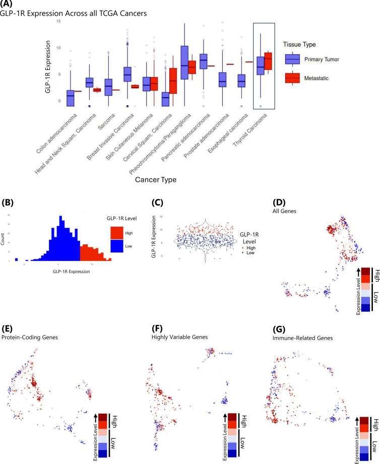
Using data obtained from The Cancer Genome Atlas (TCGA), median GLP-1R expression was compared across all TCGA cancer types, split by primary and metastatic tissue, and found to be highest in metastatic papillary thyroid carcinoma (f1), despite normal thyroid tissue being ranked 6th highest overall among TCGA cancers (Supplementary Figure 2). Using TCGA Thyroid Carcinoma samples, (n = 572), we identified a clear division of populations between samples with high GLP-1R expression and samples with low GLP-1R (f1). Within all thyroid samples, high expression was determined to be any mRNA expression greater than 300 (approximately top 25th percentile of samples). Low expression was any mRNA expression less than that (approximately bottom 75th percentile of samples). Using TumorMap’s two-dimensional reduction visualization, we aimed to understand whether GLP-1R expression led to transcriptomic grouping of high versus low samples (ref. 25). Importantly, we found that GLP-1R high samples clustered together relatively strongly across all coding and non-coding genes (36,327 genes), suggesting overarching trends in tumor biology and underlying transcriptomic similarities (f1). Further, this trend was similarly observed in three distinct genetic subsets: in only protein-coding genes (18,713 genes) (f1), in the highest variable genes (2,000 genes) (f1), and in a set of previously identified (ref. 31) pan-immune-related genes (2,565 genes) (f1). This division speaks to the possibility of distinct clusters of tumor environments, in part differentiated by GLP-1R expression level.
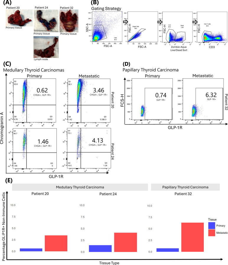
Primary patient tumor samples (f2) were analyzed via flow cytometry to examine differences in GLP-1R protein levels between primary and metastatic tumors. The general gating strategy is shown in f2. Positive and negative GLP-1R cutoffs were established using a PANC-1 cell line (Supplementary Figure 1). This cell line is known to express GLP-1R and was therefore used as a positive GLP-1R control. GLP-1R positive and negative gates were made using PANC-1 flow read outs as a guide. We found a marked increase in the percentage of chromogranin A (CHGA+), a parafollicular C-cell marker, and GLP-1R double-positive cells as disease progressed from primary to metastatic (f2). Further analysis revealed that the CD3- compartment (non-T cell compartment) in PTC patient 32 followed a similar pattern, with the percentage of GLP-1R+ cells increasing in metastatic tissue compared to primary tissue (f2). Across all three patients, there is a notable and consistent increase in the proportion of GLP-1R+ cells in the metastatic sample as compared to the primary (f2).
Differential Gene Expression (DGE) was performed, and subsequent Gene Set Enrichment Analysis (GSEA) was executed on DGE results to better understand pathway enrichment discrepancies between sample types. Samples were grouped by tissue type (primary or metastatic) and again by GLP-1R expression level (high or low). GSEA revealed clear differences in immune pathway enrichment between populations. Within all primary thyroid carcinoma samples, GLP-1R-low samples had increased immune-related pathway enrichment when compared to GLP-1R-high samples (f3). Similarly, metastatic thyroid tissue samples in the GLP-1R-low category had increased immune-related pathway enrichment when compared to samples in the GLP-1R-high category (f3).
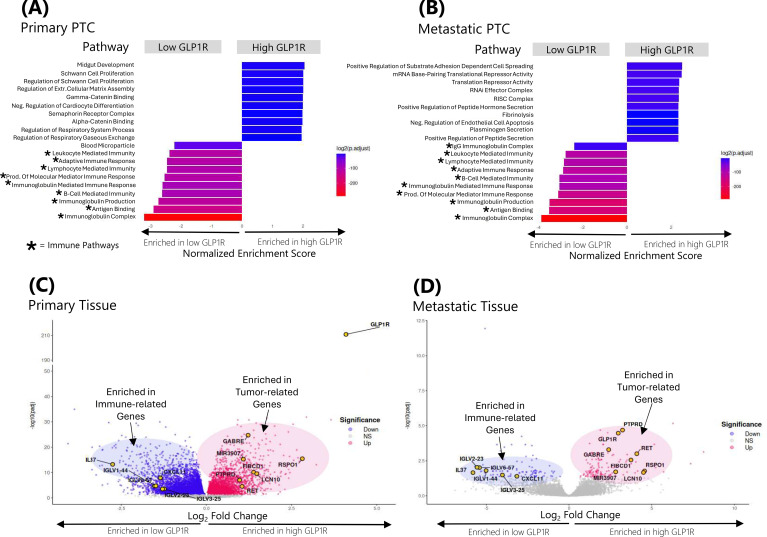
In an effort to better understand gene profiles between high and low GLP-1R groups, we examined overlapping upregulated genes between primary and metastatic tissue in both expression groups. Samples with GLP-1R-high levels in both primary and metastatic tissue were enriched in genes involved in proliferation, tumor development, and multiple prognostic genes associated with other cancers (Supplementary Figure 3a; Supplementary Table 1). Conversely, we observed that primary and metastatic tissue in the GLP-1R-low group shared several immune-related genes (Supplementary Figure 3b; Supplementary Table 2). Volcano plots (f3) were constructed to highlight differentially expressed genes (DEGs) between both comparisons, and genes that overlapped between primary and metastatic tissue were highlighted.
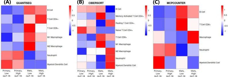
To better understand how immune cell type populations change as disease progresses from primary to metastatic for GLP-1R-high and GLP-1R-low patients, we leveraged data from the TIMER2.0 database and examined changes in immune cell type enrichment across tissue and GLP-1R level for three different Immune Deconvolution Methods: quanTIseq (ref. 27), CIBERSORT (ref. 28), and MCP-counter (ref. 29) (f4). For primary tissue, all three deconvolution methods reflected lower Myeloid Dendritic Cell enrichment in GLP-1R-high patients compared to GLP-1R-low (f4). Additionally, CIBERSORT data showed lower CD8+ T Cell levels in primary GLP-1R-High compared to GLP-1R-Low (f4). In metastatic tissue, we observed a decrease in B Cell enrichment in GLP-1R-high tissue compared to GLP-1R-low tissue in all three methods, as well (f4). In the CIBERSORT dataset, memory activated CD4+ T cells decreased in GLP-1R-high primary and metastatic tissue when compared to both GLP-1R-low groups, while resting CD4+ T cells increased in the GLP-1R-high groups (f4). For all three deconvolution methods, CD8+ T cells were also observed to decrease in the GLP-1R-high metastatic tissue when compared to that of GLP-1R-low tissue (f4).
Discussion
With millions of Americans prescribed and taking GLP-1 RAs, a clear understanding of the long-term risk of GLP-1R agonism as it pertains to the thyroid is critical. While some studies do not identify an increase in the development of thyroid carcinomas (ref. 32), continued investigation into the effects of sustained GLP-1RA exposure is paramount for broadening our depth of understanding the potential risks associated with these therapies. The lack of clarity surrounding the risk of associated thyroid carcinoma development demonstrates a clear gap in the literature and a necessity to better derive these mechanisms.
The computational findings of this study demonstrate a prominent increase in GLP-1R expression in metastatic thyroid carcinoma tissue when compared to all other TCGA cancer tissues. Following stratification of high and low GLP-1R expression, we found distinct differences in the transcriptional and immunologic landscapes, highlighting an underlying biologically driven pattern between these two groups. Flow cytometry read outs from patient tumor samples support our computational findings, showing an increase in GLP-1R+ cell percentages in metastatic tissues in both papillary and medullary carcinomas. The increase in GLP-1R positivity in metastatic cells raises potential questions about the role of GLP-1R in thyroid carcinoma disease progression. Further analysis illuminates two distinct tumor environments in both metastatic and primary thyroid carcinoma patients on the basis of GLP-1R expression. This finding offers novel insight into thyroid carcinoma patient demographics and new potential for possible prognosticator exploration. Additionally, these observations raise questions about the differences in GLP-1R activation in metastatic disease compared to primary disease.
Based on these findings, we explored differences between GLP-1R expression groups at both a pathway and a cellular level. GSEA findings revealed immune pathway activity unregulated in the GLP-1R-low group and suppressed in the GLP-1R-high group. Further exploration using DGE analysis indicated that GLP-1R-high groups in both tissue types were similarly enriched in genes that code for cell proliferation (ref. 33, ref. 34), genes implicated in tumor development (ref. 35), and prognosticator genes (ref. 36–ref. 39). Conversely, GLP-1R-low groups in both tissue types were enriched in genes that code for interleukin-37, a cytokine protective against some cancers (ref. 40), and multiple immunoglobulins. These differences in cell type enrichment pose important questions about how GLP-1R expression level affects cancer pathology and whether GLP-1 RAs may exacerbate these phenomena.
This study’s discovery of GLP-1R-high samples displaying suppressed immune-related pathways prompted further exploration utilizing three different immune cell deconvolution methods. Heatmap results revealed decreasing immune cell enrichment in GLP-1R-high samples when compared to GLP-1R-low samples across these methods for a selection of key immune cells, further validating the phenomenon of GLP-1R-high samples having immune-cold tumor microenvironments. This difference in immune activity between primary and metastatic tissues on the basis of GLP-1R expression could be indicative of GLP-1R-induced immunosuppressive activity in the tumor environment. Given the severity of late-stage thyroid carcinoma, this finding suggests a possible pathway for GLP-1 RA-associated thyroid carcinoma progression and mechanism. One study was able to identify a relationship between a GLP-1RA and M1 macrophage polarization, alleging an antitumor effect from Semaglutide exposure (ref. 41). While this study was limited to murine models, the results point to the presence of GLP-1RA-induced immune modulation. Further studies should investigate the role of GLP-1RAs in metastatic tumor immune microenvironments.
GSEA immune pathway discrepancies between high and low GLP-1R-expressing samples, and subsequent immune cell deconvolution analysis, demonstrated a decrease in immune cell density in GLP-1R-high samples. Decreased immune activity in the tumor microenvironment could contribute to poor prognosis and an inability to mitigate tumor growth, potentially due to immunosuppression by cancer cells or other immunosuppressive populations in the tumor microenvironment (ref. 42). While a similar trend was observed between tissue type, with metastatic tissue having decreased immune-cell infiltration when compared to primary tissue, immune cell type enrichment consistently differed between primary and metastatic comparisons and high and low GLP-1R comparisons, suggesting a subgroup-specific pattern of immune modulation. Further analysis of the tumor microenvironment could help explain how GLP-1R expression functions with respect to tumor immune infiltration.
Our study is subject to several limitations. The TCGA database only includes transcriptomic data for thyroid carcinomas from PTC patients, not MTC. Transcriptomic data for MTC is scarce due to its relative rarity, and future studies may help close this gap. It is important to note that conclusions drawn from transcriptomic PTC data from TCGA-THCA cannot be directly translated to MTC and transcriptomic MTC data are needed to investigate if the findings in this study can be similarly observed in a large-scale MTC dataset. Additionally, the TCGA database does not disclose whether patients are GLP-1 RA-exposed or -naïve, thus future transcriptomic studies may comparatively investigate these patient populations across cancer types to further dissect immunogenomic differences. Further, the small cohort of PTC and MTC patient tumor samples available for our flow cytometry analyses introduced an additional limitation to the study; patient tumor samples are scarce, and we were limited by our number of precious samples. Future studies may seek to include a higher volume of patient tumor samples, should they be available. While we were able to highlight interesting patterns and phenomena in both the TCGA-THCA computational cohort and in our MTC and PTC patient samples analyzed with flow cytometry, more studies are needed in order to understand relationship between the increase in GLP-1R expression in thyroid carcinomas and any changes in immune infiltration before causality can be established. Mechanistic studies need to be performed to understand what drives GLP-1R upregulation and the increase in pro-tumorigenic transcription.
This study demonstrates elevated GLP-1R levels in metastatic thyroid cancers compared to other TCGA cancers, with distinct GLP-1R-high and GLP-1R-low expression profiles correlating with immune pathway activation. These findings suggest a possible role for exogenous GLP-1R agonism on primary and metastatic thyroid tissues warranting further investigation. Interestingly, a retrospective observational cohort study on 18 patients with PTC did identify two patients whose tumor volume increased following exposure to GLP-1RAs (ref. 43); though, the majority of patients saw no significant volume change. However, the lack of patients with MTC is a limitation of this study. With the widespread and rapidly expanding use of GLP-1 RAs, it is paramount to further solidify our understanding of the potential proliferative effects of GLP-1R signaling in the thyroid as it pertains to the development of thyroid cancer.
References
- L Collins, RA Costello. Glucagon-like peptide-1 receptor agonists. In:. Statpearls., 2025
- Antidiabetics, GLP-1 agonists.. 2025
- C-T Hong, J-H Chen, C-J Hu. Role of glucagon-like peptide-1 receptor agonists in Alzheimer’s disease and Parkinson’s disease.. J BioMed Sci. (, 2024. [DOI | PubMed]
- D Lecis, FR Prandi, L Barone, M Belli, D Sergi, S Longo. Beyond the cardiovascular effects of glucagon-like peptide-1 receptor agonists: Body slimming and plaque stabilization. Are new statins born?. Biomolecules. (, 2023. [DOI | PubMed]
- A Montero, G Sparks, G Presiado, L Hamel. KFF Health Tracking Poll May 2024: The public’s use and views of GLP-1 drugs. In:. KFF., 2024
- A Montero, A Kearney, M Mulugeta, A Kirzinger, L Hamel. KFF Health Tracking Poll: Prescription drug costs, views on Trump administration actions, and GLP-1 use. In:. KFF., 2025
- V Ramos Santillan, SR Master, G Menon, B Burns. Medullary thyroid cancer. In:. Statpearls., 2025
- YS Khan, A Farhana. Histology, thyroid gland. In:. Statpearls., 2025
- J Morillo-Bernal, JM Fernández-Santos, JC Utrilla, M de Miguel, R García-Marín, I Martín-Lacave. Functional expression of the thyrotropin receptor in C cells: New insights into their involvement in the hypothalamic-pituitary-thyroid axis.. J Anat. (, 2009. [DOI | PubMed]
- GJ Cote, EG Grubbs, M-C Hofmann. Thyroid C-cell biology and oncogenic transformation.. Recent Results Cancer Res Fortschr Krebsforsch Progres Dans Rech Sur Cancer. (, 2015. [DOI | PubMed]
- Ozempic(semaglutide) injection prescribing information.. 2023
- LW Madsen, JA Knauf, C Gotfredsen, A Pilling, I Sjögren, S Andersen. GLP-1 receptor agonists and the thyroid: C-cell effects in mice are mediated via the GLP-1 receptor and not associated with RET activation.. Endocrinology. (, 2012. [DOI | PubMed]
- L Bjerre Knudsen, LW Madsen, S Andersen, K Almholt, AS de Boer, DJ Drucker. Glucagon-like peptide-1 receptor agonists activate rodent thyroid C-cells causing calcitonin release and C-cell proliferation.. Endocrinology. (, 2010. [DOI | PubMed]
- Z Zheng, Y Zong, Y Ma, Y Tian, Y Pang, C Zhang. Glucagon-like peptide-1 receptor: Mechanisms and advances in therapy.. Signal Transduct Target Ther. (, 2024. [DOI | PubMed]
- D Mossmann, S Park, MN Hall. mTOR signalling and cellular metabolism are mutual determinants in cancer.. Nat Rev Cancer. (, 2018. [DOI | PubMed]
- J Bezin, A Gouverneur, M Pénichon, C Mathieu, R Garrel, D Hillaire-Buys. GLP-1 receptor agonists and the risk of thyroid cancer.. Diabetes Care. (, 2022. [DOI | PubMed]
- ML Gild, RJ Clifton-Bligh, LJ Wirth, BG Robinson. Medullary thyroid cancer: Updates and challenges.. Endocr Rev. (, 2023. [DOI | PubMed]
- Medullary thyroid cancer – NCI. In:. cgvArticle., 2019
- MI Love, W Huber, S Anders. Moderated estimation of fold change and dispersion for RNA-seq data with DESeq2.. Genome Biol. (, 2014. [DOI | PubMed]
- G Yu, L-G Wang, Y Han, Q-Y He. clusterProfiler: An R package for comparing biological themes among gene clusters.. Omics J Integr Biol. (, 2012. [DOI | PubMed]
- K Blighe, S Rana, M Lewis. EnhancedVolcano: Publication-ready volcano plots with enhanced colouring and labeling. In:. Bioconductor., 2024
- H Wickham. ggplot2: Elegant graphics for data analysis., 2016
- G Yu, L-G Wang, G Yan, Q-Y He. DOSE: An R/Bioconductor package for disease ontology semantic and enrichment analysis.. Bioinformatics. (, 2015. [DOI | PubMed]
- G Korotkevich, V Sukhov, A Sergushichev. Fast gene set enrichment analysis.. Cold Spring Harb Labs J. (, 2019. [DOI]
- Y Newton, AM Novak, T Swatloski, DC McColl, S Chopra, K Graim. TumorMap: Exploring the molecular similarities of cancer samples in an interactive portal.. Cancer Res. (, 2017. [DOI | PubMed]
- T Li, J Fu, Z Zeng, D Cohen, J Li, Q Chen. TIMER2.0 for analysis of tumor-infiltrating immune cells.. Nucleic Acids Res. (, 2020. [DOI | PubMed]
- F Finotello, C Mayer, C Plattner, G Laschober, D Rieder, H Hackl. Molecular and pharmacological modulators of the tumor immune contexture revealed by deconvolution of RNA-seq data.. Genome Med. (, 2019. [DOI | PubMed]
- AM Newman, CL Liu, MR Green, AJ Gentles, W Feng, Y Xu. Robust enumeration of cell subsets from tissue expression profiles.. Nat Methods. (, 2015. [DOI | PubMed]
- E Becht, NA Giraldo, L Lacroix, B Buttard, N Elarouci, F Petitprez. Estimating the population abundance of tissue-infiltrating immune and stromal cell populations using gene expression.. Genome Biol. (, 2016. [DOI | PubMed]
- R Kolde. raivokolde/pheatmap. pheatmap: Pretty heatmaps. R package version 1.0.13 (. 2026
- V Thorsson, DL Gibbs, SD Brown, D Wolf, DS Bortone, T-H Ou Yang. The immune landscape of cancer.. Immunity. (, 2018. [DOI | PubMed]
- A Patrizio, A Mauguen, JA Fagin, A Farooki, SS Fish, J Flory. Exposure to GLP-1RA and risk of structural progression in differentiated thyroid cancer.. J Clin Endocrinol Metab. (, 2026. [DOI | PubMed]
- EJ ter Steege, ERM Bakker. The role of R-spondin proteins in cancer biology.. Oncogene. (, 2021. [DOI | PubMed]
- M Takahashi, K Kawai, N Asai. Roles of the RET proto-oncogene in cancer and development.. JMA J. (, 2020. [DOI | PubMed]
- V Walia, TD Prickett, J-S Kim, JJ Gartner, JC Lin, M Zhou. Mutational and functional analysis of the tumor-suppressor PTPRD in human melanoma.. Hum Mutat. (, 2014. [DOI | PubMed]
- Y Wang, M Sun, J Liu, Y Liu, C Jiang, H Zhu. FIBCD1 overexpression predicts poor prognosis in patients with hepatocellular carcinoma.. Oncol Lett. (, 2020. [DOI | PubMed]
- L Yan, Y-Z Gong, M-N Shao, G-T Ruan, H-L Xie, X-W Liao. Distinct diagnostic and prognostic values of γ-aminobutyric acid type A receptor family genes in patients with colon adenocarcinoma.. Oncol Lett. (, 2020. [DOI | PubMed]
- L Wang, H Liu, Y Feng, X Liu, Y Wang, Y Liu. Decoding the immune landscape: A comprehensive analysis of immune-associated biomarkers in cervical carcinoma and their implications for immunotherapy strategies.. Front Genet. (, 2024. [DOI | PubMed]
- C Zhang, L Zhu, X Liu, M Jiang, Q Tang, F Xu. MicroRNA-3907 promotes the proliferation and migration of sebaceous gland carcinoma of the eyelid by targeting thrombospondin 1.. Oncol Lett. (, 2021. [DOI | PubMed]
- Z Su, X Tao. Current understanding of IL-37 in human health and disease.. Front Immunol. (, 2021. [DOI | PubMed]
- L Wang, L Zhang, R Ma, Y Zhang, Q Chang, D Yin. Semaglutide reprograms macrophages via the GLP-1R/PPARG/ACSL1 pathway to suppress papillary thyroid carcinoma growth.. J Clin Endocrinol Metab. (, 2025. [DOI | PubMed]
- T Blankenstein, PG Coulie, E Gilboa, EM Jaffee. The determinants of tumour immunogenicity.. Nat Rev Cancer. (, 2012. [DOI | PubMed]
- A Patrizio, SK Newman, RM Tuttle, L Boucai. Effect of GLP-1 receptor agonists on patients with thyroid carcinomas undergoing active surveillance.. J Endocr Soc. (, 2026. [DOI | PubMed]