
SLC39A6 as a pan-cancer promising biomarker and actionable therapeutic target for CH5132799 sensitivity
Abstract
Background:
The zinc transporter SLC39A6, a member of the ZIP (Zrt-Irt-like protein) family, mediates zinc influx from the extracellular milieu into the cytosol and is indispensable for the function of numerous enzymes, transcription factors and signaling molecules. Previous studies have shown that SLC39A6 expression is associated with prognosis in esophageal squamous cell carcinoma and cervical cancer, indicating its potential impact on patient survival and tumor immunity. But a comprehensive pan-cancer analysis of SLC39A6 is still lacking. This study aimed to systematically delineate the oncogenic and prognostic relevance of SLC39A6 across multiple cancer types, to unravel its interplay with immune-infiltration patterns in the tumor micro-environment, and to preliminarily identify SLC39A6-associated therapeutic vulnerabilities.
Methods:
SLC39A6 expression profiles were retrieved from The Cancer Genome Atlas (TCGA) and cross-validated with GTEx, TIMER, HPA, cBioPortal, GEPIA2, STRING, KEGG, GO and other public repositories. Pan-cancer analyses were performed to characterize expression patterns, prognostic value, mutational landscape and functional networks. We further interrogated correlations between SLC39A6 and immune infiltration, tumor mutational burden (TMB), microsatellite instability (MSI) and immune-regulatory genes. Drug-sensitivity associations were evaluated using the CellMiner database, which facilitated molecular docking to predict binding poses and subsequent in vivo validation of lead compounds.
Results:
SLC39A6 exhibited marked dysregulation across diverse tumor types and was significantly linked to patient survival. High SLC39A6 expression is associated with reduced overall survival and progression-free survival, particularly in cervical squamous cell carcinoma (CESC), as evidenced by studies that have analyzed the gene’s expression and its impact on patient survival. Immune deconvolution revealed robust associations between SLC39A6 levels and the abundance of cytotoxic T cells, dendritic cells, macrophages and other immune subsets. CellMiner analyses demonstrated that increased levels of SLC39A6 resulted in enhanced sensitivity to the PI3Kα inhibitor CH5132799. Molecular docking studies predicted a strong affinity between CH5132799 and the zinc-binding pocket of SLC39A6, while mouse xenograft models further validated that CH5132799 effectively inhibited SLC39A6-mediated tumor growth.
Conclusion:
SLC39A6 regulates the dynamics of immune infiltration and impacts prognosis in a wide range of malignancies. It emerges as a promising biomarker for prognosis, immunology, and therapy in the field of precision oncology.
Supplementary Information:
The online version contains supplementary material available at 10.1186/s12885-026-16063-6.
Article type: Research Article
Keywords: SLC39A6, Bioinformatics, Prognosis, Immunotherapy response, CH5132799
Affiliations: https://ror.org/0152hn881grid.411918.40000 0004 1798 6427Department of Thoracic Oncology, Lung Cancer Diagnosis and Treatment Center, Tianjin Medical University Cancer Institute and Hospital, Tianjin, 300060 China; https://ror.org/0152hn881grid.411918.40000 0004 1798 6427National Clinical Research Center for Cancer, State Key Laboratory of Druggability Evaluation and Systematic Translational Medicine, Tianjin’s Clinical Research Center for Cancer, Key Laboratory of Cancer Prevention and Therapy, Tianjin, 300060 China; https://ror.org/016m2r485grid.452270.60000 0004 0614 4777Department First of Oncology, Cangzhou Central Hospital, Cangzhou, 061000 China; https://ror.org/016m2r485grid.452270.60000 0004 0614 4777Department of Traumatic Orthopedics II, Cangzhou Central Hospital, Cangzhou, 061000 China; https://ror.org/00xw2x114grid.459483.7Eighth Oncology Department, Tangshan People’s Hospital, Tangshan, 063000 China
License: © The Author(s) 2026 CC BY 4.0 Open Access This article is licensed under a Creative Commons Attribution 4.0 International License, which permits use, sharing, adaptation, distribution and reproduction in any medium or format, as long as you give appropriate credit to the original author(s) and the source, provide a link to the Creative Commons licence, and indicate if changes were made. The images or other third party material in this article are included in the article’s Creative Commons licence, unless indicated otherwise in a credit line to the material. If material is not included in the article’s Creative Commons licence and your intended use is not permitted by statutory regulation or exceeds the permitted use, you will need to obtain permission directly from the copyright holder. To view a copy of this licence, visit http://creativecommons.org/licenses/by/4.0/.
Article links: DOI: 10.1186/s12885-026-16063-6 | PubMed: 42151900 | PMC: PMC13366974
Relevance: Moderate: mentioned 3+ times in text
Full text: PDF (7.8 MB)
Introduction
Cancer continues to be one of the primary causes of illness and death globally. Breast invasive carcinoma, cholangiocarcinoma, colon adenocarcinoma, and lung adenocarcinoma together contribute to millions of new cases and deaths each year, exerting significant clinical and socioeconomic pressures [ref. 1]. Despite recent progress in surgery, cytotoxic chemotherapy, and radiotherapy, the effectiveness of treatments remains limited and frequently comes with severe side effects [ref. 2]. Consequently, there is an urgent need for reliable molecular biomarkers to guide personalized therapy.
The solute carrier family 39 member 6 (SLC39A6, also known as ZIP6) encodes a zinc transporter that regulates intracellular zinc homeostasis [ref. 3]. Dysregulated zinc signaling has been causally linked to tumorigenesis and malignant progression [ref. 4]. Over the past five years, numerous studies have documented that SLC39A6 is frequently up-regulated in breast, colorectal, and lung cancers, where higher expression correlates with advanced stage and shorter overall survival [ref. 5–ref. 7]. These findings support the candidacy of SLC39A6 as a pan-cancer prognostic marker.
Beyond prognosis, the therapeutic implications of SLC39A6 remain underexplored. We conducted an integrative drug sensitivity analysis to identify cytotoxic and targeted agents whose efficacy is significantly associated with SLC39A6 expression. This in-silico drug-sensitivity screen has nominated candidate compounds that may preferentially benefit SLC39A6-high tumors and provides a mechanistic rationale for repositioning existing drugs, thereby accelerating translational applications.
Recent pan-cancer studies have explored various molecular markers and analytical tools, providing valuable paradigms and methodologies for investigating potential cancer biomarkers and therapeutic targets, such as the research on ZNF165 as a pan-cancer prognostic biomarker and the development of the DrugSurvPlot platform for drug sensitivity and survival analysis. These studies, along with single-cell pan-cancer analyses and integrated analyses of cancer-related genes, offer important references for systematically exploring the role of SLC39A6 in pan-cancer progression and its potential as a therapeutic target [ref. 8–ref. 11].To systematically characterize SLC39A6 across malignancies, we conducted a comprehensive pan-cancer study using The Cancer Genome Atlas (TCGA) dataset accessed via TCGA biolinks [ref. 12]. We employed Cox regression, Kaplan–Meier analyses, immune-infiltration profiling, and drug-sensitivity modeling to delineate the associations among SLC39A6 expression, clinical outcomes, and therapeutic response. The overarching goal is to clarify the expression patterns of SLC39A6 across cancer types, to validate its prognostic value, and to nominate druggable vulnerabilities for SLC39A6-driven tumors.
Materials and methods
Data download
By R package TCGA biolinks from Cancer Genome project (The Cancer Genome Atlas, TCGA) (https://portal.gdc.cancer.gov/) to download 33 Cancer data set, The expression of matrix, respectively is: Adrenocortical carcinoma (ACC), Bladder urothelial carcinoma (BLCA), Breast invasive carcinoma (BRCA), Cervical endocervical adenocarcinoma and squamous cell carcinoma (CESC), Cholangiocarcinoma (CHOL), Colon adenocarcinoma (COAD), Lymphoid neoplasm diffuse large b-cell lymphoma (DLBC), Esophageal carcinoma (ESCA), Glioblastoma multiforme (GBM), Head and neck squamous cell carcinoma(HNSC), Kidney chromophobe (KICH), Kidney renal clear cell carcinoma (KIRC), Kidney renal papillary cell carcinoma (KIRP), Acute myeloid leukemia (LAML), Brain lower grade glioma (LGG), Liver hepatocellular carcinoma (LIHC), Lung adenocarcinoma (LUAD), Lung squamous cell carcinoma (LUSC), Mesothelioma (MESO), Ovarian serous cystadenocarcinoma (OV), Prostate adenocarcinoma (PAAD), Pheochromocytoma and paraganglioma (PCPG), Pancreatic adenocarcinoma (PRAD), Rectum adenocarcinoma (READ), Sarcoma (SARC), Skin cutaneous melanoma (SKCM), Stomach adenocarcinoma, (STAD), Testicular germ cell tumors (TGCT), Thyroid carcinoma (THCA), Thymoma (THYM), Uterine corpus endometrial carcinoma (UCEC), Uterine carcinosarcoma (UCS). All Tumor samples and Normal samples in the above 33 TCGA cancer types dataset were included in the analysis study. The gene expression of each sample was standardized to FPKM (Fragments Per Kilobase Million) format. The corresponding clinical data were obtained through the UCSC Xena database [ref. 13] (https://xena.ucsc.edu/).
Comparative analysis of SLC39A6 pan-cancer level tumors and normal groups
Based on the sample grouping of 33 TCGA cancer datasets, samples were categorized into tumor and normal groups. Subsequently, the Wilcoxon Rank Sum Test was utilized to analyze the expression differences of SLC39A6 across the 33 TCGA cancer datasets. The R package ggplot2 (version 3.4.4) was employed to generate box plots illustrating the expression levels of SLC39A6 in both the tumor and normal groups of the 33 TCGA cancer datasets. Additionally, group comparison plots were created to highlight the differences between the two groups.
Comparison of SLC39A6 between pan-cancer level pathological groups
To investigate the relationship between the expression of the single-gene SLC39A6 and the pathological stage of cancer, the differences in SLC39A6 expression across various pathological stages were analyzed. The expression profiles of SLC39A6 from 33 TCGA cancer datasets were categorized into normal, Stage I, Stage II, Stage III, and Stage IV groups. A comparative group diagram was then created to illustrate the analysis findings.
Pan-cancer level prognostic analysis of SLC39A6
To evaluate the prognostic value of the SLC39A6 gene across 33 TCGA cancer types, a univariate Cox regression analysis was conducted using the R package ‘survival((version 3.5–7). This analysis aimed to assess the impact of SLC39A6 on prognosis and to determine if it served as an independent prognostic factor [ref. 14]. In the univariate Cox regression analysis, the prognostic value of SLC39A6 was characterized by a cancer category with a p-value less than 0.05. A pan-cancer univariate Cox regression model was constructed, and the grouped expression levels of SLC39A6 in each cancer type were visualized using a Forest Plot.
For cancer types with a P-value < 0.05 for SLC39A6, Kaplan–Meier curve analysis was conducted using the R package survival (Version 3.5–7), and a KM curve was plotted based on the expression level of SLC39A6 [ref. 14, ref. 15]. Overall survival (OS), Disease-Specific Survival (DSS), and Progression-Free Interval (PFI) were compared between groups with high and low SLC39A6 expression.
Finally, in the prognostic analysis of overall survival (OS), disease-specific survival (DSS), and progression-free interval (PFI), the key cancer types with a p-value less than 0.05 were intersected to identify the significant cancer types.
Pan-cancer ssGSEA immune infiltration analysis
Single-Sample Gene-Set Enrichment Analysis (ssGSEA), also known as single-sample gene-set enrichment analysis, quantifies the relative abundance of each immune cell infiltrate [ref. 16]. Initially, immune cell types such as ADCs, B cells, CD8 T cells, and other human immune cell subtypes were labeled. Then, the enrichment scores calculated by ssGSEA analysis were used to represent the relative abundance of each immune cell type in each sample, resulting in an immune infiltration matrix for 33 TCGA cancer types. Next, the correlation between SLC39A6 and the abundance of immune cell infiltration was calculated using the Spearman algorithm, and the R package ggplot2 (Version 3.3.4) was employed to create a correlation heat map to display the results of the correlation analysis. For the key cancer types, the R package ggplot2 (Version 3.4.4) was used to generate group comparison maps, illustrating the expression differences of immune cells between the high and low expression groups of SLC39A6 in these cancer types.
Correlation analysis of pan-cancer immune checkpoint gene (ICG) and mismatch repair (MMR) genes
Immune Checkpoint Genes (ICG) refer to ligand-receptor pairs that inhibit or stimulate immune responses. Immune checkpoint plays a crucial immunomodulatory role in maintaining immune homeostasis and preventing autoimmunity. Immune checkpoint blockade therapy has brought significant clinical benefits against several solid malignancies. Studies have published on the PubMed website (https://pubmed.ncbi.nlm.nih.gov/) literature that includes 47 (ICG) on immune checkpoint genes [ref. 17]. Subsequently, heat maps of the correlation between pan-cancer immune checkpoint genes (ICG) and SLC39A6 were drawn to display the results of correlation analysis across 33 TCGA cancer types.
Mismatch Repair (MMR) refers to the repair of DNA molecules containing mismatched bases to restore the nucleotide sequence to normal. The PubMed website (https://pubmed.ncbi.nlm.nih.gov/) has published literature indicating the presence of five MMR genes: MLH1, MSH2, MSH6, PMS2, and EPCAM. Associations between SLC39A6 and MMR genes were evaluated, and heatmaps were constructed [ref. 18].
Genetic variant analysis
CBioPortal database(https://www.cbioportal.org/) is a collection of search, download, analysis, and visualization of cancer genomics data in a database, the integration of genomic data type is very broad,The database includes somatic mutations, DNA copy number alterations (CNAs), mRNA and microRNA (miRNA) expression, DNA methylation, protein abundance, and phosphoprotein abundance [ref. 19–ref. 21]. CBioPortal can perform a variety of analyses, but the main ones are mutation-related analyses and their visualization. In addition, cBioPortal database not only supports the query of single gene and single cancer, but also can conduct the analysis of multi-gene single cancer, single-gene multiple cancer, multi-gene multiple cancer, and even cross-cancer genome projects. The differences of Tumor Mutation Burden (TMB) and Microsatellite Instability (MSI) of SLC39A6 gene in 33 TCGA cancer types were analyzed by cBioPortal database.
PPI interaction network and Gene ontology (GO) enrichment analysis
Protein–Protein Interaction Network (PPI) is composed of proteins and proteins that interact with each other and participate in all aspects of life processes such as biological signal transmission, gene expression regulation, energy and substance metabolism, and cell cycle regulation. Systematic analysis of protein–protein interactions in biological systems is of great significance for understanding the working principle of proteins in biological systems, understanding the reaction mechanism of biological signals and energy and substance metabolism under special physiological conditions such as diseases, and understanding the functional relationship between proteins. The STRING database (https://string-db.org/) is a database for searching the interactions between known and predicted proteins [ref. 22]. In this study, the STRING database was applied based on SLC39A6 gene with a minimum required interaction score greater than 0.400,and medium confidence (0.400) was used to construct the SLC39A6 gene-related PPI interaction network. The tightly connected local regions in the PPI interaction network may represent molecular complexes with specific biological functions. Interacting Genes in PPI network were selected for subsequent analysis.
Gene Ontology (GO) analysis is a common method for conducting large-scale functional enrichment studies, including Biological Process (BP), Cell Component (CC), and Molecular Function (MF) [ref. 23]. We used the R package clusterProfiler to perform gene ontology (GO) enrichment analysis of SLC39A6 gene and Interacting Genes [ref. 24]. The entry screening criteria were adj.p < 0.05 and FDR value (q value) < 0.25. The p value correction method was Benjamini-Hochberg (BH).
Drug sensitivity of SLC39A6 in pan-cancer
To analyze the drug sensitivity of SLC39A6 in pan-cancer, CellMiner™ was used to obtain NCI-60 compound activity data and RNA-seq expression profiles [ref. 25, ref. 26]. Analysis using R version 4.4.1 to selecte FDA-approved or clinical trial drugs. The structures of drugs and key target proteins were obtained from the PubChem database (https://pubchem.ncbi.nlm.nih.gov) and the PDB database (https://www.rcsb.org). The protein structures were dehydrated using PyMOL 3.0 software. Proteins and ligands were processed with AutoDockTools 1.5.7, followed by molecular docking using AutoDock Vina 1.1.2. Finally, chemical bond analysis of the results was performed using LigPlot + software, and visualization was conducted with PyMOL 3.0.
Xenograft models
All experimental procedures were performed in accordance with relevant institutional and national guidelines and approved by the Institutional Animal Care and Use Committee of the Tianjin XINRUI (permit no. XinRui-DWLL-2025012). A total of 5 × 10⁶ A549 cells that had been suspended in 150 μL serum-free culture medium were injected subcutaneously into the left flanks of female BALB/c nude mice (4 weeks old, 15 ± 3 g weight, SPF (Beijing)Biotechnology CO.,Ltd).Tumor volume (V) was calculated with the following formula: length × width2 × 1/2. The mice were randomly divided into three groups (n = 4 or 5 per group). (1) Control group: mice were injected with A549 SCL39A6-vector. (2) SLC39A6 over expression group: mice were injected with A549 SCL39A6-OE. (3) CH5132799 group: mice were injected with A549 SCL39A6-OE and CH5132799 was orally administered at 12.5 mg/kg once a day.The animals were euthanized under deep anesthesia via cervical dislocation. Anesthesia was induced by intraperitoneal injection of a combination of ketamine (100 mg/kg) and xylazine (10 mg/kg). The humane endpoint was a tumor volume of 2000 mm3 or a diameter of 15 mm, whichever was reached first.
Statistical analysis
All data processing and analysis in this article were based on R software (Version 4.2.2). If not otherwise specified, statistical significance of normally distributed variables was estimated by independent Student’s T-Test for comparisons of continuous variables between two groups. Mann–Whitney U Test (Wilcoxon Rank Sum Test) was used to analyze the differences between variables that were not normally distributed. Kruskal–Wallis test was used for comparison of three or more groups. Spearman correlation analysis was used to calculate the correlation coefficient between different molecules. All statistical p values were two-sided if not specified, and a p value of less than 0.05 was considered to indicate statistical significance Fig. 1.
Results
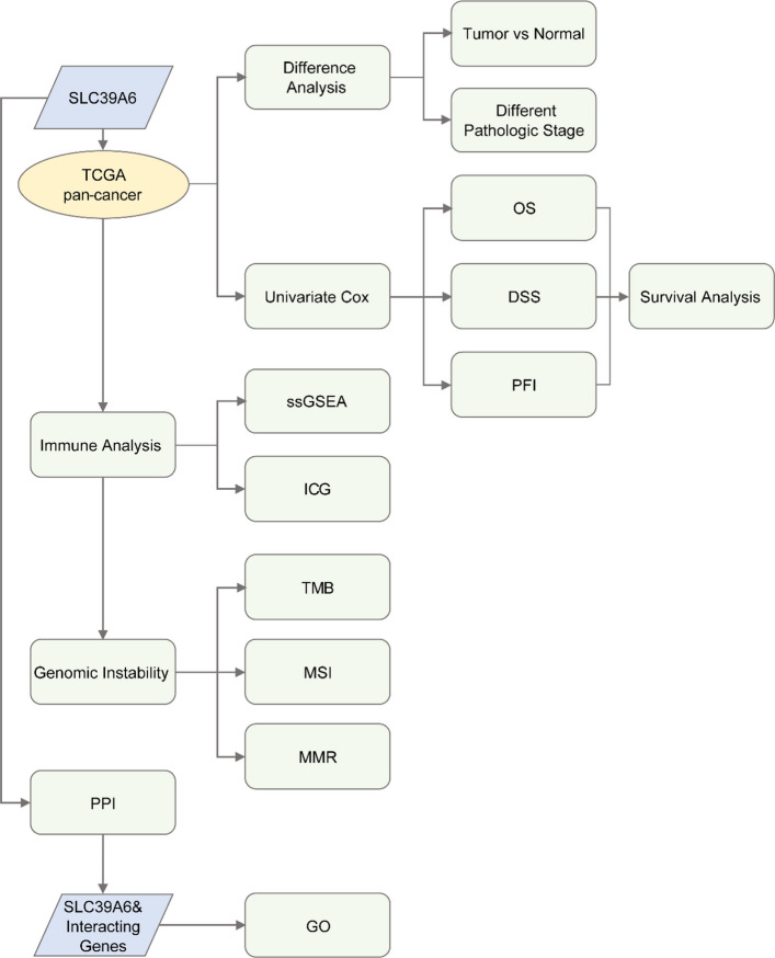
Technology roadmap
Comparison of SLC39A6 pan-cancer level tumors and normal groups
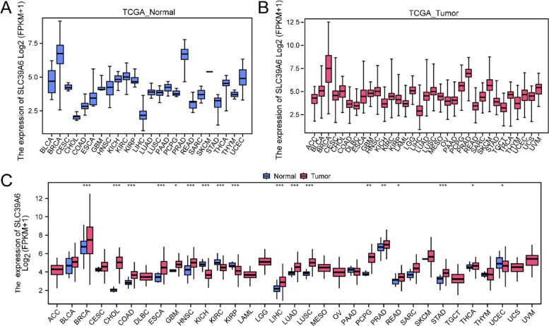
In order to explore the differential expression of SLC39A6 in 33 TCGA cancers, the expression and differential analysis results of SLC39A6 in the Tumor group and the normal group in 33 TCGA cancers were displayed by box plots (Fig. 2A-B) and group comparison plots (Fig. 2C). Differential results showed, SLC39A6 in breast invasive carcinoma (BRCA), cholangiocarcinoma (CHOL), colon cancer (COAD), Esophageal cancer (ESCA), glioblastoma multiforme (GBM), squamous cell carcinoma of head and neck (HNSC), chromophobe renal cell carcinoma (KICH), clear cell renal cell carcinoma (KIRC), squamous cell carcinoma of head and neck (HNSC), renal cell carcinoma of chromophobe (KICH), renal cell carcinoma of clear cell (KIRC). Renal papillary cell carcinoma (KIRP), Hepatocellular carcinoma (LIHC), Lung adenocarcinoma (LUAD), Lung squamous cell carcinoma (LUSC), Pheochromocytoma and paraganglioma (PCPG), Prostate cancer (PRAD), Rectal adenocarcinoma (READ), gastric adenocarcinoma (STAD), Thyroid carcinoma (THCA).There was a statistically significant difference in the expression level between the tumor group and the normal group of endometrial carcinoma (UCEC) (p < 0.05).
Comparative analysis of SLC39A6 pan-cancer level pathological groups
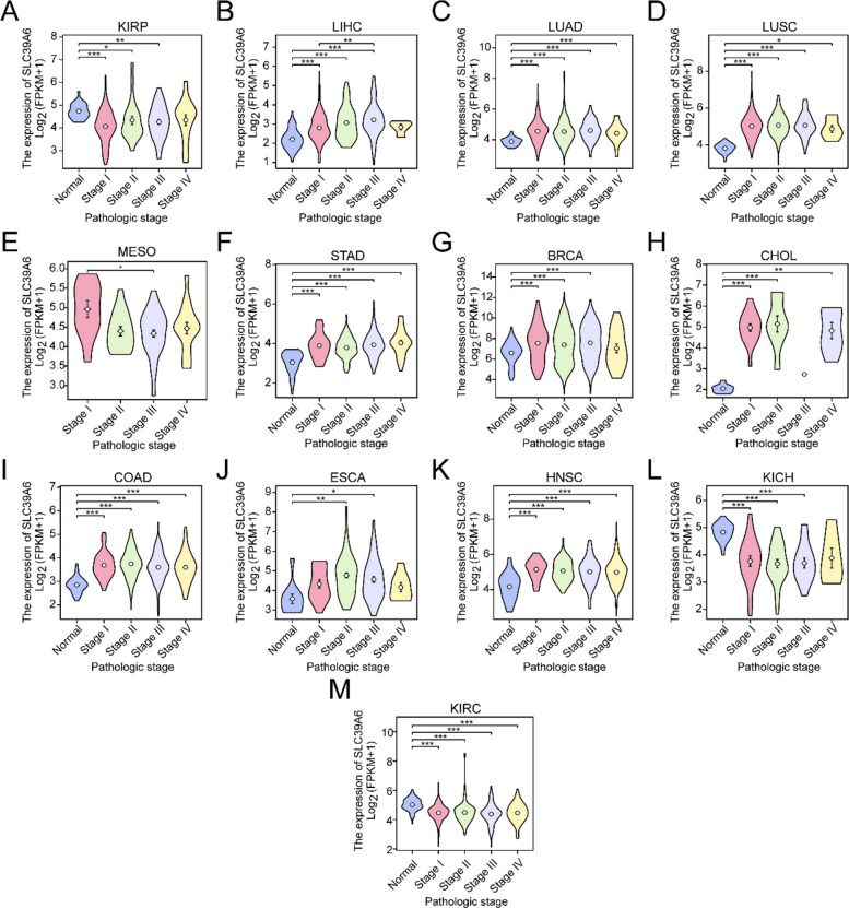
In order to explore the relationship between the expression of single-gene SLC39A6 and the pathological stage of cancer, the difference of the expression of single-gene SLC39A6 in different pathological stages of cancer was analyzed. The group comparison figure (Fig. 3A-M) shows the difference analysis results between different pathological stages of SLC39A6 in TCGA cancer types with significant differences. Differential results showed (Fig. 3A-M), SLC39A6 in breast invasive carcinoma (BRCA), cholangiocarcinoma (CHOL), colon cancer (COAD), Esophageal cancer (ESCA), squamous cell carcinoma of head and neck (HNSC), Chromophobe renal cell carcinoma (KICH), clear cell renal cell carcinoma (KIRC), papillary renal cell carcinoma (KIRP), squamous cell carcinoma of head and neck (HNSC). The expression levels of hepatocellular carcinoma (LIHC), lung adenocarcinoma (LUAD), lung squamous cell carcinoma (LUSC), mesothelioma (MESO), and gastric adenocarcinoma (STAD) were statistically significant between different cancer pathological stages (p < 0.05).
Pan-cancer level prognostic analysis of SLC39A6
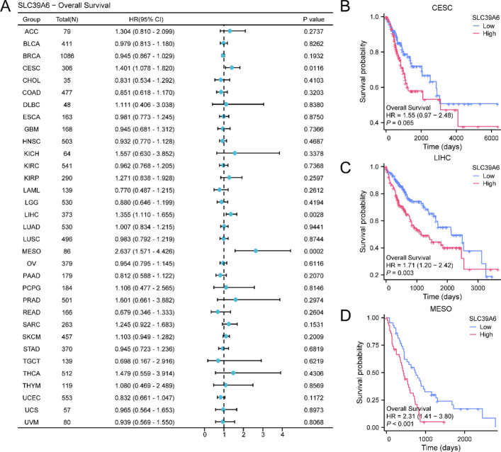
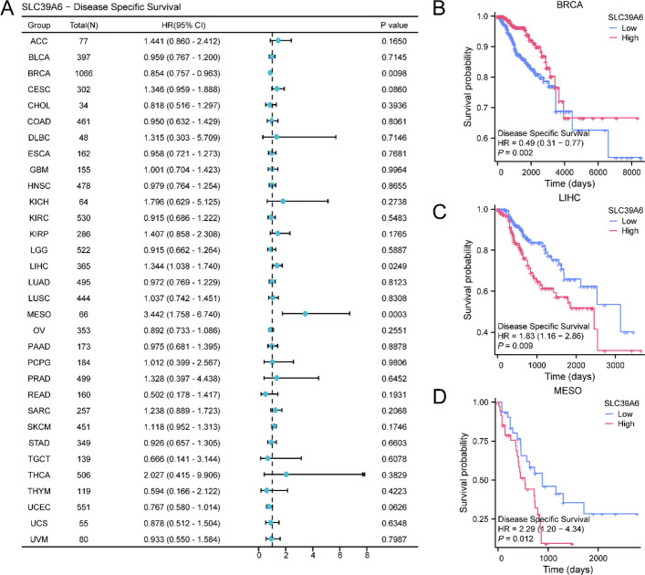
A survival analysis of SLC39A6 expression in each cancer was performed based on TCGA dataset cancer samples, and the positive results are shown in Table 1. First, the results of single-gene Cox analysis of SLC39A6 overall survival (OS) showed that SLC39A6 was significant in the single-gene Cox model of cervical squamous cell carcinoma and adenocarcinoma (CESC), hepatocellular carcinoma (LIHC), and mesothelioma (MESO) (p value < 0.05). The Kaplan–Meier (KM) curves of SLC39A6 in the overall survival (OS) of the above three cancers are shown (Fig. 4B-D), hepatocellular carcinoma (LIHC), The overall survival (OS) of mesothelioma (MESO) was significantly different between high and low SLC39A6 expression groups (p < 0.05).
Table 1: Results of univariate cox
| Tumor | Outcome | Total(N) | HR(95% CI) | P value |
|---|---|---|---|---|
| CESC | OS | 306 | 1.401 (1.078–1.820) | 0.0116 |
| LIHC | OS | 373 | 1.355 (1.110–1.655) | 0.0028 |
| MESO | OS | 86 | 2.637 (1.571–4.426) | 0.0002 |
| BRCA | DSS | 1066 | 0.854 (0.757–0.963) | 0.0098 |
| LIHC | DSS | 365 | 1.344 (1.038–1.740) | 0.0249 |
| MESO | DSS | 66 | 3.442 (1.758–6.740) | 0.0003 |
| ACC | PFI | 79 | 1.748 (1.150–2.658) | 0.0090 |
| BRCA | PFI | 1086 | 0.905 (0.828–0.990) | 0.0286 |
| CESC | PFI | 306 | 1.544 (1.174–2.030) | 0.0019 |
| LIHC | PFI | 373 | 1.235 (1.037–1.470) | 0.0178 |
| MESO | PFI | 84 | 1.882 (1.082–3.275) | 0.0253 |
ACC Adrenocortical carcinoma, BRCA Breast invasive carcinoma, CESC Cervical endocervical adenocarcinoma and squamous cell carcinoma, LIHC Liver hepatocellular carcinoma, MESO Mesothelioma, OS Overall Survival, DSS Disease Specific Survival, PFI Progress Free Interval, HR Hazard Ratio, CI Confidence Intervals
Results of single-gene Cox analysis of SLC39A6 disease-specific survival (DSS) showed that SLC39A6 was significant in single-gene Cox models of breast invasive carcinoma (BRCA), hepatocellular carcinoma (LIHC), and mesothelioma (MESO) (p value < 0.05). Kaplan–meier (KM) curves of SLC39A6 in DSS were also significantly different among the three types of cancer (Fig. 5B-D).
Results of single-gene Cox analysis of SLC39A6 progression-free interval (PFI) showed (Fig. 6A) that SLC39A6 was significantly associated with adrenal cortical carcinoma (ACC), invasive breast carcinoma (BRCA), cervical squamous and adenocarcinoma (CESC), hepatocellular carcinoma (LIHC). In the single-gene Cox model of mesothelioma (MESO), it was significant (p < 0.05). The Kaplan–Meier (KM) curves of progression-free interval (PFI) of SLC39A6 were also significantly different among the five cancers (Fig. 6B-F).
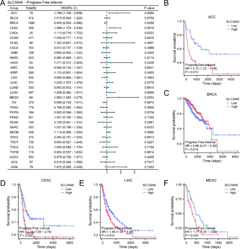
Kaplan–meier (KM) curves (OS, DSS, PFI) of SLC39A6 were significant in hepatocellular carcinoma (LIHC) and mesothelioma (MESO) (p < 0.05).
Pan-cancer ssGSEA immune infiltration analysis
ssGSEA algorithm was used to calculate the infiltration abundance of 24 immune cells in 33 TCGA cancer types. According to the results of immune infiltration analysis, the heat map of the correlation between the immune infiltration abundance of 24 immune cells and SLC39A6 gene expression in 33 TCGA cancer types was plotted (Fig. 7A).
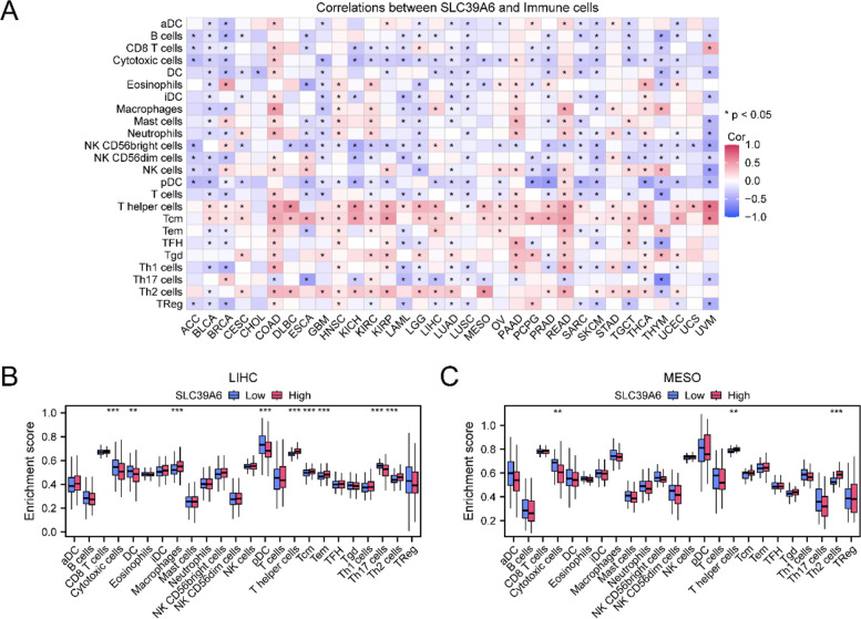
Then, the grouping comparison of immune cell infiltration abundance between high and low SLC39A6 expression groups in key cancer types hepatocellular carcinoma (LIHC), mesothelioma (MESO) (Fig. 7B-C), hepatocellular carcinoma (LIHC) (Fig. 7B) showed that the 9 immune cells, Nine kinds of immune cells, including Cytotoxic cells, DC, Macrophages, pDC, T helper cells, Tcm, Tem, Th17 cells, and Th2 cells, were statistically significant between the high and low expression groups of SLC39A6 (p < 0.05). The group comparison diagram of mesothelioma (MESO) (Fig. 7C) showed that three immune cells, including Cytotoxic cells, T helper cells and Th2 cells, had statistically significant differences between the high and low expression groups of SLC39A6 (p value < 0.05).
Correlation analysis of pan-cancer immune checkpoint gene (ICG) and mismatch repair (MMR) genes
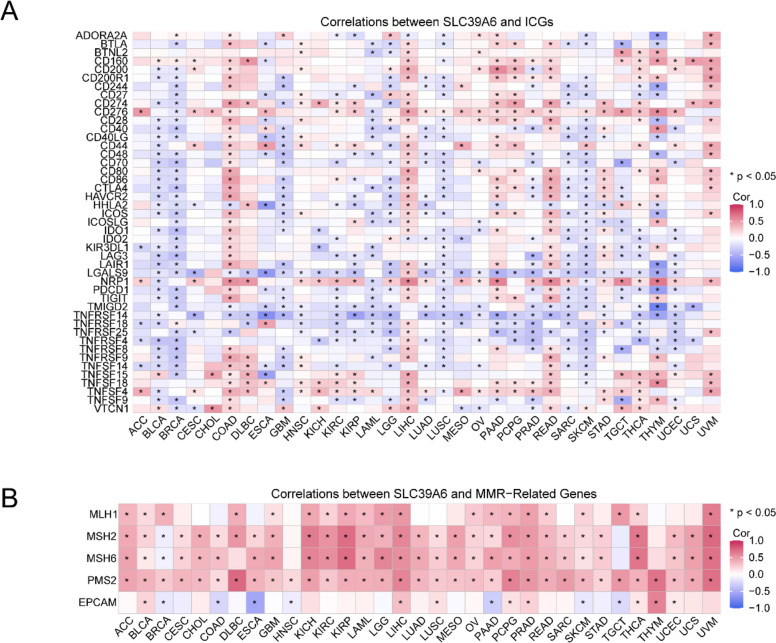
In PubMed website (https://pubmed.ncbi.nlm.nih.gov/) has been published in the literature got 47 (ICG) on immune checkpoint gene, with the TCGA extensive cancer gene expression profile contained in the intersection, got 45 genes (ICG) on immune checkpoints. The correlation between the expression of 45 immune checkpoint genes (ICG) and SLC39A6 gene expression in 33 TCGA cancer types was calculated, and the correlation heat map (Fig. 8A) was drawn. The results showed that SLC39A6 was negatively correlated with the immune checkpoint gene (ICG) in most cancer types. Then, the correlation between the expression of 5 MMR genes and SLC39A6 gene in 33 TCGA cancer types was calculated and the correlation heat map was plotted (Fig. 8B). The results showed that MLH1, MSH2, MSH6, PMS2 were mainly positively correlated with SLC39A6 gene expression. The correlation between the expression of EPCAM and SLC39A6 was similar.We supplement the complete list of 45 immune checkpoint genes (ICGs) and 5 mismatch repair (MMR) genes (see Supplement 1–3 for details).
Genetic variant analysis (TMB/MSI)
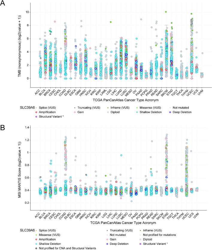
Tumor mutation burden (TMB) (Fig. 9A) and microsatellite instability (MSI) (Fig. 9B) of SLC39A6 in 33 TCGA cancer types were obtained from cBioPortal database. We have also performed relevant analyses using the cBioPortal database and further investigated the associated protein alterations (as shown in Supplementary Fig. 1–2). In this study, t-SNE (t-distributed stochastic neighbor embedding) dimensionality reduction was used for visual analysis of single-cell transcriptome data. The expression level of SLC39A6 showed a significantly heterogeneous distribution pattern among different subpopulations (as shown in Supplementary Fig. 3).
PPI interaction network and Gene ontology (GO) enrichment analysis
Firstly, protein–protein interaction analysis was performed, and the protein–protein interaction Network (PPI Network) of SLC39A6 was constructed using STRING database (Fig. 10A). The protein–protein interaction Network (PPI Network) results showed that 10 genes were related to SLC39A6, which were: SLC30A1, SLC30A2, SLC30A4, SLC30A5, SLC30A6, SLC30A7, SLC30A9, SLC39A1, SLC39A11, SLC39A9, which were recorded as the Interacting Genes of SLC39A6.
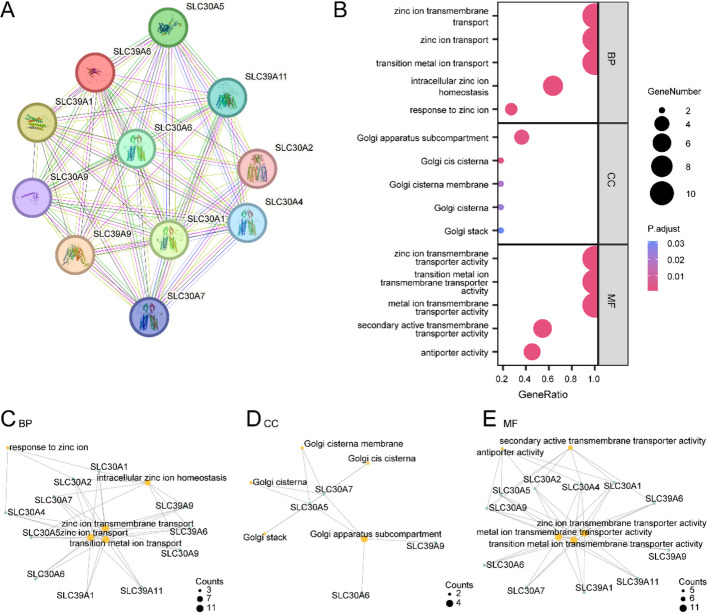
Gene ontology (GO) enrichment analysis was used to further explore the relationship between biological process (BP), cellular component (CC), and molecular function (MF) of SLC39A6 gene and Interacting Genes. SLC39A6 gene and Interacting Genes were used for gene ontology (GO) enrichment analysis, and the specific results are shown in Table 2. The results showed that SLC39A6 gene and Interacting Genes were mainly enriched in zinc ion transmembrane transport, zinc ion transport, and zinc ion transport. transition metal ion transport, intracellular zinc ion homeostasis, response to zinc ion and other biological processes (BP); Golgi apparatus subcompartment, Golgi cis cisterna, Golgi cisterna membrane, Golgi cisterna, Golgi stack; zinc ion transmembrane transporter activity, transition metal ion transmembrane transporter activity, metal ion transmembrane transporter activity, secondary active transmembrane transporter activity, antiporter activity and other molecular functions (MF). The results of Gene ontology (GO) enrichment analysis were visualized by bubble plots (Fig. 10B).
Table 2: Results of GO enrichment analysis for SLC39A6 and interacting genes
| ONTOLOGY | ID | Description | GeneRatio | BgRatio | pvalue | p.adjust | qvalue |
|---|---|---|---|---|---|---|---|
| BP | GO:0071577 | zinc ion transmembrane transport | 11/11 | 27/18870 | 4.83E-33 | 4.54E-31 | 2.97E-31 |
| BP | GO:0006829 | zinc ion transport | 11/11 | 28/18870 | 7.96E-33 | 4.54E-31 | 2.97E-31 |
| BP | GO:0000041 | transition metal ion transport | 11/11 | 100/18870 | 5.25E-26 | 1.99E-24 | 1.31E-24 |
| BP | GO:0006882 | intracellular zinc ion homeostasis | 7/11 | 36/18870 | 1.62E-17 | 4.62E-16 | 3.03E-16 |
| BP | GO:0010043 | response to zinc ion | 3/11 | 51/18870 | 3.02E-06 | 6.89E-05 | 4.52E-05 |
| CC | GO:0098791 | Golgi apparatus subcompartment | 4/11 | 382/19886 | 3.98E-05 | 1.99E-03 | 1.13E-03 |
| CC | GO:0000137 | Golgi cis cisterna | 2/11 | 31/19886 | 1.28E-04 | 3.21E-03 | 1.82E-03 |
| CC | GO:0032580 | Golgi cisterna membrane | 2/11 | 93/19886 | 1.16E-03 | 1.93E-02 | 1.10E-02 |
| CC | GO:0031985 | Golgi cisterna | 2/11 | 118/19886 | 1.85E-03 | 2.32E-02 | 1.32E-02 |
| CC | GO:0005795 | Golgi stack | 2/11 | 151/19886 | 3.01E-03 | 3.01E-02 | 1.71E-02 |
| MF | GO:0005385 | zinc ion transmembrane transporter activity | 11/11 | 24/18496 | 1.15E-33 | 3.23E-32 | 1.09E-32 |
| MF | GO:0046915 | transition metal ion transmembrane transporter activity | 11/11 | 42/18496 | 1.98E-30 | 2.77E-29 | 9.37E-30 |
| MF | GO:0046873 | metal ion transmembrane transporter activity | 11/11 | 441/18496 | 1.25E-18 | 1.17E-17 | 3.95E-18 |
| MF | GO:0015291 | secondary active transmembrane transporter activity | 6/11 | 286/18496 | 5.62E-09 | 3.93E-08 | 1.33E-08 |
| MF | GO:0015297 | antiporter activity | 5/11 | 135/18496 | 8.58E-09 | 4.80E-08 | 1.63E-08 |
GO Gene Ontology, BP Biological Process, MF Molecular Function
Meanwhile, the network maps of biological process (BP), cellular component (CC) and molecular function (MF) were drawn according to Gene ontology (GO) enrichment analysis (Fig. 10C-E). The lines show the corresponding molecules and the annotations of the corresponding entries, and the larger the nodes, the more molecules the entries contain Fig. 11.
Drug sensitivity analysis of SLC39A6
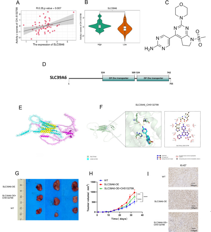
Increased drug sensitivity is critical to prevent cancer cells from developing resistance to treatment. To explore this further, we performed a correlation analysis between drug sensitivity and SLC39A6 expression levels using data from the CellMiner database. Our analysis revealed significant positive correlations between SLC39A6 and drug sensitivity to CH5132799, of which is a PI3K inhibitor (Fig. 11 A-B). Moreover, molecular docking results revealed that the binding energy of compound CH5132799 to the SLC39A6 protein was −7.6 kcal/mol, indicating its strong inhibitory potential against SLC39A6 (Fig. 11 C-F). Meanwhile, we conducted in vivo validation in animal models. The results demonstrated that CH5132799 administration significantly inhibited tumor proliferation when SLC39A6 was highly expressed (SLC39A6-OE + CH5132799 210.0 mm3 ± 83.14 mm3 vs. SLC39A6-OE 282.8 mm3 ± 109.8 mm3, p < 0.001; and vs. WT 158.5 mm3 ± 55.69 mm3, p < 0.0001 (Fig. 11 G-H)). Immunohistochemical analysis revealed that Ki67 expression in tumor tissues was consistent with these findings (Fig. 11 I). Individual tumor growth curves are provided in in Supplementary Fig. 4. H-score of Ki67 expression are provided in in Supplementary Table 1.
Discussion
Despite decades of progress, common solid malignancies continue to impose an unacceptable burden on global public health, and current therapeutic modalities still fall short of providing durable clinical benefits. By integrating pan-cancer, multi-omics analyses, we delineated a central oncogenic role for the zinc transporter SLC39A6 and identified it as a tractable target for precision oncology. We report several principal findings. First, SLC39A6 is markedly dysregulated across more than ten cancer types and independently predicts shortened overall survival (OS), disease-specific survival (DSS), and progression-free interval (PFI). Second, we revealed that sensitivity to the PI3Kα-selective inhibitor CH5132799 increases with SLC39A6 expression; in vivo studies confirmed that CH5132799 effectively suppresses SLC39A6-driven tumor cell proliferation. These results provide a robust foundation for translational development.
SLC39A6 is markedly up-regulated in hepatocellular carcinoma (HCC) tissues, and its expression increases progressively with higher TNM stages, enabling discrimination between early- and advanced-stage HCC [ref. 27]. A meta-analysis of nine GEO datasets revealed that SLC39A6 mRNA is significantly elevated in early-stage tumors [ref. 28]. In ER-positive breast cancer cases, high SLC39A6 protein expression is associated with longer disease-free survival, supporting its utility for molecular subtyping [ref. 29].
Univariate Cox modeling indicated that elevated SLC39A6 expression is significantly associated with poorer overall survival (OS), disease-specific survival (DSS), and progression-free interval (PFI), particularly in cervical squamous cell carcinoma (CESC), hepatocellular carcinoma (LIHC), and mesothelioma (MESO). This establishes its role as both a prognostic biomarker and a therapeutic target. Importantly, this association is context-dependent. In hormone-receptor-positive breast cancer, higher SLC39A6 levels surprisingly indicate a favorable prognosis, especially in node-negative or luminal A subtypes, whereas no prognostic significance is seen in basal-like or HER2-positive tumors [ref. 29]. These findings underscore the importance of molecular stratification prior to clinical application. In CESC and MESO, elevated SLC39A6 expression consistently signifies a poor prognosis irrespective of FIGO or TNM stage, suggesting its value as a stage-independent risk factor. In summary, the prognostic impact of SLC39A6 varies by cancer type and subtype, and it can exhibit a "double-edged sword" effect in hormone-dependent tumors. Moving forward, incorporating SLC39A6 into multi-gene, multi-modal prognostic scores and utilizing SLC39A6 immunohistochemistry (IHC) or quantitative polymerase chain reaction (qPCR) in multidisciplinary tumor boards will facilitate precise patient stratification before clinical implementation.
Immune-microenvironment analyses have revealed that SLC39A6 remodels tumor immunity in liver hepatocellular carcinoma (LIHC) and malignant mesothelioma (MESO). Single-sample gene set enrichment analysis (ssGSEA) demonstrated significant skewing of nine immune subsets—including cytotoxic T cells, dendritic cells, and macrophages—between SLC39A6-high and SLC39A6-low tumors. Spatial transcriptomic mapping in colorectal cancer showed inverse localization of SLC39A6 with CD8⁺ T cells and dendritic cells, but co-localization with M2-polarized macrophages. Mechanistically, SLC39A6 activates the Toll-like receptor and IL-17 signaling axes, triggering the release of CXCL8 and CCL2, which recruits M2 macrophages and dampens cytotoxic immunity [ref. 28]. In LIHC, SLC39A6-mediated zinc influx phosphorylates STAT3 and transcriptionally up-regulates PD-L1 and VEGF-A, thereby impairing dendritic-cell maturation and macrophage polarization. Knockdown of SLC39A6 in HepG2 and Hep3B cells restored CD8⁺ T-cell cytotoxicity, reduced PD-L1 by 45%, and synergized with anti-PD-1 therapy to shrink tumors by 65% in murine models [ref. 27]. Similarly, the SLC39A6-directed antibody–drug conjugate (ADC) BRY812 reduced M2 infiltration and restored CD8⁺ T-cell activity in patient-derived colorectal organoids; combination with anti-CTLA-4 improved objective response rates from 23 to 58% [ref. 28]. A pan-cancer immunogenomic cohort (n = 1,054) confirmed that SLC39A6-high tumors exhibit reduced CD8⁺ T-cell infiltration, lower tumor mutational burden, and diminished sensitivity to PD-1 blockade, suggesting that this immune-regulatory axis is conserved across multiple histologies [ref. 30].
Protein–protein interaction network analysis has identified ten proteins significantly associated with SLC39A6, including multiple members of the zinc transporter family. Collectively, these proteins participate in zinc-ion transmembrane transport and other essential biological processes. This interaction network highlights the critical role of metal-ion homeostasis and suggests that these transporters may cooperatively modulate cell proliferation and apoptosis pathways, thereby influencing tumor progression.
Beyond immunity, the disruption of the ZIP/ZnT/metallothionein axis leads to increased intracellular zinc levels, which activates the PI3K-AKT, MAPK/ERK, and STAT3 cascades. These pathways drive cell-cycle progression, epithelial-mesenchymal transition (EMT), and the evasion of apoptosis [ref. 4, ref. 27, ref. 28]. In models of breast and liver cancer, zinc influx mediated by SLC39A6 phosphorylates AKT, inactivates GSK-3β, and stabilizes Snail, thereby promoting metastasis. Iron, copper, and manganese exert similar effects through transporters such as ZIP8/SLC30A/DMT1/ATP7A, highlighting the broader role of metal-ion homeostasis in oncogenesis [ref. 31]. Targeting this circuitry is clinically viable: the SLC39A6-directed antibody–drug conjugate (ADC) ladiratuzumab vedotin is currently in phase I/II trials, and ZnO nanoparticles utilize NCOA4-mediated ferritinophagy to induce ferroptosis, offering a versatile drug-delivery platform [ref. 28, ref. 32–ref. 35].
Correlative analyses have revealed that SLC39A6 inversely correlates with immune-checkpoint gene (ICG) expression, while positively correlating with the DNA-mismatch-repair genes MLH1, MSH2, MSH6, and PMS2. To our knowledge, this is the first report linking SLC39A6 to ICG/MMR networks. Low ICG expression indicates an immune-cold microenvironment and predicts a poor response to PD-1/PD-L1 blockade, whereas high MMR gene expression signifies microsatellite stability and chemoresistance to DNA-damaging agents. Integrating SLC39A6 with MMR status may therefore refine risk stratification and guide combination regimens—for instance, CH5132799 plus platinum-free immunotherapy in SLC39A6-high/MMR-intact tumors [ref. 30].
High expression of SLC39A6 sensitizes tumors to CH5132799, a selective PI3Kα inhibitor that effectively suppresses cell proliferation driven by SLC39A6. This observation is consistent with the seminal report by Tanaka, who first demonstrated potent in vitro and in vivo antitumor activity of CH5132799 against PI3K-hyperactivated tumor cells [ref. 36]. Building on this foundation, we are the first to advance SLC39A6 from a prognostic indicator to an actionable companion-diagnostic biomarker for CH5132799. Clinically, high SLC39A6 expression can be used to identify patients most likely to benefit from this agent. Phase II basket trials are planned in cervical squamous cell carcinoma (CESC), hepatocellular carcinoma (LIHC), and mesothelioma (MESO). Given CH5132799’s exquisite selectivity for SLC39A6-high tumors, combination strategies with PD-1 inhibitors or zinc chelators could overcome immune-cold phenotypes and potentiate metal-ion-induced ferroptosis.We systematically reviewed and compared existing SLC39A6-targeted strategies (ADC, zinc chelation, RNAi) and summarized their inherent limitations, such as poor specificity, low efficacy, and high toxicity.Different from previous single-cancer or single-strategy studies, our research conducts a comprehensive pan-cancer analysis of SLC39A6, identifies it as a companion diagnostic biomarker for the PI3Kα inhibitor CH5132799, clarifies its related molecular mechanisms, and thus fills the gap in SLC39A6-targeted precision oncology.
Limitations encompass the retrospective nature of publicly available datasets, uneven sample sizes, and the lack of comprehensive confounder adjustment. Failure to integrate co-expression and interaction data is a limitation of this study. In subsequent research, we plan to combine TCGA and other public databases to construct a multi-layer network integrating expression correlation and protein interaction evidence, so as to further analyze the functional modular characteristics of oxidative stress-related genes. Further mechanistic studies and prospective validation are necessary to confirm the function of SLC39A6 and to assess its full potential as a therapeutic target and predictive biomarker.
Conclusion
In conclusion, our study determined SLC39A6 regulates the dynamics of immune infiltration and impacts prognosis in a wide range of malignancies. Besides, our results advance SLC39A6 from a prognostic indicator to an actionable companion-diagnostic biomarker for CH5132799. It emerges as a promising biomarker for prognosis, immunology, and therapy in the field of precision oncology.
References
- H Sung, J Ferlay, RL Siegel, M Laversanne, I Soerjomataram, A Jemal, F Bray. Global cancer statistics 2020: GLOBOCAN estimates of incidence and mortality worldwide. CA Cancer J Clin, 2021. [DOI | PubMed]
- RL Siegel, KD Miller, HE Fuchs, A Jemal. Cancer statistics, 2023. CA Cancer J Clin, 2023. [DOI | PubMed]
- RR Prasad, CK Singh, J Meng, S Nagamuga, S Satarug. Stage-specific differential expression of zinc transporter SLC39A6 during tumorigenesis. Mol Carcinog, 2022. [DOI | PubMed]
- 4.Fukada T, Kambe T. Molecular and genetic features of zinc transporters in physiology and pathogenesis. Metallomics. 2011;3(7):662–74. 10.1039/c1mt00011j.
- R Saravanan, KM Taylor, D Brethour. Zinc transporter LIV-1 (SLC39A6): a promising cell-surface target for triple-negative breast cancer. J Cell Physiol, 2022. [DOI]
- MY Liu, XB Cui, XX Cheng, L Zhao, W Chen, X Li. SLC39A6 promotes aggressiveness of esophageal carcinoma by activating PI3K signaling via zinc influx. Gastroenterology, 2021. [DOI | PubMed]
- H Zhou, J Wang, Y Li, Y Zhang, X Wang, Z Li. Evaluation of the prognostic value of SLC39A6 in lung adenocarcinoma using TCGA data. Aging Albany NY, 2021. [DOI | PubMed]
- G Lyu, D Li. ZNF165: a pan-cancer biomarker with prognostic and therapeutic potential. Protein Pept Lett, 2025. [DOI | PubMed]
- Y Zhang, Z Zhao, W Huang. Pan-cancer single-cell analysis revealing the heterogeneity of cancer-associated fibroblasts in skin tumors. Curr Gene Ther, 2025. [DOI | PubMed]
- 10.Shi Y, Shen Q, Jiang A, et al. DrugSurvPlot: a novel web-based platform harnessing drug sensitivity scores as molecular biomarkers for pan-cancer survival prognosis. Curr Gene Ther. 2025. 10.2174/0115665232412138250722020114.
- M Jiang, D Zhu, D Zhao. Integrated analysis of clinical outcome of mesenchymal stem cell-related genes in pan-cancer. Curr Genomics, 2024. [DOI | PubMed]
- A Colaprico, TC Silva, C Olsen, L Garofano, C Cava, D Garolini, TS Sabedot, TM Malta, SM Pagnotta, I Castiglioni, M Ceccarelli, G Bontempi, H Noushmehr. TCGAbiolinks: an R/Bioconductor package for integrative analysis of TCGA data. Nucleic Acids Res, 2016. [DOI | PubMed]
- MJ Goldman, B Craft, M Hastie, K Repečka, F McDade, A Kamath, A Banerjee, Y Luo, D Rogers, AN Brooks, J Zhu, D Haussler. Visualizing and interpreting cancer genomics data via the Xena platform. Nat Biotechnol, 2020. [DOI | PubMed]
- 14.Therneau TM. A package for survival analysis in R. R package version 3.4–0. 2022. https://CRAN.R-project.org/package=survival.
- JT Rich, JG Neely, RC Paniello, CC Voelker, B Nussenbaum, EW Wang. A practical guide to understanding Kaplan-Meier curves. Otolaryngol Head Neck Surg, 2010. [DOI | PubMed]
- B Xiao, L Liu, A Li, J Liu, Z Hu, D Xiang, H Jiao, J Li. Identification and verification of immune-related gene prognostic signature based on ssGSEA for osteosarcoma. Front Oncol, 2020. [DOI | PubMed]
- D Xu, X Liu, Y Wang, X Li, H Zhang, J Wang. Identification of immune subtypes and prognosis of hepatocellular carcinoma based on immune checkpoint gene expression profile. Biomed Pharmacother, 2020. [DOI | PubMed]
- R Chen, W Wu, SY Chen, X Chen, C Liu, L Chen, Y Liu, J Chen, H Liu, H Wang. A pan-cancer analysis reveals CLEC5A as a biomarker for cancer immunity and prognosis. Front Immunol, 2022. [DOI | PubMed]
- I de Bruijn, R Kundra, B Mastrogiacomo, H Zhang, S Jolly, A Luna, J Gao, U Dogrusoz, SO Sumer, N Schultz. Analysis and visualization of longitudinal genomic and clinical data from the AACR Project GENIE Biopharma Collaborative in cBioPortal. Cancer Res, 2023. [DOI | PubMed]
- E Cerami, J Gao, U Dogrusoz, BE Gross, SO Sumer, BA Aksoy, A Jacobsen, CJ Byrne, ML Heuer, E Larsson, Y Antipin, B Reva, AP Goldberg, C Sander, N Schultz. The cBio cancer genomics portal: an open platform for exploring multidimensional cancer genomics data. Cancer Discov, 2012. [DOI | PubMed]
- 21.Gao J, Aksoy BA, Dogrusoz U, Dresdner G, Gross B, Sumer SO, Sun Y, Jacobsen A, Sinha R, Larsson E, Cerami E, Sander C, Schultz N. Integrative analysis of complex cancer genomics and clinical profiles using the cBioPortal. Sci Signal. 2013;6(269):pl1. 10.1126/scisignal.2004088.
- D Szklarczyk, R Kirsch, M Koutrouli, KC Nastou, F Mehryary, R Hachilif, AL Gable, T Fang, NT Doncheva, S Pyysalo, P Bork, LJ Jensen, C von Mering. The STRING database in 2023: protein-protein association networks and functional enrichment analyses for any sequenced genome of interest. Nucleic Acids Res, 2023. [DOI | PubMed]
- H Mi, A Muruganujan, D Ebert, X Huang, PD Thomas. PANTHER version 14: more genomes, a new PANTHER GO-slim and improvements in enrichment analysis tools. Nucleic Acids Res, 2019. [DOI | PubMed]
- G Yu, LG Wang, Y Han, QY He. clusterProfiler: an R package for comparing biological themes among gene clusters. OMICS, 2012. [DOI | PubMed]
- UT Shankavaram, S Varma, D Kane, M Sunshine, KK Chary, WC Reinhold, Y Pommier, JN Weinstein. Cell Miner: a relational database and query tool for the NCI-60 cancer cell lines. BMC Genomics, 2009. [DOI | PubMed]
- 26.Reinhold WC, Sunshine M, Varma S, Doroshow JH, Pommier Y. Using CellMiner 1.6 for systems pharmacology and genomic analysis of the NCI-60. Clin Cancer Res. 2015;21(17):3841–3852. 10.1158/1078-0432.CCR-15-0335.
- Z Wan, X Wang. Role of SLC39A6 in the development and progression of liver cancer. Oncol Lett, 2022. [DOI | PubMed]
- X Liu, W Liu, Y Wu, Y Wang, Q Jiang, Y Li, H Li, L Hao. Investigation of the cytotoxic effects and mechanisms of the SLC39A6-targeting ADC drug BRY812 in CRC. Sci Rep, 2025. [DOI | PubMed]
- M Althobiti, KA El-Sharawy, C Joseph, M Aleskandarany, MS Toss, AR Green, EA Rakha. Oestrogen-regulated protein SLC39A6: a biomarker of good prognosis in luminal breast cancer. Breast Cancer Res Treat, 2021. [DOI | PubMed]
- P Charoentong, F Finotello, M Angelova. Pan-cancer immunogenomic analyses reveal genotype–immunophenotype relationships and predictors of response to checkpoint blockade. Cell Rep, 2017. [DOI | PubMed]
- 31.Zhou D, Lu P, Mo X, et al. Ferroptosis and metabolic syndrome and complications: association, mechanism, and translational applications. Front Endocrinol (Lausanne). 2024;14:1248934. 10.3389/fendo.2023.1248934.
- D Sussman, LM Smith, ME Anderson. SGN-LIV1A: a novel antibody-drug conjugate targeting LIV-1 for the treatment of metastatic breast cancer. Mol Cancer Ther, 2014. [DOI | PubMed]
- XB Cui, YY Shen, TT Jin. SLC39A6: a potential target for diagnosis and therapy of esophageal carcinoma. J Transl Med, 2015. [DOI | PubMed]
- 34.Qin X, Zhang J, Wang B, et al. Ferritinophagy is involved in the zinc oxide nanoparticles-induced ferroptosis of vascular endothelial cells. Autophagy. 2021;17(12):4266–4285. 10.1080/15548627.2021.1911016.
- 35.Bartos A, Sikora J. Bioinorganic modulators of ferroptosis: a review of recent findings. Int J Mol Sci. 2023;24(4):3634. 10.3390/ijms24043634.
- H Tanaka, M Yoshida, H Tanimura, T Fujii, K Sakata, Y Tachibana, J Ohwada, H Ebiike, S Kuramoto, K Morita, Y Yoshimura, T Yamazaki, N Ishii, O Kondoh, Y Aoki. The selective class I PI3K inhibitor CH5132799 targets human cancers harboring oncogenic PIK3CA mutations. Clin Cancer Res, 2011. [DOI | PubMed]