An Investigation on the Synthesis of Molybdenum Oxide and Its Silica Nanoparticle Composites for Dye Degradation
Abstract
The molybdenum oxide (MoO3) and MoO3@SiO2 nanoparticles were successfully prepared using the chemical bath deposition (CBD) method. The photocatalytic activities of molybdenum oxide (MoO3), SiO2, and MoO3@SiO2 nanoparticles composite have shown a synergistic photocatalytic effect of SiO2 combined with MoO3. The first-order degradation rate constants for MoO3, SiO2, and MoO3@SiO2 nanocomposite were 10.3 × 10⁻³ min−1, 15.1 × 10⁻³ min−1, and 16.3 × 10⁻³ min−1, respectively. The MoO3@SiO2 composite showed degradation efficiencies in the methylene blue solution close to 100% after 60 min of UV irradiation. The X-ray diffraction (XRD) showed that the MoO3 powder has a hexagonal crystal structure and the silica is the tridymite type of SiO2. The crystallite size was about 94 nm, 32 nm, and 125 nm for MoO3, silica, and MoO3@SiO2, respectively, as calculated by the Scherrer equation. The scanning electron microscopy (SEM) images revealed that the MoO3 powder consisted of a uniform hexagonal structure; the silica showed a rod-like micro-flake morphology and the MoO3@SiO2 composite had the appearance of coral-like structures.
Article type: Research Article
Keywords: MoO, silica, nanoparticle composite, structural properties, photocatalytic properties, methylene blue
Affiliations: Laboratoire de Physique de la Matière Condensée, Faculté des Sciences de Tunis, Université de Tunis El Manar, Tunis 2092, Tunisia; o.kamoun@yahoo.fr (O.K.); n.najouakamoun@gmail.com (N.T.-K.); Department of Physics, Faculty of Science, King Khalid University, P.O. Box 9004, Abha 61413, Saudi Arabia; Laboratoire Matériaux Utiles, Institut National de Recherche et d’Analyse Physico-Chimique (INRAP) Sidi Thabet, Ariana 2020, Tunisia; kouasssa@gmail.com; Nanotechnology and Advanced Materials Program, Kuwait Institute for Scientific Research, P.O. Box 24885, Safat 13109, Kuwait; bhalaili@kisr.edu.kw; Department of Electrical and Computer Engineering, University of California Davis, Davis, CA 95616, USA; Faculty of Materials Science and Engineering, University POLITEHNICA of Bucharest, 313 Splaiul Independentei, RO-060042 Bucharest, Romania
License: © 2020 by the authors. CC BY 4.0 Licensee MDPI, Basel, Switzerland. This article is an open access article distributed under the terms and conditions of the Creative Commons Attribution (CC BY) license (http://creativecommons.org/licenses/by/4.0/).
Article links: DOI: 10.3390/nano10122409 | PubMed: 33276515 | PMC: PMC7761561
Relevance: Relevant: mentioned in keywords or abstract
Full text: PDF (2.4 MB)
1. Introduction
Catalytic materials were developed over the years to purify the polluted water and air, where the heterogeneous photocatalysis plays an important role [ref. 1,ref. 2,ref. 3,ref. 4,ref. 5]. The well-known transition metal oxide photocatalysts are ZnO [ref. 6], TiO2 [ref. 7], WO3 [ref. 8], and MoO3 [ref. 9]. The oxidation process of heterogeneous photocatalysis is achieved by using light to activate the catalyst and to generate highly reactive free radicals, which then reduce particular organic compounds [ref. 6,ref. 7,ref. 9,ref. 10,ref. 11]. When the mineralization process is finished, the outcome consists of H2O and CO2.
The photocatalytic mechanism is activated when the oxide semiconductor is immersed in a liquid or placed in a gaseous medium and then irradiated with light of an energy that is equal to or greater than its bandgap [ref. 11]. In this case, electron-hole pairs are generated on the surface of the semiconductor and subsequent chemical reactions with the environmental media lead to the production of free radicals, which degrade the organic pollutants [ref. 10,ref. 11].
Photocatalysis requires a large surface area for reaction. The exterior of natural silica is covered with a network of pores to optimize the capture of light. This feature of silica has attracted the attention of nanotechnologists. Moreover, silica is a phylum of unicellular microalgae (from 2 μm to 1 mm) present in all aquatic environments, and the majorities are in biofilms (with a preference for cold water) and enveloped by a siliceous external skeleton. The degradation of MB under the visible light has been demonstrated for photocatalysts prepared using green and renewable resources [ref. 12]. Mesoporous silica impregnated with Pt-Porphyrin or PtNPs [ref. 13] and titania sensitized with porphyrin [ref. 14] or magnetic photocatalyst porphyrin [ref. 15] has also been shown visible-light-driven photocatalysis. MoO3 is one of the most promising metal oxides because of its many advantages, such as its non-toxic nature, and can be widely used in an organic light-emitting diode, gas sensing, catalysis, transistors, and solar cells [ref. 16,ref. 17,ref. 18]. The smaller the particle size of MoO3, the larger the surface area, which potentially increases the adsorption methylene blue and photoactive sites, resulting in enhanced photocatalytic activity [ref. 9,ref. 16,ref. 19]. Various methods have been used to optimize the preparation and processing technology of molybdenum oxide films, such as thermal evaporation, sol–gel deposition, and chemical vapor deposition [ref. 20,ref. 21,ref. 22]. In our work, we used a simple and inexpensive chemical bath deposition (CBD) technique because the film properties can be optimized through various deposition process parameters. The CBD technique has received great consideration from the research community for the production of low-cost semiconductor photocatalyst.
In our search for novel and sustainable photocatalysts, we used the unique architecture of silica and high surface area to increase the degradation efficiency of the organic dyes. Because MoO3 has shown good photocatalytic degradation properties [ref. 23,ref. 24,ref. 25], present in this study the synthesis of a novel MoO3 and SiO2 nanoparticle composites, labeled MoO3@SiO2, and their photocatalytic properties. The main objective of this work is to study the photocatalytic activities of the films for the photodegradation of methylene blue (MB) under UV light, as well as examine the physical properties of the MoO3 films.
2. Experimental and Characterization Details
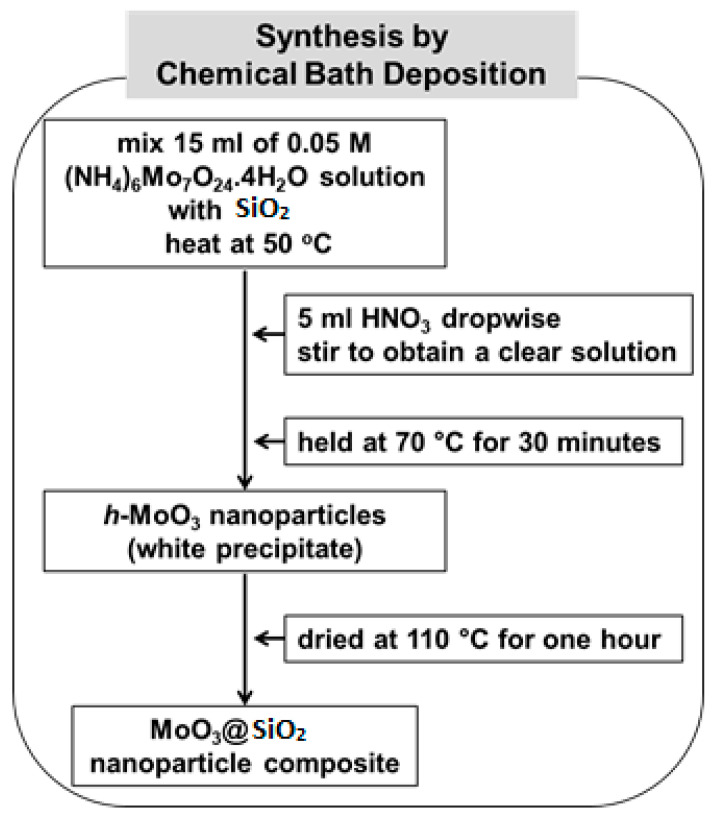
The chemical bath deposition (CBD) method was performed to synthesize nanocrystalline MoO3 and MoO3 on SiO2, as illustrated in Figure 1. In a typical synthesis, an aqueous solution of 15 mL of 0.05 M (NH4)6Mo7O24·4H2O (99%, Merck, Kenilworth, NJ, USA) solution was mixed with SiO2 in a reaction bath. The temperature of the reaction bath was slowly increased to 50 °C. Then, 5 mL of concentrated HNO3 (ACS reagent, ≥90.0%) was added dropwise with constant stirring until the pH of the solution was 2.2, and a clear solution was obtained. Then, after the solution was stirred for 15 min, the temperature of the reaction bath was raised to 70 °C, where the initial seeds started to form. The reaction bath was held at 70 °C for 30 min, during which time a white precipitate of h-MoO3 nanoparticles was observed. When the synthesis was complete, the white precipitate was filtered using deionized water and then dried in an oven at a constant temperature of 110 °C for 1 h [ref. 26,ref. 27].
The nanoparticle composites were analyzed by X-ray diffraction (XRD) scanning electron microscopy (SEM), and UV-Vis spectroscopy. The XRD patterns were recorded on an X’pert PRO X-ray diffractometer (Malvern Panalytical Ltd, Malvern, UK) with graphite monochromatized Cu Kα radiation source (1.5406 Å). Morphologies of nanopowders were examined using a JEOL-JSM-6490 LV scanning electron microscope (SEMTech Solutions, Inc., North Billerica, MA, USA) and absorbance measurements were performed using a Perkin Elmer Lambda 950 spectrometer (Perkin Elmer, Waltham, MA, USA). The UV irradiation was performed at 254 nm wavelength using an 8 W power lamp (Philips Germicidal Ultraviolet-C, Philips Lightning, Eindhoven, The Netherlands).
3. Results and Discussion
3.1. Structural Properties
Both MoO3 nanoparticles and SiO2 were analyzed by X-ray diffraction analysis and compared to the MoO3@SiO2 nanoparticles composite. Figure 2 shows the XRD patterns of the MoO3, SiO2, and MoO3@SiO2 nanoparticles grown by chemical bath deposition. The diffraction patterns correspond to the h-MoO3 phase for the MoO3 nanoparticles and to tridymite, which is the monoclinic phase corresponding to SiO2 for the silica [ref. 28].
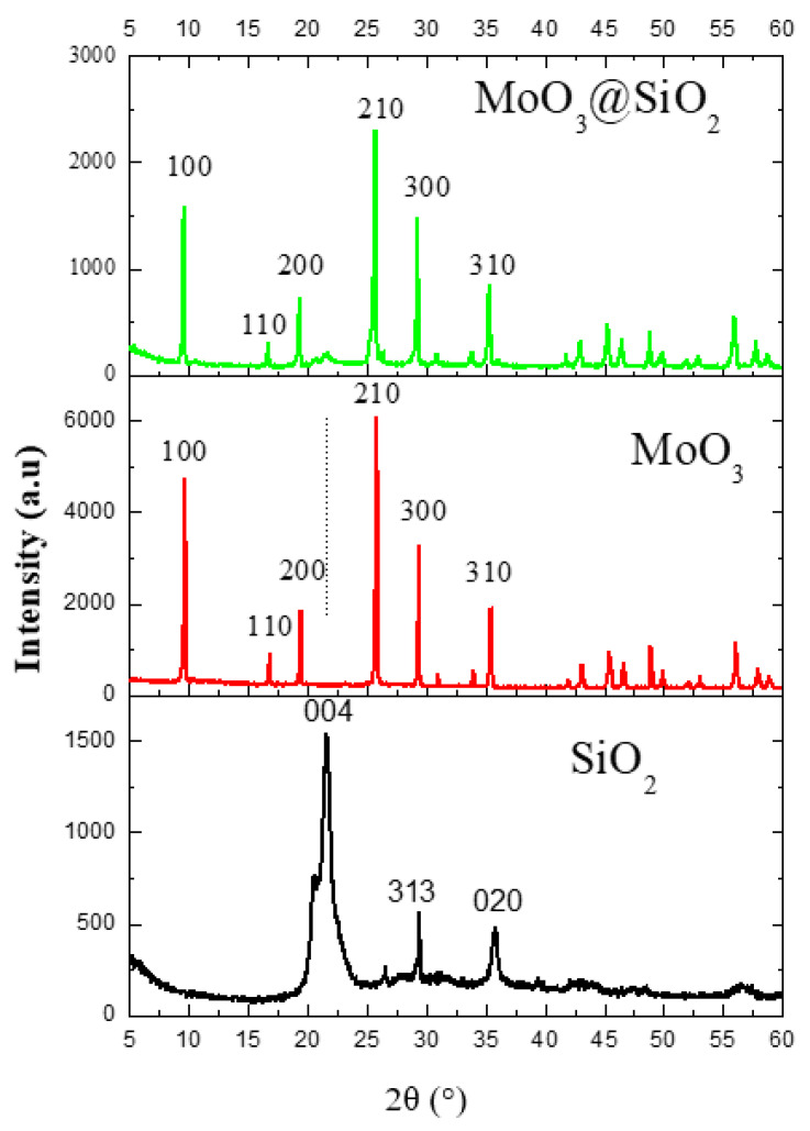
Tridymite is a species of mineral of the tectosilicate family, and one of the polymorphs of silica with quartz, coesite, cristobalite, stishovite, having the chemical formula of SiO2 and containing traces of titanium, aluminum, iron, manganese, magnesium, calcium, sodium, and potassium.
MoO3 has the following lattice parameters: a = 10.53 Å and c = 14.876 Å (JCPDS card no. 21-0569) [ref. 19]. The SiO2 has the following lattice parameters: a = 25.93 Å, b = 5.01 Å, and c = 18.54 Å, with the highest intensity at 2θ = 21.6°, matching the reference R090042 [ref. 29].
The crystallite size can be estimated from the full width half maximum (FWHM) values obtained from the predominant (210) for MoO3 and MoO3@SiO2 diffraction peak at 2θ = 25.7° according to the following Debye–Sherrer equation [ref. 30,ref. 31,ref. 32]:
where λ is the wavelength of Cu-Kα1 radiation (1.5406 Å) and θ is the Bragg diffraction angle. The crystallite sizes calculated with Equation (1) were around 94, 32, and 125 nm for MoO3, SiO2, and MoO3@SiO2, respectively. The observed broadening of the SiO2 peak(004) in the MoO3@SiO2 spectrum is attributed to the size and strain effect between MoO3 and SiO2 [ref. 19].
In the XRD spectra of the MoO3@SiO2 composite, all peaks attributed to the MoO3phase are observed, which confirms that the MoO3 nanoparticles are well grown on the SiO2 surface. It was also observed that the intensity for all MoO3 peaks decreases for MoO3@SiO2 composite compared to those of MoO3. Note the existence of the preferential SiO2 peak at 2θ = 21.6°. All MoO3@SiO2 peaks decreased in intensity, which may be due to the fact that MoO3 nanoparticles are well-formed on the surface of SiO2, but in a dispersed manner.
3.2. Morphological Analysis
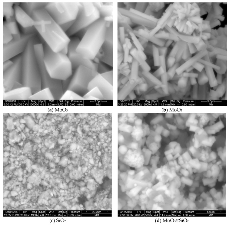
Surface morphology of the nanoparticles composite and constituent nanoparticles were investigated by using SEM analyses. The nanoparticles of MoO3 consist of a uniform hexagonal rod-like morphology. The regular faceted surface of each hexagonal rod [ref. 33] is clearly seen in Figure 3a. In Figure 3b, stems have developed out of a central point [ref. 34], with flower-like clusters of hexagonal MoO3 stem-shaped petals. The SEM images of SiO2 (Figure 3c) shows a similar morphology as SiO2 [ref. 35]. The morphology of SiO2 depicts mostly micro-flake and irregular rod-shaped with particles agglomeration. Regarding the MoO3@SiO2 composite (Figure 3d,e), we observed the appearance of coral-like structures in the form of hexagonal rods. The morphology of this composite indicates the incorporation of MoO3 into the SiO2 in the MoO3@SiO2 composite, which is in agreement with the XRD analysis in Figure 2. SEM observation shows that the specific surface area increased for the MoO3@SiO2 composite compared to those for MoO3 or SiO2. Increasing the specific surface area, especially in the case of MoO3@SiO2, could play an important role in improving sensitivity in optoelectronic applications like photocatalysis and gas sensors.
3.3. Photocatalytic Studies
MoO3 nanoparticles were used as photocatalyst for the degradation of methylene blue (MB), which was used as a model compound. It was found that there was no MB degradation in the dark and in the presence of MoO3, SiO2, and MoO3@SiO2. In this work, we have monitored the MB degradation under UV light at different times with MoO3, SiO2, and MoO3@SiO2 nanoparticle catalysts. Figure 4 presents the UV-Vis absorption spectra of MoO3 nanoparticles, SiO2 and MoO3@SiO2 nanoparticles exposed to UV light for different times.
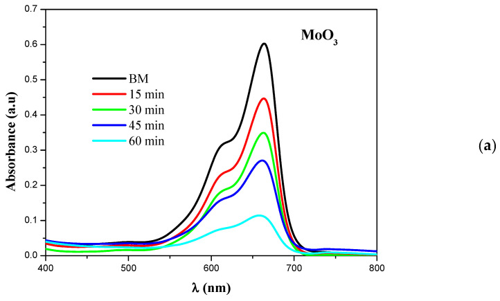
There are two different absorption bands for the aqueous cationic MB dye solution, i.e., at 293 nm (π-π∗) and 664 nm (n-π∗) [ref. 34]. In this work, the intensities of the absorption peaks at 664 nm decrease with increasing the time of irradiation, compared to the catalyst-free solution. The degradation of the MB solution containing h-MoO3 catalyst synthesized by CBD was 90% [ref. 34].
During photocatalysis, the electrons in the valence band of the oxide semiconductor are excited under UV light radiation and leave holes in the valence band after they jump to the conduction band. The holes combine with H2O to produce ∙ H and ∙ OH radicals. In the meanwhile, the electrons in the conduction band scattered towards the adsorbed O2 to generate activated ∙ O2 [ref. 36] with the consequent transformation of the water molecules into ∙ OH radicals.
The mechanism of photocatalytic degradation for MoO3nanoparticles is similar to that of a metal oxide semiconductor [ref. 37], as follows:
323−+
+−.
−22−
.*22
2−*22
These oxidizing species can degrade the MB dye into chemical forms of CO2 and H2O, which is a better solution to water remediation treatments [ref. 36]. If the photocatalytic processes do not take place, the recombination of the (e− + h+) pairs happens, and heat is generated in the materials. The photocatalytic activity depends on various factors, including the structure and the dimension of the particles, degree of crystallinity, specific surface area, adsorbed water molecules, and hydroxyl groups [ref. 38,ref. 39,ref. 40,ref. 41].
The degradation efficiency was further studied in the presence of h-MoO3, SiO2, and MoO3@SiO2 nanoparticle composite in MB dye, and the results are presented in Figure 5. The degradation efficiency was calculated using the following equation [ref. 42,ref. 43]:
Degradation efficiencyC0CC0
where C0 is the initial dye concentration in the solution, and C is the dye concentration in the solution after irradiation, for a given time interval [ref. 42].
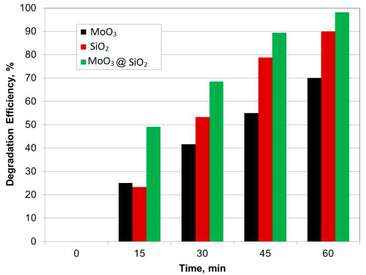
Figure 5 shows that the degradation efficiency increases with exposure time under UV-light. The MoO3@SiO2 composite showed degradation efficiencies in the MB solution close to 100% after 60 min of UV irradiation. The MoO3@SiO2 composite showed stable rates of MB photodegradation up to six cycles.
The rate kinetics analysis, an important parameter in the degradation studies, was performed to predict the rate at which MB is removed from the aqueous solution [ref. 42]. In these experiments, different amounts of MoO3, SiO2, and MoO3@SiO2 composite were used with a fixed concentration of MB. The reaction kinetics was calculated with Equation (8) [ref. 42]:
LnCC0kt
where C0 and C were defined for Equation (7). The graph of the natural logarithm, Ln(C/C0) for MB dye versus time in the presence of MoO3, SiO2, and MoO3@SiO2 nanocomposite is presented in Figure 6.
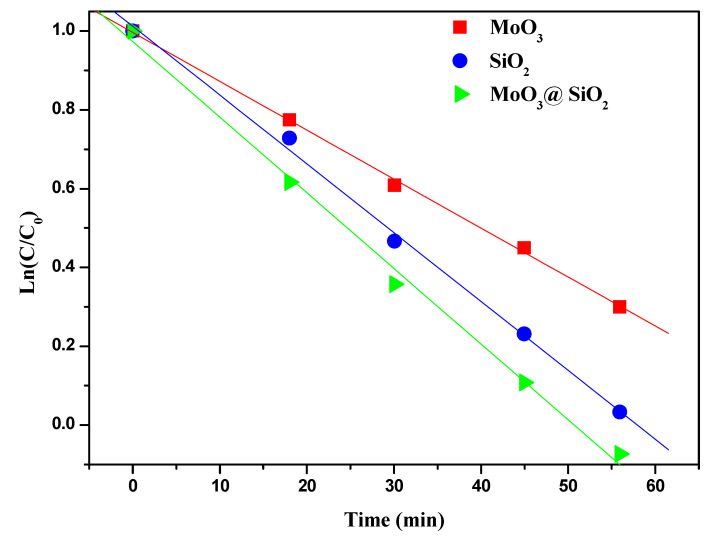
The MB concentration presented in log scale in Figure 6 varies practically linearly with time, indicating that the photodegradation of MB dye follows the first-order kinetics [ref. 42]. The kinetic rate constants (k) were determined from the slope of fitted curves. The first-order degradation rate constants for MoO3, SiO2, and MoO3@SiO2 nanocomposite were 10.3 × 10⁻³ min−1, 15.1 × 10⁻³ min−1, and 16.3 × 10⁻³ min−1, respectively. Table 1 presents the rate constants for MB degradation obtained in this work in comparison to other literature data. The degradation rate of MB is faster for MoO3@SiO2 nanocomposite compared to MoO3 or SiO2.
Table 1: Comparative rate constants for different photocatalysts, including our work.
| Material | Rate Constants × 10−3 min−1 | References |
|---|---|---|
| MoO3 (CBD) | 10.3 | this work |
| SiO2 | 15.1 | this work |
| MoO3@SiO2 | 16.3 | this work |
| MoO3 | 0.334 | [ref. 44] |
| ZnO | 15.15 | [ref. 6] |
| α-Fe2O3 | 2.01 | [ref. 45] |
| SnS2 | 4.43 | [ref. 45] |
| SrFe12O19 | 13.6 | [ref. 46] |
| TiO2 | 35.58 | [ref. 47] |
4. Conclusions
We have synthesized MoO3@SiO2 nanoparticle composite using chemical bath deposition. The diffraction patterns are in good agreement with the hexagonal phase MoO3 with the lattice parameters of a = 10.53 Å and c = 14.876 Å. The SiO2 has the following lattice parameters: a = 25.93 Å, b = 5.01 Å, and c = 18.54 Å. The XRD analysis showed that MoO3, silica, and MoO3@SiO2 nanoparticle composite have crystalline characteristics phase with an average crystallite size of about 94 nm, 32 nm, and 125 nm, respectively. SiO2 showed micro-flakes morphology with agglomeration as confirmed by SEM analysis, irregular rod-shaped for MoO3, and coral-like structure for MoO3@SiO2. The optimum photocatalytic activity was found for MoO3@SiO2 nanoparticles, with an efficiency of about 100% after 60 min of exposure to the UV-light, while the degradation efficiency for the same UV exposure time was about 90% and 70% for SiO2 and MoO3, respectively. The degradation rate constants for MoO3, SiO2, and MoO3@SiO2 nanocomposite were 10.3 × 10⁻³ min−1, 15.1 × 10⁻³ min−1, and 16.3 × 10⁻³ min−1, respectively. These results show that SiO2 particles have a beneficial photocatalytic effect combined with MoO3 in the MoO3@SiO2 composite in the photocatalytic processes.
References
- S. Bolisetty, M. Peydayesh, R. Mezzenga. Sustainable technologies for water purification from heavy metals: Review and analysis. Chem. Soc. Rev., 2019. [DOI | PubMed]
- J.B. DeCoste, G.W. Peterson. Metal–organic frameworks for air purification of toxic chemicals. Chem. Rev., 2014. [DOI | PubMed]
- P. Pichat, J. Disdier, C. Hoang-Van, D. Mas, G. Goutailler, C. Gaysse. Purification/deodorization of indoor air and gaseous effluents by TiO2 photocatalysis. Catal. Today, 2000. [DOI]
- G. Shen, L. Pan, R. Zhang, S. Sun, F. Hou, X. Zhang, J.-J. Zou. Low-spin-state hematite with superior adsorption of anionic contaminations for water purification. Adv. Mater., 2020. [DOI]
- J. Zhao, X. Yang. Photocatalytic oxidation for indoor air purification: A literature review. Build. Environ., 2003. [DOI]
- S. Bhatia, N. Verma. Photocatalytic activity of zno nanoparticles with optimization of defects. Mater. Res. Bull., 2017. [DOI]
- H. Li, P. Wang, X. Yi, H. Yu. Edge-selectively amidated graphene for boosting h2-evolution activity of TiO2 photocatalyst. Appl. Catal. B Environ., 2020. [DOI]
- A.B.D. Nandiyanto, R. Zaen, R. Oktiani. Correlation between crystallite size and photocatalytic performance of micrometer-sized monoclinic WO3 particles. Arab. J. Chem., 2020. [DOI]
- J. Peña-Bahamonde, C. Wu, S.K. Fanourakis, S.M. Louie, J. Bao, D.F. Rodrigues. Oxidation state of Mo affects dissolution and visible-light photocatalytic activity of MoO3 nanostructures. J. Catal., 2020. [DOI]
- R. Mimouni, A. Souissi, A. Madouri, K. Boubaker, M. Amlouk. High photocatalytic efficiency and stability of chromium-indium codoped ZnO thin films under sunlight irradiation for water purification development purposes. Curr. Appl. Phys., 2017. [DOI]
- M. Ponce-Mosso, M. Pérez-González, P.E. García-Tinoco, H. Crotte-Ledesma, M. Morales-Luna, S.A. Tomás. Enhanced photocatalytic activity of amorphous MoO3 thin films deposited by rf reactive magnetron sputtering. Catal. Today, 2020. [DOI]
- G.S. Das, J.P. Shim, A. Bhatnagar, K.M. Tripathi, T. Kim. Biomass-derived carbon quantum dots for visible-light-induced photocatalysis and label-free detection of Fe(iii) and ascorbic acid. Sci. Rep., 2019. [DOI | PubMed]
- D. Anghel, A. Lascu, C. Epuran, I. Fratilescu, C. Ianasi, M. Birdeanu, E. Fagadar-Cosma. Hybrid materials based on silica matrices impregnated with pt-porphyrin or ptnps destined for CO2 gas detection or for wastewaters color removal. Int. J. Mol. Sci., 2020. [DOI]
- J.M. Wan, Z.Z. Wu, H.G. Wang, X.M. Zheng. Visible-light photocatalytic degradation of methylene blue with porphyrin-sensitized TiO2. Adv. Mater. Res., 2012. [DOI]
- E. Gholamrezapor, A. Eslami. Sensitization of magnetic TiO2 with copper(ii) tetrahydroxylphenyl porphyrin for photodegradation of methylene blue by visible led light. J. Mater. Sci. Mater. Electron., 2019. [DOI]
- C.I. Fernandes, S.C. Capelli, P.D. Vaz, C.D. Nunes. Highly selective and recyclable MoO3 nanoparticles in epoxidation catalysis. Appl. Catal. A Gen., 2015. [DOI]
- C. Hanmandlu, C.-Y. Chen, K.M. Boopathi, H.-W. Lin, C.-S. Lai, C.-W. Chu. Bifacial perovskite solar cells featuring semitransparent electrodes. ACS Appl. Mater. Interfaces, 2017. [DOI | PubMed]
- P. Schulz, J.O. Tiepelt, J.A. Christians, I. Levine, E. Edri, E.M. Sanehira, G. Hodes, D. Cahen, A. Kahn. High-work-function molybdenum oxide hole extraction contacts in hybrid organic–inorganic perovskite solar cells. ACS Appl. Mater. Interfaces, 2016. [DOI | PubMed]
- A. Manivel, G.-J. Lee, C.-Y. Chen, J.-H. Chen, S.-H. Ma, T.-L. Horng, J.J. Wu. Synthesis of MoO3 nanoparticles for azo dye degradation by catalytic ozonation. Mater. Res. Bull., 2015. [DOI]
- Y.J. Lee, W.T. Nichols, D.-G. Kim, Y.D. Kim. Chemical vapour transport synthesis and optical characterization of MoO3 thin films. J. Phys. D Appl. Phys., 2009. [DOI]
- F. Liu, S. Shao, X. Guo, Y. Zhao, Z. Xie. Efficient polymer photovoltaic cells using solution-processed MoO3 as anode buffer layer. Sol. Energy Mater. Sol. Cells, 2010. [DOI]
- I. Navas, R. Vinodkumar, K.J. Lethy, A.P. Detty, V. Ganesan, V. Sathe, V.P. Mahadevan Pillai. Growth and characterization of molybdenum oxide nanorods by rf magnetron sputtering and subsequent annealing. J. Phys. D Appl. Phys., 2009. [DOI]
- O. Kamoun, A. Boukhachem, S. Alleg, B. Jeyadevan, M. Amlouk. Physical study of nano-structured MoO3 films codoped with cobalt and nickel in which there is a ferro-diamagnetic transition. J. Alloys Compd., 2018. [DOI]
- O. Kamoun, A. Boukhachem, M. Amlouk, S. Ammar. Physical study of Eu doped MoO3 thin films. J. Alloys Compd., 2016. [DOI]
- O. Kamoun, A. Mami, M.A. Amara, R. Vidu, M. Amlouk. Nanostructured Fe,Co-codoped MoO3 thin films. Micromachines, 2019. [DOI]
- N. Desai, S. Mali. Chemically grown MoO3 nanorods for antibacterial activity study. J. Nanomed. Nanotechnol., 2015. [DOI]
- A. Dhara, G. Hodes, S.K. Sarkar. Two stage chemical bath deposition of MoO3 nanorod films. RSC Adv., 2014. [DOI]
- J.H. Konnert, D.E. Appleman. The crystal structure of low tridymite. Acta Crystallogr., 1978. [DOI]
- R. Tridymite
- R. Edy, G. Huang, Y. Zhao, Y. Guo, J. Zhang, Y. Mei, J. Shi. Influence of reactive surface groups on the deposition of oxides thin film by atomic layer deposition. Surf. Coat. Technol., 2017. [DOI]
- O. Kamoun, A. Boukhachem, C. Mrabet, A. Yumak, P. Petkova, K. Boubaker, M. Amlouk. Effect of europium content on physical properties of In2O3 thin films for sensitivity and optoelectronic applications. Bull. Mater. Sci., 2016. [DOI]
- X.-L. Li, J.-F. Liu, Y.-D. Li. Low-temperature synthesis of large-scale single-crystal molybdenum trioxide (MoO3) nanobelts. Appl. Phys. Lett., 2002. [DOI]
- R. Senthilkumar, G. Anandhababu, T. Mahalingam, G. Ravi. Photoelectrochemical study of MoO3 assorted morphology films formed by thermal evaporation. J. Energy Chem., 2016. [DOI]
- C.V. Ramana, V.V. Atuchin, I.B. Troitskaia, S.A. Gromilov, V.G. Kostrovsky, G.B. Saupe. Low-temperature synthesis of morphology controlled metastable hexagonal molybdenum trioxide (MoO3). Solid State Commun., 2009. [DOI]
- I.M. Joni, L. Nulhakim, M. Vanitha, C. Panatarani. Characteristics of crystalline silica (SiO2) particles prepared by simple solution method using sodium silicate (Na2SiO3) precursor. J. Phys. Conf. Ser., 2018. [DOI]
- P. Wongkrua, T. Thongtem, S. Thongtem. Synthesis of h- and α-MoO3 by refluxing and calcination combination: Phase and morphology transformation, photocatalysis, and photosensitization. J. Nanomater., 2013. [DOI]
- R. Mimouni, B. Askri, T. Larbi, M. Amlouk, A. Meftah. Photocatalytic degradation and photo-generated hydrophilicity of methylene blue over ZnO/ZnCr2O4 nanocomposite under stimulated UV light irradiation. Inorg. Chem. Commun., 2020. [DOI]
- Y. Chen, C. Lu, L. Xu, Y. Ma, W. Hou, J.-J. Zhu. Single-crystalline orthorhombic molybdenum oxide nanobelts: Synthesis and photocatalytic properties. CrystEngComm, 2010. [DOI]
- Y. Ku, Y.-H. Huang, Y.-C. Chou. Preparation and characterization of ZnO/TiO2 for the photocatalytic reduction of Cr(vi) in aqueous solution. J. Mol. Catal. A Chem., 2011. [DOI]
- L.X. Song, J. Xia, Z. Dang, J. Yang, L.B. Wang, J. Chen. Formation, structure and physical properties of a series of α-MoO3 nanocrystals: From 3d to 1d and 2d. CrystEngComm, 2012. [DOI]
- M. Vijay, V. Selvarajan, K.P. Sreekumar, J. Yu, S. Liu, P.V. Ananthapadmanabhan. Characterization and visible light photocatalytic properties of nanocrystalline TiO2 synthesized by reactive plasma processing. Sol. Energy Mater. Sol. Cells, 2009. [DOI]
- K. Mageshwari, S.S. Mali, R. Sathyamoorthy, P.S. Patil. Template-free synthesis of mgo nanoparticles for effective photocatalytic applications. Powder Technol., 2013. [DOI]
- N. Tariq, R. Fatima, S. Zulfiqar, A. Rahman, M.F. Warsi, I. Shakir. Synthesis and characterization of MoO3/CoFe2O4 nanocomposite for photocatalytic applications. Ceram. Int., 2020. [DOI]
- R.B. Anjaneyulu, B.S. Mohan, G.P. Naidu, R. Muralikrishna. Visible light enhanced photocatalytic degradation of methylene blue by ternary nanocomposite, MoO3/Fe2O3/rGO. J. Asian Ceram. Soc., 2018. [DOI]
- S. Balu, K. Uma, G.-T. Pan, T.C.-K. Yang, S.K. Ramaraj. Degradation of methylene blue dye in the presence of visible light using SiO2@α-Fe2O3 nanocomposites deposited on SnS2 flowers. Materials, 2018. [DOI]
- D.D. Mishra, G. Tan. Visible photocatalytic degradation of methylene blue on magnetic SrFe12O19. J. Phys. Chem. Solids, 2018. [DOI]
- Y.-H. Xu, D.-H. Liang, M.-L. Liu, D.-Z. Liu. Preparation and characterization of Cu2O–TiO2: Efficient photocatalytic degradation of methylene blue. Mater. Res. Bull., 2008. [DOI]