
Advances in phytochemical-derived nanotherapeutics for multimodal eradication of multidrug-resistant Staphylococcus aureus
Abstract
The past decade, particularly the post-pandemic period, has intensified the challenge of managing bacterial infections. Multidrug-resistant Staphylococcus aureus (MDR-SA) has emerged as a dominant cause of hospital-acquired infections, creating a sustained public health emergency and underscoring the need for alternative antimicrobial strategies. Plant-derived secondary metabolites have gained attention as promising antibacterial agents; however, their therapeutic potential is constrained by poor solubility, limited target selectivity, low drug-loading capacity, rapid metabolism, and reduced systemic bioavailability. Nanoparticle carriers provide a corrective platform by improving physicochemical stability, enhancing solubility, enabling controlled release, and strengthening pharmacokinetic and pharmacodynamic behavior. Phytochemical-based nanoparticles (phyto-NPs) form a multitarget antibacterial architecture capable of weakening bacterial defense networks through efflux pump interference, disruption of metal-ion homeostasis, and alteration of membrane permeability. These systems also induce reactive oxygen species, leading to DNA damage, protein denaturation, mitochondrial impairment, and peptidoglycan disruption. In parallel, phyto-NPs inhibit biofilm formation and quorum-sensing pathways, reducing virulence and limiting dissemination. Their ability to penetrate the extracellular matrix enhances antibiotic access and restores susceptibility in resistant strains. Recent investigations demonstrate strong activity of phyto-NPs both as independent therapeutics and as synergistic partners to conventional antibiotics. Microenvironment-responsive release, intracellular targeting, and improved delivery efficiency further strengthen their translational relevance. Curcumin-loaded nanosystems disrupt MRSA membranes and impair biofilm formation, while quercetin-loaded liposomes penetrate S. aureus biofilms more effectively than free quercetin. These examples illustrate the capacity of nanoscale engineering to overcome the pharmacological constraints of phytochemicals. This review examines recent advances in phyto-NP strategies targeting MDR-SA, with emphasis on phytochemical selection, nanoscale design principles, and the multifunctional antibacterial mechanisms underpinning next-generation antimicrobial development.
Article type: Review Article
Keywords: antimicrobial resistance, multidrug-resistant, next-generation antimicrobial therapies, pharmacokinetics, phytochemical-based nanoparticles, phytometabolites
Affiliations: Department of Crop Sciences and Agroforestry, Faculty of Tropical AgriSciences, Czech University of Life Sciences Prague, Prague, Czechia; Department of Economic Theories, Faculty of Economics and Management, Czech University of Life Sciences Prague, Prague, Czechia; Department of Agroecology and Crop Production, Faculty of Agrobiology, Food and Natural Resources, Czech University of Life Sciences Prague, Prague, Czechia; Department of Health Sciences, University of Basilicata, Potenza, Italy; Centre for Natural Products Discovery, School of Pharmacy and Biomolecular Sciences, Liverpool John Moores University, Liverpool, United Kingdom; Laboratory of Growth Regulators, Palacký University and Institute of Experimental Botany, The Czech Academy of Sciences, Olomouc, Czechia
License: Copyright © 2026 Gupta, Bhattacharya, Lela, Dutta, Faraone, Fernández-Cusimamani, Sarker, Milella, Nahar and Leuner. CC BY 4.0 This is an open-access article distributed under the terms of the Creative Commons Attribution License (CC BY). The use, distribution or reproduction in other forums is permitted, provided the original author(s) and the copyright owner(s) are credited and that the original publication in this journal is cited, in accordance with accepted academic practice. No use, distribution or reproduction is permitted which does not comply with these terms.
Article links: DOI: 10.3389/fphar.2026.1866492 | PubMed: 42344795 | PMC: PMC13290124
Relevance: Moderate: mentioned 3+ times in text
Full text: PDF (4.7 MB)
Introduction
The global rise of multidrug-resistant bacterial infections has created a sustained threat to healthcare systems and reduced the effectiveness of frontline antibiotics. Multidrug-resistant Staphylococcus aureus (MDR-SA) has emerged as a major contributor to this burden due to its ability to accumulate resistance determinants and cause severe infections ranging from pneumonia to bacteraemia (ref. Sultan et al., 2018; ref. Murray et al., 2022; ref. Abebe and Birhanu, 2023). Increasing resistance to methicillin, vancomycin, tetracycline, and linezolid has further complicated clinical management and intensified the need for alternative therapeutic strategies (ref. Jones et al., 2007; ref. Lee et al., 2014). Additional resistance to fluoroquinolones, macrolides, aminoglycosides, and glycopeptides has reduced the effectiveness of conventional antibiotics and increased the burden of difficult-to-treat infections (ref. Khameneh et al., 2019; ref. Makarewicz et al., 2021; ref. Díaz-Puertas et al., 2023).
The clinical prominence of MRSA reflects its capacity to combine extensive resistance with high virulence potential. Disease manifestations range from superficial skin infections to invasive conditions such as osteomyelitis, endocarditis, and necrotizing pneumonia, contributing to significant morbidity and mortality across healthcare settings (ref. Abebe and Birhanu, 2023; ref. Murray et al., 2022). Additional phenotypes, including vancomycin-resistant S. aureus (VRSA) and linezolid-resistant strains, further restrict therapeutic options and increase the complexity of treatment decisions (ref. Jones et al., 2007; ref. Lee et al., 2014; ref. Sultan et al., 2018).
The success of S. aureus as a resistant pathogen stems from a diverse repertoire of molecular defense mechanisms. mecA-mediated β-lactam resistance, enzymatic detoxification pathways, and activation of multidrug efflux pumps such as NorA and MepA collectively strengthen bacterial survival under antibiotic pressure (ref. Vestergaard et al., 2019; ref. Abebe and Birhanu, 2023). Additional mechanisms include structural modification of cell-wall components, regulation of intracellular metal homeostasis, and induction of the SOS stress response, each contributing to enhanced adaptability and persistence (ref. Vestergaard et al., 2019; ref. Zhao et al., 2020; ref. Douglas, 2022). Biofilm formation provides further protection by limiting antibiotic penetration and enabling long-term colonization on host tissues and medical devices (ref. Monte et al., 2014; ref. Yadav et al., 2015; ref. Bogdanova et al., 2018).
Plant-derived phytochemicals have gained increasing attention as antibacterial agents capable of countering MDR-SA through structurally diverse and mechanistically distinct pathways. Major classes such as alkaloids, polyphenols, terpenoids, coumarins, and cannabinoids demonstrate activity against resistant strains through membrane disruption, efflux-pump inhibition, toxin suppression, and interference with quorum-sensing networks (ref. Appendino et al., 2008; ref. Miklasińska-Majdanik et al., 2018; ref. Khameneh et al., 2021). Representative compounds including berberine, quercetin, curcumin, caffeic acid, and sabinene exhibit inhibitory effects across multiple MDR-SA lineages, highlighting the therapeutic potential of phytochemical scaffolds (ref. Chung et al., 2014; ref. Hua et al., 2018; ref. Sinsinwar and Vadivel, 2020). Their ability to act on multiple bacterial systems reduces the likelihood of rapid resistance development and broadens their therapeutic relevance.
Despite their mechanistic diversity, phytochemicals face significant translational barriers. Poor aqueous solubility, rapid metabolic degradation, and low systemic bioavailability limit therapeutic performance and restrict clinical applicability (ref. Huang et al., 2019; ref. Makarewicz et al., 2021; ref. Paul-Chima et al., 2024). Variability in plant sources and extraction methods introduces challenges in standardization and reproducibility, while high concentrations required for antibacterial activity raise concerns regarding toxicity and off-target effects (ref. Khameneh et al., 2019; ref. Kongkham et al., 2020; ref. Patil et al., 2023).
Nanotechnology-based delivery systems provide a rational framework for overcoming these limitations by encapsulating phytochemicals within engineered nanocarriers. Improved solubility, enhanced stability, controlled release, and targeted delivery have been demonstrated across liposomes, polymeric nanoparticles, metallic nanoparticles, and solid-lipid nanoparticles (ref. Jeetah et al., 2014; ref. Naseri et al., 2015; ref. Cano et al., 2020). Stimuli-responsive nanocarriers, including pH-sensitive and enzyme-responsive systems, enable selective release within infected microenvironments and improve clearance of intracellular MRSA (ref. Mohammed et al., 2023; ref. Zou et al., 2023; ref. Ali Khan et al., 2024). Preclinical studies, including curcumin-loaded nanoparticles and quercetin-loaded liposomes, demonstrate enhanced membrane disruption, improved biofilm penetration, and superior bactericidal activity compared with free compounds (ref. Cui et al., 2016; ref. Kumar et al., 2022; ref. Zhang et al., 2023). This review integrates phytochemical classes, nanocarrier design considerations, and antibacterial mechanisms to frame how phyto-nanotherapeutic strategies can address persistent resistance challenges in MDR-SA infections.
Phytochemicals: Promising antibacterial agents against MDR S. aureus
Phytochemicals have emerged as promising antibacterial agents in response to the growing limitations of conventional antibiotics against multidrug-resistant bacterial pathogens. Their structural diversity and capacity to act on multiple bacterial targets provide mechanistic advantages over single-target antibiotics, particularly in the context of MDR-SA. Unlike traditional antimicrobials, phytochemicals interfere with bacterial physiology through a broad range of molecular mechanisms, thereby reducing susceptibility to rapid resistance development (ref. Khameneh et al., 2021).
The antibacterial activities of phytochemicals encompass disruption of bacterial cell walls and membranes, leading to increased permeability and leakage of intracellular contents. Several compounds impair protein synthesis, interfere with nucleic acid replication, induce DNA damage, and promote oxidative stress through reactive oxygen species generation. Phytochemicals also suppress quorum-sensing and virulence-associated signaling pathways, thereby attenuating toxin production, adhesion, and biofilm formation in MDR-SA strains (ref. Khameneh et al., 2016; ref. Fazly Bazzaz et al., 2018; ref. Fatemi et al., 2020).
A defining feature of phytochemicals is their complex chemical architecture, which limits bacterial enzymatic degradation and reduces the efficiency of resistance acquisition. Many phytochemicals retain activity against resistant strains by bypassing classical resistance pathways, including altered penicillin-binding proteins and target-modification mechanisms. In addition, several phytochemicals function as resistance-modifying agents by inhibiting efflux pumps and restoring bacterial susceptibility to co-administered antibiotics (ref. Miklasińska-Majdanik et al., 2018; ref. Khameneh et al., 2021).
Beyond antibacterial efficacy, phytochemicals have demonstrated synergistic interactions with conventional antibiotics, enhancing bactericidal effects and reducing required antibiotic dosages. These interactions are particularly relevant in MDR-SA infections, where efflux-pump inhibition, membrane destabilization, and biofilm disruption collectively improve drug penetration and intracellular accumulation (ref. Khameneh et al., 2019; ref. Kongkham et al., 2020). Certain phytochemicals, including capsaicin, codeine, colchicine, and paclitaxel, have received approval from the U.S. Food and Drug Administration for various therapeutic applications, underscoring their clinical relevance (ref. Kongkham et al., 2020).
Despite these advantages, the clinical translation of phytochemicals remains constrained by pharmacokinetic limitations. Poor aqueous solubility, chemical instability, rapid metabolic degradation, and limited bioavailability compromise systemic exposure and therapeutic consistency. Variability in plant sources, extraction procedures, and compound composition further complicates standardization and reproducibility across studies (ref. Huang et al., 2019; ref. Makarewicz et al., 2021; ref. Paul-Chima et al., 2024). High concentrations required for antibacterial activity raise concerns regarding toxicity and off-target effects, reinforcing the need for improved delivery strategies (ref. Khameneh et al., 2019; ref. Kongkham et al., 2020; ref. Patil et al., 2023).
Advances in high-throughput screening, analytical chemistry, and phytochemical classification have accelerated the identification of bioactive compounds and clarified their structure-activity relationships. Structural classification into alkaloids, phenolic compounds, terpenoids, coumarins, and cannabinoids provides a mechanistic framework for understanding antibacterial activity and guiding rational therapeutic development. Each phytochemical class exhibits distinct physicochemical properties and antibacterial mechanisms relevant to MDR-SA infections, as outlined in the following subsections and accompanying structural representation.
Alkaloids
Alkaloids are heterocyclic nitrogen-containing compounds with structurally diverse frameworks and significant antimicrobial activity. Their ability to act as hydrogen-bond donors or acceptors contributes to strong target-ligand interactions and underpins their broad antibacterial potential against MDR-SA (ref. Kittakoop et al., 2014; ref. Kumar et al., 2022; ref. Díaz-Puertas et al., 2023). Many alkaloids function as efflux-pump inhibitors (EPIs), targeting transport systems within the major facilitator superfamily and resistance-nodulation-division families.
Reserpine reverses Bmr-mediated multidrug resistance and inhibits drug transport in resistant strains (ref. Gibbons and Udo, 2000; ref. Wink et al., 2012; ref. Díaz-Puertas et al., 2023). Berberine suppresses the MexXY-OprM efflux system in imipenem-resistant bacteria and demonstrates potent activity against MRSA, with reported MIC values of 32–128 mg/L and 90% growth inhibition at concentrations ≤64 mg/L (ref. Yu et al., 2005; ref. Laudadio et al., 2019; ref. Díaz-Puertas et al., 2023). Structural diversity within alkaloid scaffolds has inspired the synthesis of derivatives incorporating indole, isoindole, imidazole, and oxazolidinone motifs, many of which exhibit broad-spectrum antibacterial activity (ref. Kittakoop et al., 2014; ref. Kumar et al., 2022; ref. Díaz-Puertas et al., 2023).
Canthin-6-one isolated from Allium neapolitanum inhibits MRSA and tetracycline-resistant S. aureus (TRSA) with MIC values of 8 mg/L and 32 mg/L, respectively (ref. O’Donnell and Gibbons, 2007; ref. Kumar et al., 2022; ref. Díaz-Puertas et al., 2023). Piperine, derived from Piper nigrum, exhibits anti-MRSA activity at 100 mg/L and enhances gentamicin efficacy through efflux-pump interference (ref. Mohtar et al., 2009; ref. Khameneh et al., 2015; ref. Díaz-Puertas et al., 2023). Harmaline isolated from Peganum harmala reduces MRSA MIC values by four-to eight-fold, with effective concentrations ranging from 1.95 to 62.5 mg/L (ref. Mohtar et al., 2009; ref. Kumar et al., 2022; ref. Díaz-Puertas et al., 2023). Evocarpine from Tetradium ruticarpum enhances oxacillin efficacy by sixteen-fold and demonstrates an MIC of 8 mg/L (ref. Pan et al., 2014; ref. Kumar et al., 2022; ref. Díaz-Puertas et al., 2023).
Indirubin isolated from Isatis tinctoria exhibits activity against ciprofloxacin-resistant S. aureus with a reported MIC of 12.5 mg/L (ref. Ponnusamy et al., 2010; ref. Kumar et al., 2022; ref. Díaz-Puertas et al., 2023). Additional alkaloids with significant activity against MRSA and TRSA include sanguinarine, tetrandrine, tryptanthrin, and galegine (ref. Williams et al., 2010; ref. Coqueiro et al., 2014; ref. Costa et al., 2017). Indole-3-carbinol derived from cruciferous vegetables also demonstrates antibacterial activity against MDR-SA and enhances the efficacy of other antimicrobial agents (ref. Monte et al., 2014; ref. Kumar et al., 2022; ref. Díaz-Puertas et al., 2023).
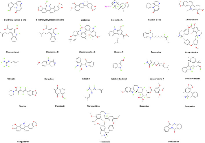
The structural diversity of alkaloid scaffolds underpins their broad antibacterial activity against MDR-SA. Variations in heterocyclic ring systems, nitrogen positioning, and functional-group composition influence membrane interaction, efflux-pump inhibition, and bacterial target binding. Representative alkaloid structures with reported anti-MRSA activity are illustrated to highlight chemical features associated with antibacterial efficacy (ref. Kittakoop et al., 2014; ref. Díaz-Puertas et al., 2023). Figure 1 outlines representative alkaloid scaffolds with reported anti-MRSA activity, illustrating structural features associated with antibacterial efficacy.
Phenolic compounds and polyphenols
Phenolic compounds are characterized by hydroxyl groups attached to aromatic rings and represent a structurally diverse class of plant-derived metabolites with significant pharmacological activity against multidrug-resistant microorganisms. This group includes flavonoids, non-flavonoids, phenolic acids, and tannins, each contributing distinct chemical scaffolds and antibacterial mechanisms relevant to MDR-SA (ref. Souza et al., 2019). Antibacterial activity arises from multiple interactions with bacterial systems, including disruption of membrane integrity, inhibition of virulence-associated enzymes and toxins, and suppression of biofilm formation, thereby positioning phenolics as promising natural antimicrobial agents (ref. Miklasińska-Majdanik et al., 2018; ref. Makarewicz et al., 2021).
Aspidinol isolated from Dryopteris fragrans demonstrates potent anti-MRSA activity, with MIC values ranging from 0.5 to 2 mg/L, and significantly improves survival outcomes in MRSA-infected murine models, achieving an 80% survival rate at a dose of 25 mg/kg (ref. Hua et al., 2018). Galangin derived from Alpinia officinarum exhibits antibacterial activity against penicillin-resistant S. aureus (PRSA) with reported MIC values of 100–300 mg/L (ref. Eumkeb et al., 2010). Hyperforin demonstrates strong activity against both MRSA and EMRSA at concentrations in the range of 0.5–2 mg/L (ref. Schiavone et al., 2013). α-Mangostin from Garcinia mangostana also exhibits antibacterial efficacy, with MIC values between 1.57 and 12.5 mg/L against MRSA strains (ref. Iinuma et al., 1996).
Isovalerylshikonin isolated from Arnebia euchroma suppresses antimicrobial resistance by targeting the MsrA efflux pump in MRSA, thereby enhancing intracellular drug retention (ref. He et al., 2019). Rutin, when combined with carbon dots, exhibits antibacterial activity with an MIC of 32 mg/L against MRSA, highlighting the potential of phenolic compounds within hybrid nanoscale systems (ref. Lang et al., 2024).
Additional phenolic compounds demonstrate broad antibacterial activity against MRSA, including gallic acid, catechin, quercetin, caffeic acid, eugenol, kaempferol, and epigallocatechin gallate (ref. Xu and Lee, 2001; ref. Luís et al., 2014; ref. Yadav et al., 2015; ref. Randhawa et al., 2016; ref. Sinsinwar and Vadivel, 2020; ref. Knidel et al., 2021). Baicalein exhibits activity against ciprofloxacin-resistant S. aureus, with MIC values ranging from 64 to 256 mg/L (ref. Chan et al., 2011). Curcumin, glabridin, humulone, salicylic acid, and tannic acid demonstrate antibacterial effects across multiple MDR-SA strains, acting through combined mechanisms that include membrane disruption, metabolic interference, efflux modulation, and virulence suppression (ref. Tsuchiya et al., 1996; ref. Monte et al., 2014; ref. Bogdanova et al., 2018; ref. Kırmusaoğlu, 2019; ref. Pinheiro et al., 2022). Figure 2 displays representative phenolic and polyphenolic structures with documented anti-MRSA activity, illustrating chemical motifs linked to antibacterial potency.
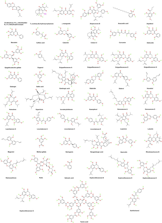
The structural diversity and multitarget mechanisms of phenolic compounds highlight their potential as effective antibacterial agents against MDR-SA. Their ability to disrupt membranes, inhibit virulence pathways, suppress efflux activity, and interfere with biofilm formation provides a strong mechanistic foundation for phenolic-based therapeutic strategies. The chemical diversity of phenolic and polyphenolic scaffolds contributes directly to variation in antibacterial potency and mechanism of action. Differences in hydroxylation patterns, ring conjugation, and molecular size influence membrane affinity, redox activity, and virulence modulation. Representative phenolic compounds with reported anti-MRSA activity are illustrated to highlight structural features associated with antibacterial efficacy (ref. Makarewicz et al., 2021; ref. Díaz-Puertas et al., 2023).
Terpenoids
Terpenoids constitute a major class of bioactive natural products derived from isoprene units and encompass monoterpenes, sesquiterpenes, diterpenes, and related derivatives. Their extensive structural diversity and frequent incorporation of oxygenated functional groups contribute to broad antibacterial activity against multidrug-resistant pathogens. The lipophilic nature of terpenoids facilitates interaction with microbial cell envelopes, resulting in membrane destabilization, increased permeability, and loss of cytoplasmic integrity (ref. Trindade et al., 2015; ref. Sil et al., 2020; ref. Sharma et al., 2020; ref. Musayeib et al., 2022). Antibacterial mechanisms include membrane disruption, inhibition of essential enzymes, interference with protein synthesis, induction of reactive oxygen species, and modulation of quorum-sensing and adhesion pathways (ref. Mahizan et al., 2019; ref. Adoui and Lahouel, 2019).
Guaianolide, a sesquiterpene lactone isolated from Artemisia gilvescens, demonstrates potent anti-MRSA activity with a reported MIC of 1.95 mg/L (ref. Elmaidomy et al., 2022). Horminone, a diterpene quinone derived from Salvia deserta, exhibits antibacterial activity against MRSA with MIC values ranging from 7.81 to 15.63 mg/L (ref. Gaspar-Marques et al., 2006). Mansonone F, a sesquiterpene isolated from Ulmus davidiana var. japonica, exhibits antibacterial activity against nineteen MRSA strains, with a reported MIC of 2 mg/mL (ref. Park et al., 2018). In comparison, totarol from Podocarpus nagi demonstrates strong antibacterial activity against MRSA strains, with MIC values ranging from 1.56 to 3.12 mg/L (ref. Yu et al., 2015).
Additional terpenoid-derived compounds with activity against MRSA include 18β-glycyrrhetinic acid, 6-O-isobutyroylplenolin, betulin, betulinaldehyde, betulinic acid, dehydroepingaione, dehydroleucodine, dehydromyoporone, lupeol, xanthatin, zerumbol, and α-amyrin (ref. Sato et al., 1997; ref. Taylor and Towers, 1998; ref. Ordóñez et al., 2011; ref. Chung et al., 2014; ref. Wang et al., 2015; ref. Wang et al., 2016; ref. Long et al., 2013; ref. Dong et al., 2018; ref. Siddique et al., 2019). Compounds such as dehydroabietic acid, isopimaric acid, and sabinene also demonstrate antibacterial activity against MDR-SA strains (ref. Smith et al., 2005; ref. Leandro et al., 2014; ref. Matias et al., 2016).
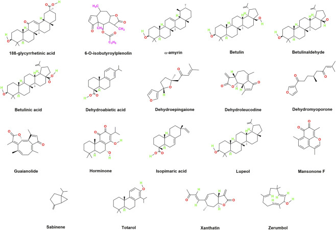
The structural diversity and multitarget mechanisms of terpenoids highlight their potential as broad-spectrum antibacterial agents against resistant S. aureus. Their demonstrated efficacy across multiple MRSA lineages supports continued investigation into terpenoid-based therapeutic applications. Structural variation across terpenoid scaffolds contributes directly to differences in antibacterial potency and mechanism of action. Modulation of ring systems, degree of oxidation, and substituent positioning influences membrane affinity, intracellular target engagement, and oxidative-stress induction. Representative terpenoid structures with reported anti-MRSA activity are illustrated to highlight chemical features associated with antibacterial efficacy (ref. Mahizan et al., 2019; ref. Musayeib et al., 2022). Figure 3 presents representative terpenoid scaffolds with reported activity against MDR-SA, illustrating structural elements associated with antibacterial mechanisms.
Coumarins
Coumarins form a prominent class of heterocyclic benzopyrone derivatives widely distributed in plant species and recognized for diverse pharmacological activities, including antibacterial, antiviral, antioxidant, anti-inflammatory, and antitumor effects. Their antibacterial activity is primarily attributed to inhibition of bacterial DNA gyrase, a critical enzyme involved in DNA supercoiling, replication, and cell division (ref. Garg et al., 2020). This mechanism confers broad activity against MDR-SA strains.
A highly active coumarin, 5,7-dihydroxy-8-(2-methylbutanoyl)-6-(3-methylbut-2-enyl)-4-phenyl-2H-chromen-2-one, isolated from Mesua ferrea, demonstrates potent antibacterial activity against methicillin-resistant S. aureus (MRSA) and fluoroquinolone-resistant S. aureus (FRSA), with reported MIC values of 6.25 mg/L and 12.5 mg/L, respectively (ref. Roy et al., 2013). Additional coumarins, including 5-geranyloxy-7-methoxycoumarin, artanin, isopimpinellin, and phellopterin, exhibit notable antibacterial activity against MRSA strains such as MRSA098, with MIC values ranging from 8 to 64 mg/L (ref. Zuo et al., 2016). These compounds exert antibacterial effects through combined mechanisms that include DNA gyrase inhibition, membrane destabilization, and interference with resistance-associated pathways.
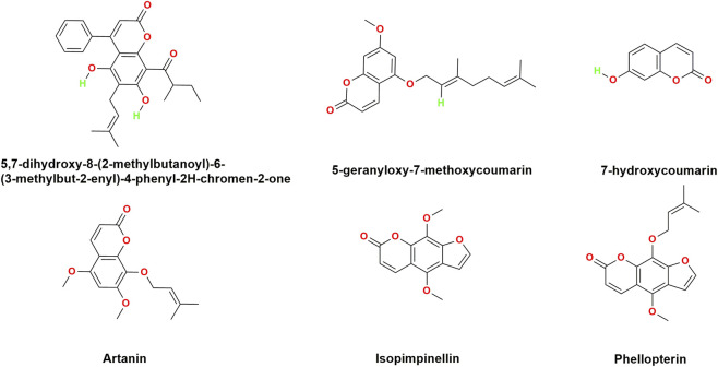
The structural diversity and mechanistic breadth of coumarins highlight their potential as natural antibacterial agents against MDR-SA. Their demonstrated efficacy against resistant S. aureus lineages supports continued investigation into coumarin-based therapeutic strategies. Variation within coumarin scaffolds influences antibacterial potency and spectrum of activity. Substituent position, degree of prenylation, and ring functionalization modulate target interaction, membrane affinity, and enzyme inhibition. Representative coumarin structures with reported antibacterial activity against MDR-SA are presented to illustrate structure-activity relationships relevant to antimicrobial efficacy (ref. Zuo et al., 2016; ref. Garg et al., 2020). Figure 4 outlines representative coumarin structures with anti-MRSA activity, highlighting substituent patterns relevant to antibacterial performance.
Cannabinoids
Cannabinoids comprise a structurally diverse group of bioactive compounds that include phytocannabinoids, endocannabinoids, and synthetic analogues. These molecules are well recognized for psychoactive, anticonvulsive, analgesic, and neuroprotective properties, mediated through cannabinoid receptor-dependent pathways involving CB1 and CB2, as well as receptor-independent mechanisms. Beyond neuromodulatory effects, several cannabinoids exhibit antibacterial activity against MDR-SA, expanding their therapeutic relevance beyond neurological applications (ref. Appendino et al., 2008).
Cannabichromene, cannabidiol, cannabigerol, and cannabinol isolated from Cannabis sativa L. demonstrate strong antibacterial activity against multiple MRSA strains. Reported minimum inhibitory concentration values range from 1 to 2 mg/L for cannabichromene, 0.5–1 mg/L for cannabidiol, 1–2 mg/L for cannabigerol, and approximately 1 mg/L for cannabinol (ref. Appendino et al., 2008). These findings indicate substantial inhibitory capacity across structurally distinct phytocannabinoids and highlight their potential as candidates for the development of alternative therapeutic strategies targeting resistant S. aureus infections.
The demonstrated antibacterial activity of cannabinoids reinforces their relevance as natural antimicrobial agents against MDR-SA. Their efficacy across multiple resistant strains supports continued investigation into cannabinoid-bacteria interactions, structure-activity relationships, and potential integration into novel antimicrobial platforms (ref. Appendino et al., 2008). Structural variation among cannabinoid scaffolds contributes to differences in antibacterial potency and spectrum of activity. Modulation of aromatic substitution patterns, alkyl side-chain length, and hydroxyl-group distribution influences lipophilicity, membrane interaction, and bacterial target engagement. Representative cannabinoid structures with reported anti-MRSA activity are illustrated to highlight chemical features associated with antibacterial efficacy (ref. Appendino et al., 2008). Figure 5 showcases representative cannabinoid scaffolds with reported anti-MRSA activity, emphasizing structural determinants of antibacterial efficacy. Table 1 summarises phytochemicals with reported antibacterial activity against MDR-SA, grouped by chemical class and supported by MIC values.
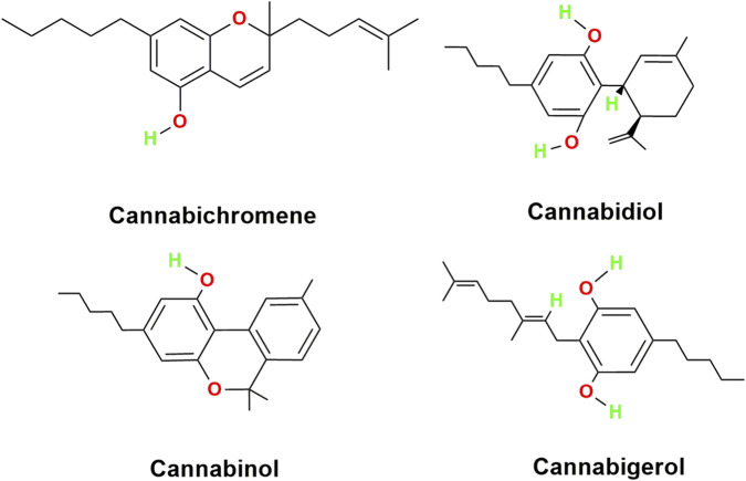
TABLE 1: Phytochemicals showing potential antibacterial efficacy against MDR-SA.
| Chemical class | Phytochemical | Bacterial species | MIC value | References |
|---|---|---|---|---|
| Alkaloids | 8-Hydroxy-canthin-6-one | TetRSA and MRSA | 8 and 64 mg/L, respectively | ref. O’Donnell and Gibbons (2007) |
| 8-Hydroxydihydrosanguinarine | MRSA | 1.95mg/L | ref. Zuo et al. (2008) | |
| Berberine | MRSA | 32–128mg/L | ref. Yu et al. (2005) | |
| Caesanine A | MRSA | 12.5mg/L | ref. Zhang et al. (2013) | |
| Canthin-6-one | TetRSA and MRSA | 8 and 32mg/L, respectively | ref. O’Donnell and Gibbons (2007) | |
| Chelerythrine | MRSA | 2–4mg/L | ref. Wang et al. (2021) | |
| Clausamine A | MRSA | 8mg/L | ref. Maneerat et al. (2012) | |
| Clausamine B | MRSA | 0.25mg/L | ref. Maneerat et al. (2012) | |
| Clausenawalline E | MRSA | 8mg/L | ref. Maneerat et al. (2012) | |
| Clausine F | MRSA | 4mg/L | ref. Maneerat et al. (2012) | |
| Evocarpine | MRSA strain 33591 | 8mg/L | ref. Ponnusamy et al. (2010) | |
| Fangchinoline | MRSA strain 13366 | 160mg/L | ref. Fu et al. (2017) | |
| Galegine | Epidemic-MRSA-15 strain | 4 mg/L | ref. Coqueiro et al. (2014) | |
| Harmaline | Two clinical strains of MRSA | MIC at 1.95 and 62.5mg/L, respectively | ref. Mohtar et al. (2009) | |
| Indirubin | Ciprofloxacin-resistant S. aureus | 12.5mg/L | ref. Ponnusamy et al. (2010) | |
| Indole-3-carbinol | MDR strain of S. aureus | 400mg/L | ref. Monte et al. (2014) | |
| Myoporumine A | MRSA | 6.25mg/L | ref. Dong et al. (2018) | |
| Pentacyclindole | MRSA-105 strain | 8mg/L | ref. Lopez and Osada (2020) | |
| Piperine | MRSA | 100mg/L | ref. Khameneh et al. (2015) | |
| Plumbagin | MRSA | 4–8mg/L | ref. Periasamy et al. (2019) | |
| Pterogynidine | Epidemic-MRSA-15 strain | 4mg/L | ref. Coqueiro et al. (2014) | |
| Reserpine | MRSA | 100mg/L | ref. Randhawa et al. (2016) | |
| Roemerine | MRSA | 32–64mg/L | ref. Yin et al. (2015) | |
| Sanguinarine | MRSA | 3.12–6.25mg/L | ref. Obiang-Obounou et al. (2011) | |
| Tetrandrine | MRSA strain 13366 | 80mg/L | ref. Fu et al. (2017) | |
| Tryptanthrin | MRSA strain BMB9393 | 62.5mg/L | ref. Costa et al. (2017) | |
| Cannabinoids | Cannabichromene | MRSA XU212 strain | 1–2mg/L | ref. Appendino et al. (2008) |
| Cannabidiol | MRSA XU212 strain | 0.5–1mg/L | ref. Appendino et al. (2008) | |
| Cannabigerol | MRSA XU212 strain | 1–2mg/L | ref. Appendino et al. (2008) | |
| Cannabinol | MRSA XU212 strain | 1mg/L | ref. Appendino et al. (2008) | |
| Coumarin | 5,7-dihydroxy-8-(2-methylbutanoyl)-6-(3-methylbut-2-enyl)-4-phenyl-2H-chromen-2-one | Fluoroquinolone-resistant mutant of SA-1199 strain and MRSA strain 831 | 12.5 and 6.25mg/L, respectively | ref. Roy et al. (2013) |
| 5-geranyloxy-7-methoxycoumarin | MRSA | 8–64mg/L | ref. Zuo et al. (2016) | |
| | 7-hydroxycoumarin | MDR strain of S. aureus | 200mg/L | ref. Monte et al. (2014) |
| Artanin | MRSA098 | 8–64mg/L | ref. Zuo et al. (2016) | |
| Isopimpinellin | MRSA | 32–64mg/L | ref. Zuo et al. (2016) | |
| Phellopterin | MRSA | 8–32mg/L | ref. Zuo et al. (2016) | |
| Phenols | 3,9-Dihyroxy- 10-γ, γ-dimethylallyl- 6α,11α- dehydropterocarpan | MRSA G47 strain | 12.5mg/L | ref. Tanaka et al. (2004) |
| 7α-acetoxy-6β-hydroxyroyleanone | MRSA | 3.91–15.63mg/mL | ref. Gaspar-Marques et al. (2006) | |
| Alopecurone B | MRSA | 3.13–6.25mg/L | ref. Sato et al. (1995) | |
| Anacardic acid | MRSA | >100μmol/L | ref. Saedtler et al. (2020) | |
| Aspidinol | Clinical isolate of MRSA strain and MRSA (in vivo) | 0.5–2mg/L. An in vivo study of aspidinol against an MRSA strain in mice found that 80% of mice survived for 5 days at a dose of 25 mg/kg | ref. Hua et al. (2018) | |
| Baicalein | Ciprofloxacin-resistant S. aureus | 64–256mg/L | ref. Chan et al. (2011) | |
| Caffeic acid | MRSA | 62.5–250mg/L | ref. Luís et al. (2014) | |
| Catechin | MRSA | 78.1mg/L | ref. Sinsinwar and Vadivel (2020) | |
| Coleon U | MRSA | 0.98–31.25mg/mL | ref. Gaspar-Marques et al. (2006) | |
| Curcumin | MDR strains of S. aureus | 125–250mg/L | ref. Mun et al. (2013) | |
| Datiscetin | MRSA strain 9247922 | 512mg/L | ref. Xu and Lee (2001) | |
| Epigallocatechin gallate | MRSA | 7.81–62.5mg/L | ref. Knidel et al. (2021) | |
| Eugenol | MRSA (in vitro and in vivo) | 0.1%–0.4%. In vivo analysis showed that sub-MIC eugenol inhibited growth by 88% in the rat middle ear | ref. Yadav et al. (2015) | |
| Exiguaflavanone A | MRSA | 6.25mg/L | ref. Tsuchiya et al. (1996) | |
| Exiguaflavanone C | MRSA | 12.5mg/L | ref. Tsuchiya et al. (1996) | |
| Exiguaflavanone D | MRSA | 3.13–6.25mg/L | ref. Tsuchiya et al. (1996) | |
| Exiguaflavanone G | MRSA | 12.5mg/L | ref. Tsuchiya et al. (1996) | |
| Galangin | Penicillin-resistant S. aureus | 100–300mg/L | ref. Eumkeb et al. (2010) | |
| Gallic acid | MRSA | 4mg/L | ref. Luís et al. (2014) | |
| Gambogic acid | MRSA strains | 0.5–4mg/L | ref. Hua et al. (2019) | |
| Glabridin | MDR strain of S. aureus | 3.12–25mg/L | ref. Singh et al. (2015) | |
| Glabrol | MRSA strain T144 | 2mg/L | ref. Wu et al. (2019) | |
| Honokiol | MRSA | 16–32mg/L | ref. Zuo et al. (2015) | |
| Humulone | MDR strain of S. aureus | 15mg/L | ref. Bogdanova et al. (2018) | |
| Hyperforin | Clinical MRSA strains XU212 and Ig5 and EMRSA-15 strain | 0.5–2mg/L | ref. Schiavone et al. (2013) | |
| Isovalerylshikonin | MRSA strain RN4220 | 16mg/L | ref. He et al. (2019) | |
| Kaempferol | Fluoroquinolone-resistant S. aureus and MRSA | 250mg/L and 125mg/L, respectively | ref. Randhawa et al. (2016) | |
| Kenusanone A | MRSA | 6.25–12.5mg/L | ref. Tsuchiya et al. (1996) | |
| | Kenusanone D | MRSA | 3.13–12.5mg/L | ref. Tsuchiya et al. (1996) |
| Leachianone G | MRSA | 12.5mg/L | ref. Tsuchiya et al. (1996) | |
| Licochalcone A | MRSA strain T144 | 4mg/L | ref. Wu et al. (2019) | |
| Licochalcone C | MRSA strain T144 | 4mg/L | ref. Wu et al. (2019) | |
| Licochalcone E | MRSA strain T144 | T4mg/L | ref. Wu et al. (2019) | |
| Lupulone | MRSA | 0.5mg/L | ref. Bogdanova et al. (2018) | |
| Luteolin | MRSA strain 9247922 | 512mg/L | ref. Xu and Lee (2001) | |
| Magnolol | MRSA | 8–64mg/L | ref. Zuo et al. (2015) | |
| Methyl gallate | MRSA strain ATCC33591 | 250mg/L | ref. Chew et al. (2018) | |
| Naringenin | MRSA | 200–400mg/L | ref. Tsuchiya et al. (1996) | |
| Neogambogic acid | MRSA ATCC33591 strain | 0.5–4mg/L | ref. Hua et al. (2019) | |
| Quercetin | MRSA strain 9247922 | 256mg/L | ref. Xu and Lee (2001) | |
| Rhodomyrtosone B | MRSA (In vitro and In vivo) | 0.62–1.25mg/L. An in vivo study in mice showed that it attenuated skin ulceration in a murine model of MRSA infection at a single dose of 40µg per mouse | ref. Zhao et al. (2019) | |
| Rubraxanthone | MRSA | 0.31–1.25mg/L | ref. Iinuma et al. (1996) | |
| Rutin | MRSA | 32mg/L | ref. Lang et al. (2024) | |
| Salicylic acid | MDR strain of S. aureus | 1600mg/L | ref. Monte et al. (2014) | |
| Sophoraflavanone B | MRSA | 15.60–31.25mg/L | ref. Mun et al. (2014) | |
| Sophoraflavanone D | MRSA | 3.13–12.5mg/L | ref. Tsuchiya et al. (1996) | |
| Sophoraflavanone E | MRSA | 6.25–12.5mg/L | ref. Tsuchiya et al. (1996) | |
| Sophoraflavanone G | MRSA | 3.13–6.25mg/L | ref. Tsuchiya et al. (1996) | |
| Tannic acid | Five MDR strains of S. aureus | 4–256mg/L by the authors | ref. Kırmusaoğlu (2019) | |
| Xanthohumol | MRSA | 4mg/L | ref. Bogdanova et al. (2018) | |
| α-mangostin | MRSA | 1.57–12.5mg/L | ref. Iinuma et al. (1996) | |
| Terpenoids | 18β-glycyrrhetinic acid | MRSA USA400 strain | 60mg/L | ref. Long et al. (2013) |
| 6-O-isobutyroylplenolin | MRSA | 300mg/L | ref. Taylor and Towers (1998) | |
| Betulin | MRSA ATCC43300 strain | 128mg/L | ref. Wang et al. (2016) | |
| Betulinaldehyde | MRSA ATCC43300 strain | 8–512mg/L | ref. Chung et al. (2014) | |
| Betulinic acid | MRSA | 4–64mg/L | ref. Chung et al. (2014) | |
| Dehydroabietic acid | MDR strains of S. aureus | 6.25 and 50mg/L | ref. Leandro et al. (2014) | |
| Dehydroepingaione | MRSA | 25mg/L | ref. Costa et al. (2017) | |
| Dehydroleucodine | MRSA | 49–147mg/L | ref. Ordóñez et al. (2011) | |
| Dehydromyoporone | MRSA | 50mg/L | ref. Dong et al. (2018) | |
| Guaianolide | MRSA | 1.95mg/L | ref. Elmaidomy et al. (2022) | |
| Horminone | MRSA | 7.81–15.63mg/L | ref. Gaspar-Marques et al. (2006) | |
| | Isopimaric acid | MDR strains of S. aureus | 32–64mg/L | ref. Smith et al. (2005) |
| Lupeol | MRSA | 4mg/L | ref. Wang et al. (2016) | |
| Mansonone F | MRSA strain | 2mg/L | ref. Park et al. (2018) | |
| Sabinene | MDR strains of S. aureus | 1024mg/L | ref. Matias et al. (2016) | |
| Totarol | MRSA strains NRS-1, NRS-70, NRS-100, NRS-108 and NRS-271 | 1.56–3.12mg/L | ref. Yu et al. (2015) | |
| Xanthatin | MRSA | 7.8–15.6mg/L | ref. Sato et al. (1997) | |
| Zerumbol | MRSA | 32–128mg/L | ref. Siddique et al. (2019) | |
| α-Amyrin | MRSA | 2–64mg/L | ref. Chung et al. (2014) |
Advancement of nanotechnology platforms to counter MDR S. aureus infections
Nanotechnology provides a strategic framework for overcoming key limitations of conventional antibiotics, particularly poor cellular uptake, restricted penetration across bacterial membranes, and limited intracellular bioavailability (ref. Mba and Nweze, 2021). Nanoparticles (NPs) enhance intracellular delivery through endocytosis-mediated uptake and direct interactions with membrane lipids, thereby improving internalization and therapeutic exposure. Encapsulation within nanosystems offers physical protection to antimicrobial agents, reducing susceptibility to enzymatic degradation and mitigating efflux pump-mediated resistance (ref. Huh and Kwon, 2011; ref. Wang et al., 2017). In addition to delivery enhancement, many nanosystems exhibit intrinsic antibacterial activity through combined mechanisms that include disruption of bacterial cell walls, inhibition of biofilm formation, induction of reactive oxygen species (ROS), and intracellular DNA and protein damage (ref. Wang et al., 2017; ref. Baptista et al., 2018). These properties establish nanotechnology as a transformative platform for managing persistent MDR-SA infections.
Recent advances in nanosystem engineering have led to platforms that interact directly with bacterial surfaces while simultaneously serving as drug-delivery vehicles. Such dual functionality enables both physical destabilization of bacterial membranes and improved accumulation of antimicrobial agents at infection sites. Figure 6 summarises representative nanosystems developed for combating MDR-SA, highlighting nanoparticle-membrane interactions and key drug-delivery mechanisms relevant to antimicrobial efficacy (ref. Wang et al., 2017; ref. Mba and Nweze, 2021).
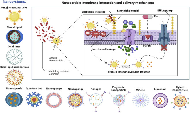
Nanoparticles exert broad-spectrum antibacterial activity against MDR-SA through synergistic physicochemical mechanisms that circumvent canonical resistance pathways. These mechanisms include ROS-mediated oxidative stress, disruption of membrane integrity and fluidity, pore formation, and electrostatic destabilization through interactions with negatively charged cell wall components such as lipoteichoic acid, ultimately leading to cytoplasmic leakage and cell lysis (ref. Wang et al., 2017; ref. Baptista et al., 2018). Concurrently, NPs perturb ionic homeostasis by inducing potassium and magnesium efflux, impairing protein function, and increasing membrane permeability. Nanosystems further bypass efflux pump-mediated resistance by facilitating intracellular delivery of antimicrobial cargo or through direct interference with efflux activity (ref. Huh and Kwon, 2011). These coordinated mechanisms underpin the development of diverse nanoplatforms engineered to enhance antibacterial efficacy, biofilm penetration, and therapeutic durability against MDR-SA infections.
Liposomes
Liposomes, owing to their biomimetic phospholipid bilayer structure, enable dual encapsulation of hydrophilic and hydrophobic therapeutics and efficient intracellular delivery (ref. Akbarzadeh et al., 2013; ref. Bozzuto and Molinari, 2015). Advanced systems such as TLR4-targeted, hyaluronic-acid-modified vancomycin-loaded liposomes (∼121 nm; 49.97% encapsulation efficiency) exhibited a fivefold increase in antibacterial and antibiofilm activity compared with free drug, attributed to enhanced stability and targeted delivery (ref. Ismail et al., 2024). Integration of vancomycin-loaded liposomes within PLGA-PEG-PLGA hydrogels co-delivering DNase I enabled sequential release (77.2% DNase I in 72 h; 82.6% vancomycin over 14 days), effectively disrupting MRSA biofilms and promoting bone regeneration (ref. Li et al., 2023). Oral tetraether-lipid-stabilized liposomes functionalized with cell-penetrating peptides for delivery of a vancomycin derivative (FU002) further demonstrated significant reduction in hepatic MRSA burden at an MIC of 1 mg/L, underscoring translational potential (ref. Werner et al., 2024).
Solid lipid nanoparticles (SLNPs)
Solid lipid nanoparticles (SLNPs) provide enhanced physicochemical stability, controlled release, and responsiveness to infection-specific microenvironments (ref. Naseri et al., 2015; ref. Scioli Montoto et al., 2020). Enzyme-responsive vancomycin-loaded SLNPs (VCM-AS-SLNs; ∼102.2 nm) achieved superior antibacterial activity (MIC 1.56 mg/L vs. 3.125 mg/L for free drug), complete bacterial eradication within 12 h at elevated concentrations, and fivefold higher biofilm removal via lipase-triggered release (∼80%) (ref. Mohammed et al., 2023). DNase I- or Tween-80-modified gentamicin-loaded SLNPs (∼293 nm; 16.85% encapsulation efficiency) demonstrated enhanced antibiofilm efficacy (86.28% vs. 65.34% for free drug) with MIC/MBC values of 3.14/6.28 mg/L (ref. Maurya et al., 2024).
Micelles
Micellar systems, formed through amphiphilic self-assembly, enable targeted delivery and biofilm disruption by modulating bacterial surface hydrophobicity and facilitating penetration (ref. Torchilin, 2007; ref. Perumal et al., 2022). Stimuli-responsive micelles, including fluorinated and light-activated systems, enhance ROS-mediated killing and biofilm eradication (ref. Wan et al., 2023; ref. Lu et al., 2025; ref. Wang et al., 2025). Mercaptosuccinic-acid-decorated micelles (DCSMs) reduced ciprofloxacin MIC fourfold (6.25 mg/L at pH 5.5 vs. 25 mg/L) and achieved ∼85% biofilm inhibition, whereas Magainin II-modified pH-sensitive micelles co-loaded with azithromycin and luteolin (∼40.17 nm; encapsulation efficiencies 93%–94%) demonstrated potent antibacterial activity (MIC 0.625 mg/L) and 86.5% biofilm inhibition (ref. Yang L. et al., 2024; ref. Wang et al., 2025).
Nanodroplets
Nanodroplet-based systems introduce a complementary physical modality via ultrasound-triggered cavitation, enhancing antibiotic penetration and mechanically disrupting biofilms (ref. Argenziano et al., 2017; ref. Banche et al., 2022). High-pressure nanodroplets (∼100 nm) induced peptidoglycan leakage even at 5 cm under 75 MPa, while phase-shift acoustic nanodroplets combined with vancomycin reduced residual biofilm to 7.43% following ultrasound activation, confirming synergistic efficacy (ref. Guo et al., 2017; ref. Tamura et al., 2024).
Dendrimers
Dendrimers, defined by their highly branched architecture and tunable surface functionality, facilitate efficient drug encapsulation and strong electrostatic interactions with bacterial membranes (ref. Tülü and Erturk, 2012). Cationic V-3-mPEA dendrimers (∼52.48 nm) achieved a 16-fold reduction in MIC (0.98 mg/L vs. 15.65 mg/L for vancomycin) and ∼90% reduction in biofilm biomass through membrane disruption and intracellular leakage (ref. Omolo et al., 2018). Nitric-oxide-releasing N-diazeniumdiolate-functionalized PPI dendrimers further demonstrated concentration-dependent antibacterial activity, with G5 variants achieving 3-log MRSA reduction at 2.5–5 µM (ref. Sun et al., 2012).
Polymeric nanoparticles (PNPs)
Polymeric nanoparticles (PNPs), particularly those based on PLGA and PLA, offer high structural integrity, biodegradability, and scalable drug delivery (ref. Cano et al., 2020; ref. Spirescu et al., 2021; ref. Yu et al., 2023; ref. Ali Khan et al., 2024; ref. Naik et al., 2024). Biguanide-derived FTP-NPs produced via polyphenol-assisted aggregation exhibited markedly enhanced antibiofilm activity (up to 5.2 log10 reduction) and reduced biofilm thickness (3.6 µm vs. 12.8 µm for free polymer), alongside superior in vivo efficacy compared with vancomycin (ref. Li et al., 2020). Neutrophil-targeted combinatorial nanosystems (NTCNs) further integrated immunomodulation, reducing TNF-α and IL-6 levels by ∼60% and bacterial load by 85%, outperforming conventional therapy (ref. Chang et al., 2024).
Polymersomes
Polymersomes extend these capabilities through stimuli-responsive vesicular systems, enabling sustained release and multimodal therapy (ref. Discher and Ahmed, 2006; ref. Lee and Feijen, 2012; ref. Zhang and Zhang, 2017). A copper-infused zinc-porphyrin polymersome (CU-PPS) induced localized heating (50.2 °C) and hydroxyl-radical generation, achieving 93% biofilm reduction and ∼80% bacterial killing under near-infrared irradiation, with complete wound closure in vivo within 14 days (ref. Yang N. et al., 2024). Hyaluronic-acid-grafted oleylamine polymersomes maintained prolonged antibacterial activity, with consistent MIC values (1.95 mg/L) over 72 h, compared with rapid loss of efficacy for free vancomycin (ref. Walvekar et al., 2019).
Nanogels
Nanogels, composed of crosslinked hydrophilic polymer networks, enable high drug-loading capacity and stimuli-responsive release (ref. Coll Ferrer et al., 2014; ref. Anooj et al., 2021; ref. Keskin et al., 2021). Polypeptide nanogels synthesized via N-carboxyanhydride polymerization exhibited strong electrostatic binding (6.02 x 10⁵ M-1) and effective MRSA inhibition (MIC 4–16 mg/L), while multifunctional ROS-scavenging nanogels incorporating superoxide dismutase, catalase, and vancomycin achieved 55.1% bacterial inhibition and 92.26% wound healing in hyperglycaemic models (ref. Wang et al., 2025).
Nanosponges
Nanosponges, particularly red-blood-cell (RBC)-membrane-coated systems, provide toxin-neutralizing and biomimetic delivery capabilities (ref. Sherje et al., 2017; ref. Ma et al., 2022; ref. Song et al., 2023). RBC-coated nanosponges (∼100 nm; −28.7 mV) effectively sequestered α-haemolysin and achieved 100% survival in toxin-challenged mice at 28 mg/kg, while redox-responsive nanosponge systems reduced intracellular MRSA burden to <2 CFU per 100 macrophages and exhibited an MIC of 2.5 mg/L (ref. Zagami et al., 2023). Cyclodextrin-based nanosponges loaded with porphyrins further demonstrated photodynamic antibiofilm activity (ref. Zagami et al., 2023).
Quantum dots (QDs)
Quantum dots (QDs), owing to their unique optical and redox properties, provide both diagnostic and therapeutic functionality. Nitrogen-doped QDs achieved >99% MRSA eradication, while graphene QD-Ag hybrids (MIC 11.4 mg/L) and MoS2 QDs (MIC 0.65 mg/L) exhibited enhanced antibacterial activity via ROS generation, glutathione depletion (77%), and membrane depolarisation, alongside low cytotoxicity and accelerated wound healing (ref. Zhong et al., 2020; ref. Ali and De, 2023).
Metallic nanoparticles and hybrid systems
Metallic nanosystems, encompassing metal, metal-oxide, and hybrid nanocomposites, exert antibacterial effects through ROS generation, membrane disruption, DNA damage, and metabolic inhibition (ref. Jan et al., 2014; ref. Perelshtein et al., 2015; ref. Stanić and Tanasković, 2020; ref. Shabatina et al., 2023; ref. Sklodowski et al., 2023). Ag@ZnO-incorporated PVA/PVP hydrogels demonstrated strong antibacterial activity and wound healing, while Ag/CaO nanocomposites (MIC 25 mg/L; MBC 150 mg/L) achieved up to 85% biofilm reduction with reduced cytotoxicity (ref. Khan et al., 2020; ref. Khan et al., 2021). Hybrid GA@TiO2 nanoparticles embedded in GO/CMCh hydrogels further enhanced antibacterial efficacy (MIC 75 mg/L vs. 312.5 mg/L for TiO2 alone) (ref. Salama et al., 2024). Additional metallic systems, including Au, Zn, Cu, and ZnO nanoparticles, reinforce broad-spectrum activity against MDR-SA (ref. Modi et al., 2023).
Emerging nanocomposites and hybrid platforms
Emerging nanosystems such as ceramic nanocomposites (3C-SiC@g-C3N4; MIC 2 mg/mL), curcumin-loaded gold nanocapsules, essential-oil nanocapsules (MIC 31.25 mL/L), Ag-containing TiO2 nanocapsules with sustained release (>70 days), biogenic Ag-Au nanoparticles with quercetin (20 mg/L), and selenium-chitosan hybrids (MIC/MBC 3.9 mg/L) highlight the expanding diversity of nanotherapeutic strategies (ref. Bhatia and Banerjee, 2020; ref. Hérault et al., 2020; ref. Baig et al., 2021; ref. Thorat et al., 2021; ref. Bouaouina et al., 2022; ref. Gamal et al., 2024). Collectively, these interconnected nanosystems demonstrate a paradigm shift towards multifunctional, mechanism-driven platforms capable of overcoming resistance, disrupting biofilms, and enabling sustained, targeted therapy for MDR-SA infections.
Phytochemical-based nanoparticles: Advanced approaches against MDR S. aureus
Pharmacokinetics and formulation advantages
The rising incidence of antibiotic resistance necessitates the development of novel antimicrobial strategies that enhance therapeutic efficacy while minimizing adverse effects. Phytochemical-based nanoparticles (phyto-NPs), which integrate bioactive plant-derived compounds with nanoscale delivery systems, have emerged as a promising antibacterial platform. These systems leverage the inherent antimicrobial properties of phytochemicals while improving their solubility, stability, bioavailability, and intracellular accumulation. Beyond these formulation advantages, the therapeutic performance of phyto-NPs is closely linked to their modified pharmacokinetic profile. Free phytochemicals are often constrained by low aqueous solubility, limited permeability, and rapid presystemic metabolism, resulting in poor systemic exposure. Nanoencapsulation overcomes these barriers by enhancing apparent solubility and facilitating epithelial transport via endocytic pathways, thereby improving bioavailability. Following systemic absorption, nanoparticle size, surface charge, and interfacial properties govern protein corona formation, which in turn modulates circulation time, immune recognition, and biodistribution. Optimized phyto-NPs can reduce rapid clearance, lower required doses, prolong circulation, and enable preferential accumulation at infection sites through personalized delivery, particularly under inflammatory conditions characteristic of MDR-SA infections (ref. Díaz-Puertas et al., 2023; ref. Guedes et al., 2024).
Encapsulation improves phytochemical stability by protecting bioactive compounds from premature degradation, thereby prolonging circulation time and sustaining therapeutic concentrations (ref. Devi et al., 2014). These effects are further reinforced by controlled release kinetics, which sustain drug levels within the therapeutic window while limiting systemic toxicity. Clearance pathways are dependent on carrier composition and size, with biodegradable systems supporting efficient elimination and reduced long-term accumulation. For example, apigenin-loaded liposomes enhanced membrane interaction and intracellular accumulation, reducing the MIC from 32 mg/L (free apigenin) to 8 mg/L (ref. Banerjee et al., 2015). Essential-oil nanoformulations also demonstrate improved stability and permeability: stearic-acid solid-lipid nanoparticles loaded with Eugenia caryophyllata essential oil reduced the MIC from 0.5 mg/L to 0.25 mg/L (ref. Fazly et al., 2018). The physicochemical properties of nanoparticles, particularly size and shape, critically influence antibacterial performance. Smaller nanoparticles exhibit a larger surface-area-to-volume ratio, enabling stronger bactericidal effects (ref. Kvitek et al., 2008). Antibacterial activity declines with increasing particle size (ref. Li K et al., 2013). Nanoparticles within the 5–15 nm range demonstrate enhanced antimicrobial efficacy due to improved membrane attachment, increased permeability, and accelerated cell death (ref. Sondi and Salopek-Sondi, 2004). Shape also modulates antimicrobial potency, spherical silver nanoparticles derived from Euphorbia hirta L. exhibited potent anti-S. aureus activity with an MIC of 9 mg/L (ref. Devi et al., 2014), while cubical silver nanoparticles outperformed spherical forms (ref. Yousaf et al., 2020). Rod-shaped ZnO nanoparticles demonstrated superior antibacterial activity compared with plate-like structures (ref. Ann et al., 2014; ref. Jones et al., 2007). Collectively, these pharmacokinetic enhancements drive improved pharmacodynamic outcomes, notably increased intracellular delivery and enhanced penetration into bacterial biofilms, both of which are critical for overcoming resistance mechanisms in MDR-SA.
Mechanistic antibacterial activity of phyto-NPs
The multifaceted antibacterial mechanisms position phyto-NPs as robust candidates for combating MDR-SA. Phyto-NPs exert bactericidal activity through coordinated extracellular and intracellular mechanisms driven by the synergistic interplay between the nanocarrier and the bioactive phytochemical cargo. Extracellularly, nanoparticles adsorb onto the bacterial surface, where electrostatic interactions and surface reactivity disrupt membrane integrity and interfere with nutrient transport and signaling pathways. In Gram-positive bacteria such as S. aureus, the anionic peptidoglycan matrix promotes nanoparticle adhesion and facilitates localized accumulation, enhancing membrane destabilization (ref. Sarwar et al., 2015). However, unlike conventional nanoparticles, phyto-NPs introduce an additional biochemical dimension. Surface-bound and released phytochemicals can directly interact with membrane lipids and proteins, altering fluidity, inducing lipid peroxidation, and inhibiting membrane-associated enzymes. Following membrane interaction, phyto-NPs can translocate into the cytoplasm via membrane perturbation or endocytic-like processes. Intracellularly, their activity extends beyond passive disruption (ref. Baptista et al., 2018). A central mechanism is the amplification of ROS generation, driven both by the redox-active nanoparticle core and the pro-oxidant or redox-cycling properties of specific phytochemicals. This dual ROS induction results in oxidative damage to DNA, proteins, and membrane components, while simultaneously overwhelming bacterial antioxidant defense systems (ref. Rudramurthy et al., 2016). Positively charged nanoparticles, including Ag-NPs and Cu-NPs, form electrostatic interactions with bacterial membranes, causing wall rupture, leakage of intracellular contents, impaired transport, and energy depletion (ref. Wang et al., 2017). Rice-shaped CuO nanoparticles synthesized using Caesalpinia bonducella L. extract effectively disrupted S. aureus membranes (ref. Sukumar et al., 2020), while Fe-NPs combined with Hibiscus rosa-sinensis L. extract induced ROS generation and altered membrane permeability (ref. Buarki et al., 2022). In parallel, phytochemicals released from the nanocarrier can target critical intracellular pathways, including inhibition of DNA gyrase and topoisomerases, disruption of protein synthesis, and interference with metabolic enzymes involved in energy production (ref. Khameneh et al., 2021). Chitosan-based nanoformulations provide intrinsic antimicrobial activity through membrane disruption and metabolic interference (ref. Zou et al., 2016). Functionalization with cationic or hydrophobic components enhances penetration through negatively charged bacterial membranes (ref. Díaz-Puertas et al., 2023; ref. Bekmukhametova et al., 2020). Liposomal formulations of carvacrol and thymol increased membrane disruption, producing inhibition zones of 16 mm and 15.7 mm compared with 15.0 mm and 13.2 mm for free compounds (ref. Liolios et al., 2009).
Importantly, phyto-NPs also target bacterial resistance mechanisms, including efflux pumps and biofilm formation, which act as protective barriers that enhance antibiotic resistance and immune evasion (ref. Kungwani et al., 2024). Nanoparticle penetration into the extracellular polymeric substance (EPS) matrix is facilitated by their size and surface properties, while phytochemicals disrupt quorum sensing (QS), inhibit EPS synthesis, and destabilize established biofilm architecture (ref. Zhao et al., 2020). Liposomal resveratrol demonstrated potent anti-adherence and antibiofilm activity against resistant S. aureus strains (ref. Prevete et al., 2023). Efflux-pump inhibition is another critical mechanism, in which liposomal piperine reduced the MIC against MRSA to 3.125 mg/L compared with 100 mg/L for free piperine (ref. Khameneh et al., 2015; ref. Seukep et al., 2020). Xylitol, which is normally unable to penetrate biofilms and is degraded by β-lactamases, exhibited enhanced antibiofilm activity when delivered via PLGA nanoparticles (ref. Arunachalam et al., 2023).
A particularly important and mechanistically distinct aspect of phyto-NP activity is their ability to interfere with QS, a central regulatory system governing virulence, toxin production, and biofilm maturation in S. aureus. In this organism, the accessory gene regulator (Agr) system coordinates population-density-dependent expression of key pathogenic determinants via autoinducing peptides (ref. LaSarre and Federle, 2013). Phyto-NPs may disrupt this signaling axis at multiple levels. Surface-associated and released phytochemicals, especially phenolics and flavonoids, have been shown to inhibit AIP synthesis, block ligand-receptor interactions at AgrC, and suppress downstream activation of the AgrA response regulator, thereby attenuating transcription of RNAIII and associated virulence genes (ref. Jakobsen et al., 2017). In addition to direct receptor-level interference, phyto-NPs can indirectly impair QS through membrane perturbation and oxidative stress.
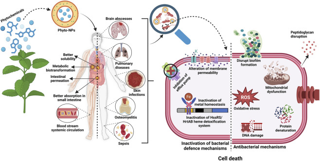
Disruption of membrane integrity alters the localization and functionality of membrane-bound QS components, while ROS overproduction can damage regulatory proteins and signaling peptides, further dampening QS signal propagation (ref. Subramaniyan et al., 2021). Nanoparticle-mediated delivery also enhances the local concentration and stability of QS-inhibitory phytochemicals within biofilms, overcoming diffusion limitations that typically reduce the efficacy of free compounds. This is particularly relevant in mature biofilms, where QS gradients are spatially organized and difficult to disrupt (ref. Lu et al., 2019). Importantly, QS inhibition by phyto-NPs does not necessarily exert strong bactericidal pressure, but instead reduces virulence and biofilm robustness, thereby sensitizing bacterial populations to host immune responses and conventional antibiotics. This antivirulence strategy reduces the selective pressure for resistance development while complementing the nanoplatform’s direct bactericidal mechanisms. Figure 7 presents a schematic representation of the antibacterial mechanisms of phyto-NPs against MDR-SA, highlighting their ability to enhance phytochemical efficacy by improving solubility, stability, circulation time, and targeted delivery.
Critical assessment of evidence and methodological limitations
Although many phyto-NPs have demonstrated promising antibacterial efficacy in vitro, it is difficult to draw direct comparisons across studies due to substantial methodological variability. The observed MICs of identical phytochemicals, such as curcumin and quercetin, can vary widely due to factors including nanoparticle type, size, surface charge, loading capacity, release profile, bacterial strain, inoculum concentration, and assay conditions. Additionally, the solvent system, culture medium, and incubation conditions introduce ambiguity and reduce the reproducibility of research outcomes. Furthermore, comparisons between free compounds and their NP-based counterparts are not always conducted under identical conditions. Since free phytochemicals often exhibit poor water solubility and limited dispersibility in aqueous solutions, NP-based formulations may contain additional substances, such as surfactants and stabilizers, which can affect their antibacterial performance. Consequently, the increase in antibacterial efficacy from nanosizing might sometimes be exaggerated if proper controls are not used. Moreover, many reported in vitro MIC values may not directly translate into clinically achievable concentrations in vivo, underscoring the need for pharmacokinetic and pharmacodynamic validation of these systems.
Synergy and multimodal activity
The multimodal antibacterial activity of phyto-NPs arises from interactions between the nanocarrier and bioactive phytochemicals, enabling them to target multiple bacterial functions simultaneously. Unlike conventional antibiotics that target single pathways, phyto-NPs act on multiple bacterial systems simultaneously, reducing the likelihood of resistance development (ref. Díaz-Puertas et al., 2023). Resistance in S. aureus may arise from intrinsic factors, spontaneous mutations, or horizontal gene transfer (ref. Sultan et al., 2018; ref. Sun et al., 2019). Alterations in penicillin-binding proteins reduce antibiotic affinity (ref. Lee et al., 2014). Phyto-NP formulations enhance receptor binding, reduce required dosages, and minimize off-target toxicity (ref. Anand et al., 2022).
The synergistic integration of conventional antibiotics with phyto-NPs represents a particularly promising strategy to restore antibiotic efficacy while reducing total antibiotic dosage and selective pressure. Silver nanoparticles synthesized using Urtica dioica L. extract increased inhibition zones from 19 mm to 39 mm when combined with antibiotics (ref. Jyoti et al., 2016). Silver nanoparticles derived from Zea mays L. waste enhanced rifampicin and kanamycin activity, increasing inhibition zones across multiple pathogens (ref. Patra and Baek, 2017). However, the defining feature of phyto-NPs lies not merely in combinatorial use but in the intrinsic mechanistic synergy between the nanoparticle core and the phytochemical cargo, which is not recapitulated by nanoparticles or phytochemicals alone. These two modalities are functionally integrated, generating a biofunctionalized system that directly underpins the observed potentiation of conventional antibiotics. Increased membrane permeability and biofilm penetration facilitate antibiotic entry, while phytochemical-mediated inhibition of efflux systems and metabolic adaptation enhances intracellular drug retention and target susceptibility (ref. Baptista et al., 2018). In addition, ROS amplification and disruption of the proton motive force further compromise bacterial defense systems, sensitizing cells to antibiotic-induced damage (ref. Hemeg, 2017). Crucially, the nanocarrier enables co-localized and sustained delivery, which may further improve therapeutic selectivity while reducing systemic exposure (ref. Baptista et al., 2018). For example, hyaluronic-acid-coated cinnamaldehyde nanoparticles exploit bacterial hyaluronidase activity for site-specific release against MRSA (ref. Sun et al., 2018). It is also necessary to differentiate between true synergy and additive/parallel activity based on standard pharmacological parameters (ref. Tamma et al., 2012). The fractional inhibitory concentration index (FICI), typically calculated by checkerboard microdilution, remains one of the most reliable approaches for assessing antimicrobial synergy. Typically, FICI scores ≤0.5 indicate synergistic interactions, 0.5 to one denote additive effects, one to four imply indifference, and >4 signify antagonism (ref. Odds, 2003). Nevertheless, despite numerous claims of synergistic activity in phyto-NP research, surprisingly few studies use reliable quantitative assessments of synergism, and most conclusions are based solely on reduced MIC values or expanded inhibition zones. Nevertheless, the current evidence base remains heterogeneous, and direct comparison across studies is complicated by variability in nanoparticle composition, phytochemical loading, bacterial strains, and susceptibility-testing methodologies.
Overall, the synergistic integration of physicochemical disruption and biochemical targeting distinguishes phyto-NPs from their individual components and provides a mechanistic basis for multimodal potentiation of antibiotic activity against MDR-SA. Table 2 details the antibacterial activities of phytochemical-based nanoparticles against MDR-SA, highlighting mechanisms of action and MIC values.
TABLE 2: Effects of nanoparticles loaded with phytochemicals against MDR-SA.
| Phytochemical-based NPs | Mechanism of action | MIC value | Bacterial species | References |
|---|---|---|---|---|
| Tribulus terrestris-AgNPs | Inhibition of bacterial growth and membrane disruption | Not determined | MDR S. aureus | ref. Gopinath et al. (2012) |
| Artemisia haussknechtii-Ag-Cu-TiO2NPs | Inhibition of bacterial growth | 40mg/L | MDR S. aureus | ref. Alavi and Karimi (2018) |
| Punica granatum extract–Au-NPs | Inhibition of bacterial growth | 15.6mg/L | MRSA | ref. Hussein et al. (2021) |
| Cinnamon essential oil–liposome | Inhibition of biofilm formation | 0.25 mg/mL | MRSA | ref. Cui et al. (2016) |
| Red propolis-polymeric NPs | Inhibition of biofilm formation | 100–125 mg/L | MRSA | ref. De Mélo Silva et al. (2020) |
| Rutin and benzamide-polymeric NPs | Inhibition of biofilm formation and disruption of cell membrane | 420 mg/L and 250 mg/L | MDR S. aureus | ref. Deepika et al. (2020) |
| Capsicum annuum-Ag-NPs | Inhibition of biofilm formation, generation of superoxide radicals and ROS | 80 mg/L | MSSA, MRSA | ref. Ahmed et al. (2018) |
| Quercetin-Ag-NPs | Inhibition of biofilm formation, generation of superoxide radicals and ROS in MRSA | 50 mg/L | MSSA, MRSA | ref. Sadeghi et al. (2024) |
| Berberine-Au-NPs | Disruption of cell membrane integrity, reduction of viability, ROS production, biofilm inhibition | 27.37 mg/L for S. aureus, 109.5 mg/L for MRSA | S. aureus, MRSA | ref. Sinsinwar and Vadivel (2021) |
| Anacardium occidentale-Ag-NPs | Membrane disruption, ROS and malondialdehyde generation, nucleotide leakage, biofilm inhibition | 7.81–31.25 mg/L | S. aureus, MSSA, MRSA | ref. Jamil et al. (2016) |
| Rutin-carbon quantum dots | Cell membrane destruction and increased membrane permeability | 32 mg/L | MRSA | ref. He et al. (2019) |
| Elettaria cardamomum essential oil–chitosan NPs | Inhibition of bacterial growth | 10% v/v | MRSA | ref. Jamil et al. (2016) |
| Epigallocatechin gallate-liposome | Inhibition of bacterial growth | 16 mg/L | MRSA | ref. Gharib et al. (2013) |
| Cotyledon orbiculate-Ag-NPs | Inhibition of bacterial growth | 40 mg/L | MRSA | ref. Díaz-Puertas et al. (2023) |
| Oleic acid-liposome | Inhibition of bacterial growth | Not determined | MRSA | ref. Huang et al. (2011) |
| Aloe vera-Te-NPs | Inhibition of bacterial growth | 11.61 mg/L | MRSA | ref. Medina-Cruz et al. (2021) |
| Silybum marianum-liposome | Inhibition of bacterial growth | 125 mg/L | MRSA | ref. Faezizadeh et al. (2015) |
| Periploca hydaspidis-Ag-NPs | Inhibition of bacterial growth | 10–20 mg/L | MRSA | ref. Ali et al. (2021) |
| Syzygium cumini-Fe-NPs | Inhibition of bacterial growth | 11 mg/L | MRSA | ref. Asghar et al. (2020) |
| Stenocereus queretaroensis-Ag-NPs | Inhibition of bacterial growth | 0.313 mg/L | MRSA | ref. Padilla-Camberos et al. (2021) |
* NPs: Nanoparticles, Au-NPs: Gold nanoparticles, Ag-NPs: Silver nanoparticles, Fe-NPs: Iron nanoparticles, Te-NPs: Tellurium nanoparticles, TiO2-NPs: Titanium dioxide nanoparticles, MDR: Multi-drug resistant, MRSA: Methicillin-resistant S. aureus, MSSA: Methicillin-sensitive S. aureus.
Challenges and future directions
Antibiotic resistance continues to pose a major challenge to modern healthcare, with S. aureus bacteraemia representing a particularly severe clinical problem. Despite substantial efforts in antimicrobial development, the pace of resistance evolution continues to outstrip the introduction of new antibiotics, underscoring the urgent need for alternative and complementary therapeutic strategies (ref. Sultan et al., 2018; ref. Vestergaard et al., 2019).
Phytochemicals and plant-derived nanomaterials have emerged as promising adjuvants and alternatives to conventional antibiotics. Rather than acting solely as physical antimicrobial agents, phyto-NPs function as hybrid systems in which nanoscale delivery enhances the stability, localization, and controlled release of phytochemicals, while the phytochemicals themselves introduce specific biochemical targeting. This convergence of physicochemical and molecular mechanisms underpins their superior efficacy against MDR-SA (ref. Anand et al., 2022; ref. Prevete et al., 2023; ref. Lang et al., 2024).
Despite these advances, substantial challenges remain before phyto-NPs can transition from promising laboratory systems to clinically viable therapies. One major obstacle is the inherent variability of plant extracts, which depends on botanical source, geographical origin, cultivation conditions, and extraction protocols, leading to inconsistent phytochemical composition and biological activity (ref. Patil et al., 2023; ref. Guedes et al., 2024). This variability complicates standardization, reproducibility, and regulatory approval. A major limitation in the current research landscape of phyto-NPs is the poor reproducibility and comparability of antimicrobial efficacy data. Reported MIC values for common phytochemicals vary widely across studies, primarily due to non-standardized experimental parameters. In addition, the physicochemical behavior of phytochemicals, such as low aqueous solubility, instability, and aggregation, can significantly influence apparent activity, leading to inconsistent potency assessments. The absence of standardized protocols and reporting frameworks limits cross-study validation and obscures structure-activity relationships.
Equally important is the limited pharmacological relevance of many in vitro findings. Antimicrobial effects are often reported at concentrations that may not be achievable in vivo due to constraints related to absorption, metabolic instability, and systemic clearance. While nanoformulation is proposed to enhance bioavailability and tissue distribution, few studies integrate pharmacokinetic-pharmacodynamic considerations or assess whether effective concentrations can be attained and sustained at infection sites, particularly in biofilm-associated infections. This gap restricts the translational interpretation of in vitro efficacy. In addition, safety, long-term toxicity, biodistribution, and immunogenicity of complex nanomaterials require far more comprehensive evaluation through well-designed in vivo and preclinical studies (ref. Anand et al., 2022; ref. Hulme, 2022).
Another critical limitation in current research is the lack of uniform criteria for assessing antibacterial efficacy and mechanisms of action of nanoparticles. Nanomaterials may exert bactericidal effects through overlapping pathways, including ROS generation, membrane disruption, efflux pump inhibition, and biofilm degradation, making cross-study comparisons difficult (ref. Rudramurthy et al., 2016; ref. Modi et al., 2023). While oxidative stress is widely recognized as a central antibacterial mechanism, the broader impacts of nanoparticles on bacterial metabolism, gene regulation, protein expression, and resistance pathways remain insufficiently characterized (ref. Sun et al., 2019; ref. Zhao et al., 2020).
Further harmonized experimental protocols are required to address these limitations, including standardized susceptibility testing conditions and reporting criteria (ref. Wiegand et al., 2008). Future studies should incorporate pharmacokinetic modeling, tissue distribution analysis, and clinically relevant dosing strategies to bridge the gap between in vitro activity and in vivo applicability. Rigorous comparative designs using equivalent molar concentrations, controlled formulation variables, and validated synergy metrics are essential to accurately define the contribution of nanoformulation. The integration of advanced models such as biofilm systems, organotypic cultures, and in vivo infection models will provide more realistic assessments of therapeutic potential. Such systematic and quantitatively grounded approaches are critical for establishing the true clinical value of phyto-NPs and enabling their rational translation into antimicrobial therapies.
Future progress in this field will require integrating advanced analytical and systems-level approaches. Omics-based strategies, including transcriptomics, proteomics, metabolomics, and genome-wide association studies, offer powerful tools to elucidate nanoparticle-bacteria interactions, adaptive stress responses, and resistance modulation at the molecular level (ref. Vestergaard et al., 2019; ref. Douglas, 2022). Such approaches should be prioritized to identify predictive biomarkers of efficacy, optimize nanoparticle design, and guide rational combination therapies. However, their successful clinical translation depends on rigorous mechanistic elucidation, standardization of phytochemical composition, comprehensive safety and toxicity validation, and the establishment of robust translational research frameworks (ref. Anand et al., 2022; ref. Díaz-Puertas et al., 2023; ref. Guedes et al., 2024). Addressing these challenges through integrated nanotechnology, microbiology, and systems-level approaches will be essential to realize the full potential of phyto-nanotechnologies in the post-antibiotic era (ref. Douglas, 2022; ref. Modi et al., 2023).
Conclusion
This review highlights the continuing challenge posed by MDR-SA and the growing limitations of conventional antimicrobial strategies. Evidence across phytochemistry and nanomedicine demonstrates that phytochemicals and phytochemical-based nanomaterials offer a credible and conceptually advanced direction for next-generation antibacterial development by integrating natural bioactivity with nanoscale delivery advantages. Their ability to enhance membrane interaction, disrupt biofilms, modulate bacterial defense mechanisms, and support synergistic antibiotic activity positions these systems as integral components of emerging therapeutic frameworks. Nanotechnology strengthens this potential by improving solubility, stability, and site-specific delivery, enabling phytochemicals to reach complex infection niches with greater precision and potency. Rather than relying on escalating systemic antibiotic exposure, rationally engineered nanosystems provide multimodal, mechanism-driven intervention capable of reshaping host-pathogen interactions. Phyto-nanomaterials further reduce the likelihood of resistance development by simultaneously targeting multiple bacterial pathways, enabling lower drug dosages and improved therapeutic durability. Despite these advances, successful translation requires deeper mechanistic resolution, consistent evaluation criteria, and comprehensive safety and toxicity assessment. Variability in plant-derived extracts, limited standardization, and incomplete understanding of nanoparticle-bacteria interactions continue to constrain clinical progress. Future work must prioritize rigorous in vivo validation, improved reproducibility, and integration of systems-level analytical approaches to clarify antibacterial mechanisms and guide rational design. Phytochemical-based nanosystems represent a forward-looking, mechanism-centered strategy for managing MDR-SA infections. Their continued development will depend on sustained interdisciplinary research that can transform experimental promise into clinically reliable tools for the post-antibiotic era.
References
- A. A. Abebe, A. G. Birhanu. Methicillin resistant staphylococcus Aureus: molecular mechanisms underlying drug resistance development and novel strategies to combat.. Infect. Drug Resist., 2023. [DOI | PubMed]
- M. Adoui, M. Lahouel. Caractérisation des souches de Staphylococcus aureus résistantes à la méticilline (SARM) et évaluation de leur sensibilité à la propolis.. 2019
- B. Ahmed, A. Hashmi, M. S. Khan, J. Musarrat. ROS mediated destruction of cell membrane, growth and biofilms of human bacterial pathogens by stable metallic AgNPs functionalized from bell pepper extract and quercetin.. Adv. Powder Technol., 2018. [DOI]
- A. Akbarzadeh, R. Rezaei-Sadabady, S. Davaran, S. W. Joo, N. Zarghami, Y. Hanifehpour. Liposome: classification, preparation, and applications.. Nanoscale Res. Lett., 2013. [DOI | PubMed]
- M. Alavi, N. Karimi. Characterization, antibacterial, total antioxidant, scavenging, reducing power and ion chelating activities of green synthesized silver, copper and titanium dioxide nanoparticles using Artemisia haussknechtii leaf extract.. Artif. Cells Nanomed. Biotechnol., 2018. [DOI | PubMed]
- S. R. Ali, M. De. Defect-engineered functionalized MoS2 quantum dots with enhanced antibacterial activity.. ACS Appl. Nano Mat., 2023. [DOI]
- S. Ali, M. R. Khan, R. Khan. Green synthesized AgNPs from Periploca hydaspidis Falc. and its biological activities.. Microsc. Res. Tech., 2021. [DOI | PubMed]
- S. Ali Khan, N. Ahmed, D. Khan, A. Bibi, A. u. Rehman. pH-responsive mupirocin-loaded hybrid nanoparticles in hydrogel and film forming spray against resistant bacterial wound infections.. J. Drug Deliv. Sci. Technol., 2024. [DOI]
- U. Anand, M. Carpena, M. Kowalska-Góralska, P. Garcia-Perez, K. Sunita, E. Bontempi. Safer plant-based nanoparticles for combating antibiotic resistance in bacteria: a comprehensive review on its potential applications, recent advances, and future perspective.. Sci. Total Environ., 2022. [DOI | PubMed]
- L. C. Ann, S. Mahmud, S. K. M. Bakhori, A. Sirelkhatim, D. Mohamad, H. Hasan. Antibacterial responses of zinc oxide structures against Staphylococcus aureus, Pseudomonas aeruginosa and Streptococcus pyogenes .. Ceram. Int., 2014. [DOI]
- E. Anooj, M. Charumathy, V. Sharma, B. Vibala, S. Gopukumar, S. B. Jainab. Nanogels: an overview of properties, biomedical applications, future research trends and developments.. J. Mol. Struct., 2021. [DOI]
- G. Appendino, S. Gibbons, A. Giana, A. Pagani, G. Grassi, M. Stavri. Antibacterial cannabinoids from cannabis sativa: a structure-activity study.. J. Nat. Prod., 2008. [DOI | PubMed]
- M. Argenziano, G. Banche, A. Luganini, N. Finesso, V. Allizond, G. R. Gulino. Vancomycin-loaded nanobubbles: a new platform for controlled antibiotic delivery against methicillin-resistant Staphylococcus aureus infections.. Int. J. Pharm., 2017. [DOI | PubMed]
- K. Arunachalam, G. P. Krishnan, S. Sethuraman, S. V. P. I. Abraham, S. T. Krishnan, A. Venkateswar. Exploring possible ways to enhance the potential and use of natural products through nanotechnology in the battle against biofilms of foodborne bacterial pathogens.. Pathogens, 2023. [DOI | PubMed]
- M. A. Asghar, E. Zahir, M. A. Asghar, J. Iqbal, A. A. Rehman. Facile, one-pot biosynthesis and characterization of iron, copper and silver nanoparticles using Syzygium cumini leaf extract as effective antimicrobial and aflatoxin B1 adsorption agents.. PLoS One, 2020. [DOI | PubMed]
- U. Baig, M. A. Gondal, M. A. Ansari, M. Dastageer, M. Sajid, W. Falath. Rapid synthesis and characterization of advanced ceramic-polymeric nanocomposites for efficient photocatalytic decontamination of hazardous organic pollutant under visible light and inhibition of microbial biofilm.. Ceram. Int., 2021. [DOI]
- G. Banche, V. Allizond, N. Mandras, N. Finesso, A. Luganini, T. Genova. Antimicrobial oxygen-loaded nanobubbles as promising tools to promote wound healing in hypoxic human keratinocytes.. Toxicol. Rep., 2022. [DOI | PubMed]
- K. Banerjee, S. Banerjee, S. Das, M. Mandal. Probing the potential of apigenin liposomes in enhancing bacterial membrane perturbation and integrity loss.. J. Colloid Interface Sci., 2015. [DOI | PubMed]
- P. V. Baptista, M. P. McCusker, A. Carvalho, D. A. Ferreira, N. M. Mohan, M. Martins. Nano-strategies to fight multidrug resistant bacteria, “A battle of the titans”.. Front. Microbiol., 2018. [DOI | PubMed]
- A. Bekmukhametova, H. Ruprai, J. M. Hook, D. Mawad, J. Houang, A. Lauto. Photodynamic therapy with nanoparticles to combat microbial infection and resistance.. Nanoscale, 2020. [DOI | PubMed]
- E. Bhatia, R. Banerjee. Hybrid silver-gold nanoparticles suppress drug resistant polymicrobial biofilm formation and intracellular infection.. J. Mat. Chem. B, 2020. [DOI | PubMed]
- K. Bogdanova, M. Röderova, M. Kolar, K. Langova, M. Dusek, P. Jost. Antibiofilm activity of bioactive hop compounds humulone, lupulone and xanthohumol toward susceptible and resistant staphylococci.. Res. Microbiol., 2018. [DOI | PubMed]
- S. Bouaouina, A. Aouf, A. Touati, H. Ali, M. Elkhadragy, H. Yehia. Effect of nanoencapsulation on the antimicrobial and antibiofilm activities of Algerian Origanum glandulosum Desf. against multidrug-resistant clinical isolates.. Nanomaterials, 2022. [DOI | PubMed]
- G. Bozzuto, A. Molinari. Liposomes as nanomedical devices.. Int. J. Nanomedicine, 2015. [DOI | PubMed]
- F. Buarki, H. AbuHassan, F. Al Hannan, F. Z. Henari. Green synthesis of iron oxide nanoparticles using Hibiscus rosa-sinensis flowers and their antibacterial activity.. J. Nanomater., 2022. [DOI]
- A. Cano, M. Ettcheto, M. Espina, A. López-Machado, Y. Cajal, F. Rabanal. State-of-the-art polymeric nanoparticles as promising therapeutic tools against human bacterial infections.. J. Nanobiotechnol., 2020. [DOI | PubMed]
- B. C. L. Chan, M. Ip, C. B. S. Lau, S. L. Lui, C. Jolivalt, C. Ganem-Elbaz. Synergistic effects of baicalein with ciprofloxacin against NorA over-expressed methicillin-resistant Staphylococcus aureus (MRSA) and inhibition of MRSA pyruvate kinase.. J. Ethnopharmacol., 2011. [DOI | PubMed]
- Y.-T. Chang, C.-Y. Lin, C.-J. Chen, E. Hwang, A. Alshetaili, H. P. Yu. Neutrophil-targeted combinatorial nanosystems for suppressing bacteremia-associated hyperinflammation and MRSA infection to improve survival rates.. Acta Biomater., 2024. [DOI | PubMed]
- Y. L. Chew, A. M. Mahadi, K. M. Wong, J. K. Goh. Anti-methicillin-resistant Staphylococcus aureus (MRSA) compounds from Bauhinia kockiana Korth. and their mechanism of antibacterial activity.. BMC Complement. Altern. Med., 2018. [DOI | PubMed]
- P. Y. Chung, L. Y. Chung, P. Navaratnam. Potential targets by pentacyclic triterpenoids from Callicarpa farinosa against methicillin-resistant and sensitive Staphylococcus aureus .. Fitoterapia, 2014. [DOI | PubMed]
- M. C. Coll Ferrer, S. Dastgheyb, N. J. Hickok, D. M. Eckmann, R. J. Composto. Designing nanogel carriers for antibacterial applications.. Acta Biomater., 2014. [DOI | PubMed]
- A. Coqueiro, L. O. Regasini, P. Stapleton, V. da Silva Bolzani, S. Gibbons. In vitro antibacterial activity of prenylated guanidine alkaloids from Pterogyne nitens and synthetic analogues.. J. Nat. Prod., 2014. [DOI | PubMed]
- D. C. M. Costa, M. M. B. Azevedo, D. O. Silva, M. T. V. Romanos, T. C. B. S. Souto-Padrón, C. S. Alviano. In vitro anti-MRSA activity of Couroupita guianensis extract and its component tryptanthrin.. Nat. Prod. Res., 2017. [DOI | PubMed]
- H. Cui, W. Li, C. Li, S. Vittayapadung. Liposome containing cinnamon oil with antibacterial activity against methicillin-resistant Staphylococcus aureus biofilm.. Biofouling, 2016. [DOI | PubMed]
- I. S. de Mélo Silva, L. M. do Amorim Costa Gaspar, A. M. O. Rocha, L. P. da Costa, D. B. Tada, E. Franceschi. Encapsulation of red propolis in polymer nanoparticles for the destruction of pathogenic biofilms.. AAPS PharmSciTech, 2020. [DOI | PubMed]
- M. S. Deepika, R. Thangam, S. Sundarraj, T. S. Sheena, S. Sivasubramanian, J. Kulandaivel. Co-delivery of diverse therapeutic compounds using PEG-PLGA nanoparticle cargo against drug-resistant bacteria: an improved anti-biofilm strategy.. ACS Appl. Bio Mat., 2020. [DOI]
- G. D. Devi, K. Murugan, C. P. Selvam. Green synthesis of silver nanoparticles using Euphorbia hirta (Euphorbiaceae) leaf extract against crop pest of cotton bollworm, Helicoverpa armigera .. J. Biopestic., 2014. [DOI]
- R. Díaz-Puertas, F. J. Álvarez-Martínez, A. Falco, E. Barrajón-Catalán, R. Mallavia. Phytochemical-based nanomaterials against antibiotic-resistant bacteria: an updated review.. Polymers, 2023. [DOI | PubMed]
- D. E. Discher, F. Ahmed. Polymersomes.. Annu. Rev. Biomed. Eng., 2006. [DOI | PubMed]
- L.-M. Dong, L.-L. Huang, H. Dai, Q. L. Xu, J. K. Ouyang, X. C. Jia. Anti-MRSA sesquiterpenes from the semi-mangrove plant Myoporum bontioides A.. Gray. Mar. Drugs, 2018. [DOI | PubMed]
- E. J. A. Douglas. The Use of Genome Wide Association Studies to Understand Two Key Aspects of Staphylococcus aureus Pathogenesis: Serum Resistance and Toxicity., 2022
- A. H. Elmaidomy, N. H. Shady, K. M. Abdeljawad, M. B. Elzamkan, H. H. Helmy, E. A. Tarshan. Antimicrobial potentials of natural products against multidrug resistance pathogens: a comprehensive review.. RSC Adv., 2022. [DOI | PubMed]
- G. Eumkeb, S. Sakdarat, S. Siriwong. Reversing β-lactam antibiotic resistance of Staphylococcus aureus with galangin from Alpinia officinarum Hance and synergism with ceftazidime.. Phytomedicine, 2010. [DOI | PubMed]
- Z. Faezizadeh, A. Gharib, M. Godarzee. In vitro and in vivo evaluation of silymarin nanoliposomes against isolated methicillin-resistant Staphylococcus aureus .. Iran. J. Pharm. Res., 2015. [PubMed]
- N. Fatemi, M. R. Sharifmoghadam, M. Bahreini, B. Khameneh, H. Shadifar. Antibacterial and synergistic effects of herbal extracts in combination with amikacin and imipenem against multidrug-resistant isolates of Acinetobacter .. Curr. Microbiol., 2020. [DOI | PubMed]
- B. S. Fazly Bazzaz, B. Khameneh, N. Namazi, M. Iranshahi, D. Davoodi, S. Golmohammadzadeh. Solid lipid nanoparticles carrying Eugenia caryophyllata essential oil: the novel nanoparticulate systems with broad-spectrum antimicrobial activity.. Lett. Appl. Microbiol., 2018. [DOI | PubMed]
- S. Y. Fu, H. Q. Yang, J. Tu, Q. Meng, S. Xiao, M. Zhang. Separation and activity against drug-resistant bacteria of tetrandrine and fangchinoline in lipophilic alkaloids from Stephania tetrandra .. Der Chem. Sin., 2017
- A.A.-R. Gamal, M. A. M. Hussein, H. A. E. Sayed, E. S. M. El-Sayed, A. M. Youssef, I. M. El-Sherbiny. Hybrid nanoparticles combining nanoselenium-mediated Carica papaya extract and trimethyl chitosan for combating clinical multidrug-resistant bacteria.. Int. J. Biol. Macromol., 2024. [DOI | PubMed]
- S. S. Garg, J. Gupta, S. Sharma, D. Sahu. An insight into the therapeutic applications of coumarin compounds and their mechanisms of action.. Eur. J. Pharm. Sci., 2020. [DOI | PubMed]
- C. Gaspar-Marques, P. Rijo, M. F. Simões, M. A. Duarte, B. Rodriguez. Abietanes from Plectranthus grandidentatus and P. hereroensis against methicillin- and vancomycin-resistant bacteria.. Phytomedicine, 2006. [DOI | PubMed]
- A. Gharib, Z. Faezizadeh, M. Godarzee. Therapeutic efficacy of epigallocatechin gallate-loaded nanoliposomes against burn wound infection by methicillin-resistant Staphylococcus aureus .. Skin. Pharmacol. Physiol., 2013. [DOI | PubMed]
- S. Gibbons, E. E. Udo. The effect of reserpine, a modulator of multidrug efflux pumps, on the in vitro activity of tetracycline against clinical isolates of methicillin resistant staphylococcus aureus (MRSA) possessing the tet(K) determinant.. Phytother. Res., 2000. [DOI | PubMed]
- V. Gopinath, D. MubarakAli, S. Priyadarshini, N. M. Priyadharsshini, N. Thajuddin, P. Velusamy. Biosynthesis of silver nanoparticles from Tribulus terrestris and its antimicrobial activity: a novel biological approach.. Colloids Surf. B Biointerfaces, 2012. [DOI | PubMed]
- B. N. Guedes, K. Krambeck, A. Durazzo, M. Lucarini, A. Santini, M. B. P. P. Oliveira. Natural antibiotics against antimicrobial resistance: sources and bioinspired delivery systems.. Braz. J. Microbiol., 2024. [DOI | PubMed]
- H. Guo, Z. Wang, Q. Du, P. Li, A. Wang. Stimulated phase-shift acoustic nanodroplets enhance vancomycin efficacy against methicillin-resistant Staphylococcus aureus biofilms.. Int. J. Nanomedicine, 2017. [DOI | PubMed]
- J. M. He, S. C. Sun, Z. L. Sun, J. T. Chen, Q. Mu. Isovalerylshikonin, a new resistance-modifying agent from arnebia Euchroma, suppresses antimicrobial resistance of drug-resistant Staphylococcus aureus .. Int. J. Antimicrob. Agents, 2019. [DOI | PubMed]
- H. A. Hemeg. Nanomaterials for alternative antibacterial therapy.. Int. J. Nanomedicine., 2017. [DOI | PubMed]
- N. Hérault, J. Wagner, S. L. Abram, J. Widmer, L. Horvath, D. Vanhecke. Silver-containing titanium dioxide nanocapsules for combating multidrug-resistant bacteria.. Int. J. Nanomedicine, 2020. [DOI | PubMed]
- X. Hua, Q. Yang, W. Zhang, Z. Dong, S. Yu, S. Schwarz. Antibacterial activity and mechanism of action of aspidinol against multi-drug-resistant methicillin-resistant Staphylococcus aureus .. Front. Pharmacol., 2018. [DOI | PubMed]
- X. Hua, Y. Jia, Q. Yang, W. Zhang, Z. Dong, S. Liu. Transcriptional analysis of the effects of gambogic acid and neogambogic acid on methicillin-resistant Staphylococcus aureus .. Front. Pharmacol., 2019. [DOI | PubMed]
- C.-M. Huang, C.-H. Chen, D. Pornpattananangkul, L. Zhang, M. Chan, M. F. Hsieh. Eradication of drug-resistant Staphylococcus aureus by liposomal oleic acids.. Biomaterials, 2011. [DOI | PubMed]
- Y. Huang, X. Zhao, Y. Zu, L. Wang, Y. Deng, M. Wu. Enhanced solubility and bioavailability of apigenin via preparation of solid dispersions of mesoporous silica nanoparticles.. Iran. J. Pharm. Res., 2019. [PubMed]
- A. J. Huh, Y. J. Kwon. “Nanoantibiotics”: a new paradigm for treating infectious diseases using nanomaterials in the antibiotic-resistant era.. J. Control. Release, 2011. [DOI | PubMed]
- J. Hulme. Application of nanomaterials in the prevention, detection, and treatment of methicillin-resistant Staphylococcus aureus (MRSA).. Pharmaceutics, 2022. [DOI | PubMed]
- M. A. M. Hussein, M. Grinholc, A. S. A. Dena, I. M. El-Sherbiny, M. Megahed. Boosting the antibacterial activity of chitosan-gold nanoparticles against antibiotic-resistant bacteria by Punica granatum L. extract.. Carbohydr. Polym., 2021. [DOI | PubMed]
- M. Iinuma, H. Tosa, T. Tanaka, F. Asai, Y. Kobayashi, R. Shimano. Antibacterial activity of xanthones from guttiferous plants against methicillin-resistant Staphylococcus aureus .. J. Pharm. Pharmacol., 1996. [DOI | PubMed]
- E. A. Ismail, C. A. Omolo, M. A. Gafar, R. Khan, V. O. Nyandoro, E. S. Yakubu. Novel peptide and hyaluronic acid coated biomimetic liposomes for targeting bacterial infections and sepsis.. Int. J. Pharm., 2024. [DOI | PubMed]
- T. H. Jakobsen, A. N. Warming, R. M. Vejborg, J. A. Moscoso, M. Stegger, F. Lorenzen. A broad range quorum sensing inhibitor working through sRNA inhibition.. Sci. Rep., 2017. [DOI | PubMed]
- B. Jamil, R. Abbasi, S. Abbasi, M. Imran, S. U. Khan, A. Ihsan. Encapsulation of cardamom essential oil in chitosan nano-composites: in vitro efficacy on antibiotic-resistant bacterial pathogens and cytotoxicity studies.. Front. Microbiol., 2016. [DOI | PubMed]
- T. Jan, J. Iqbal, M. Ismail, N. Badshah, Q. Mansoor, A. Arshad. Synthesis, physical properties and antibacterial activity of metal oxides nanostructures.. Mat. Sci. Semicond. process., 2014. [DOI]
- R. Jeetah, A. Bhaw-Luximon, D. Jhurry. Nanopharmaceutics: phytochemical-Based controlled or sustained drug-delivery systems for cancer treatment.. J. Biomed. Nanotechnol., 2014. [DOI | PubMed]
- R. N. Jones, T. R. Fritsche, H. S. Sader, J. E. Ross. LEADER surveillance program results for 2006: an activity and spectrum analysis of linezolid using clinical isolates from the United States (50 medical centers).. Diagn. Microbiol. Infect. Dis., 2007. [DOI | PubMed]
- K. Jyoti, M. Baunthiyal, A. Singh. Characterization of silver nanoparticles synthesized using Urtica dioica linn. leaves and their synergistic effects with antibiotics.. J. Radiat. Res. Appl. Sci., 2016. [DOI]
- D. Keskin, G. Zu, A. M. Forson, L. Tromp, J. Sjollema, P. van Rijn. Nanogels: a novel approach in antimicrobial delivery systems and antimicrobial coatings.. Bioact. Mat., 2021. [DOI]
- B. Khameneh, M. Iranshahy, M. Ghandadi, D. Ghoochi Atashbeyk, B. S. Fazly Bazzaz, M. Iranshahi. Investigation of the antibacterial activity and efflux pump inhibitory effect of co-loaded piperine and gentamicin nanoliposomes in methicillin-resistant Staphylococcus aureus .. Drug Dev. Ind. Pharm., 2015. [DOI | PubMed]
- B. Khameneh, R. Diab, K. Ghazvini, B. S. Fazly Bazzaz. Breakthroughs in bacterial resistance mechanisms and the potential ways to combat them.. Microb. Pathog., 2016. [DOI | PubMed]
- B. Khameneh, M. Iranshahy, V. Soheili, B. S. Fazly Bazzaz. Review on plant antimicrobials: a mechanistic viewpoint.. Antimicrob. Resist. Infect. Control, 2019. [DOI | PubMed]
- B. Khameneh, N. A. M. Eskin, M. Iranshahy, B. S. Fazly Bazzaz. Phytochemicals: a promising weapon in the arsenal against antibiotic-resistant bacteria.. Antibiotics, 2021. [DOI | PubMed]
- M. I. Khan, A. Mazumdar, S. Pathak, P. Paul, S. Kumar Behera, A. J. Tamhankar. Biogenic Ag/CaO nanocomposites kill Staphylococcus aureus with reduced toxicity towards mammalian cells.. Colloids Surf. B Biointerfaces, 2020. [DOI | PubMed]
- M. I. Khan, P. Paul, S. K. Behera, B. Jena, S. K. Tripathy, C. Stålsby Lundborg. To decipher the antibacterial mechanism and promotion of wound healing activity by hydrogels embedded with biogenic ag@zno core-shell nanocomposites.. Chem. Eng. J., 2021. [DOI]
- S. Kırmusaoğlu. Sensitizing of β-lactam resistance by tannic acid in methicillin-resistant Staphylococcus aureus .. World J. Microbiol. Biotechnol., 2019. [DOI | PubMed]
- P. Kittakoop, C. Mahidol, S. Ruchirawat. Alkaloids as important scaffolds in therapeutic drugs for the treatments of cancer, tuberculosis, and smoking cessation.. Curr. Top. Med. Chem., 2014. [DOI | PubMed]
- C. Knidel, M. F. Pereira, D. H. F. Barcelos, D. C. d. O. Gomes, M. C. C. Guimarães, R. P. Schuenck. Epigallocatechin gallate has antibacterial and antibiofilm activity in methicillin-resistant and susceptible Staphylococcus aureus of different lineages in non-cytotoxic concentrations.. Nat. Prod. Res., 2021. [DOI | PubMed]
- B. Kongkham, D. Prabakaran, H. Puttaswamy. Opportunities and challenges in managing antibiotic resistance in bacteria using plant secondary metabolites.. Fitoterapia, 2020. [DOI | PubMed]
- S. Kumar, A. Kumar, N. Kumar, P. Singh, T. U. Singh, B. R. Singh. In vivo therapeutic efficacy of Curcuma longa extract loaded ethosomes on wound healing.. Vet. Res. Commun., 2022. [DOI | PubMed]
- N. A. Kungwani, J. Panda, A. K. Mishra, N. Chavda, S. Shukla, K. Vikhe. Combating bacterial biofilms and related drug resistance: role of phyto-derived adjuvant and nanomaterials.. Microb. Pathog., 2024. [DOI | PubMed]
- L. Kvítek, A. Panáček, J. Soukupová, M. Kolář, R. Večeřová, R. Prucek. Effect of surfactants and polymers on stability and antibacterial activity of silver nanoparticles (NPs).. J. Phys. Chem. C, 2008. [DOI]
- F. Lang, Q. Zhao, Z. Sun, M. Zhong, L. Xiong, B. Liu. Rutin-loaded carbon dots for management of methicillin-resistant Staphylococcus aureus lung infection.. ACS Appl. Nano Mat., 2024. [DOI]
- B. LaSarre, M. J. Federle. Exploiting quorum sensing to confuse bacterial pathogens.. Microbiol. Mol. Biol. Rev., 2013. [DOI | PubMed]
- E. Laudadio, N. Cedraro, G. Mangiaterra, B. Citterio, G. Mobbili, C. Minnelli. Natural alkaloid berberine activity against Pseudomonas aeruginosa MexXY-mediated aminoglycoside resistance: in silico and in vitro studies.. J. Nat. Prod., 2019. [DOI | PubMed]
- L. F. Leandro, M. J. O. Cardoso, S. D. C. Silva, M. G. M. Souza, R. C. S. Veneziani, S. R. Ambrosio. Antibacterial activity of Pinus elliottii and its major compound, dehydroabietic acid, against multidrug-resistant strains.. J. Med. Microbiol., 2014. [DOI | PubMed]
- J. S. Lee, J. Feijen. Polymersomes for drug delivery: design, formation and characterization.. J. Control. Release, 2012. [DOI | PubMed]
- S. Lee, K. T. Kwon, H. I. Kim, H. H. Chang, J. M. Lee, P. G. Choe. Clinical implications of cefazolin inoculum effect and β-lactamase type on methicillin-susceptible Staphylococcus aureus bacteremia.. Microb. Drug Resist., 2014. [DOI | PubMed]
- K.-K. Li, S.-W. Yin, Y.-C. Yin, C. H. Tang, X. Q. Yang, S. H. Wen. Preparation of water-soluble antimicrobial zein nanoparticles by a modified antisolvent approach and their characterization.. J. Food Eng., 2013. [DOI]
- J. Li, W. Zhong, K. Zhang, D. Wang, J. Hu, M. B. Chan-Park. Biguanide-derived polymeric nanoparticles kill MRSA biofilm and suppress infection in vivo .. ACS Appl. Mat. Interfaces, 2020. [DOI | PubMed]
- J. Li, S. S. Y. Leung, Y. L. Chung, S. K. H. Chow, V. Alt, M. Rupp. Hydrogel delivery of DNase I and liposomal vancomycin to eradicate fracture-related methicillin-resistant Staphylococcus aureus infection and support osteoporotic fracture healing.. Acta Biomater., 2023. [DOI | PubMed]
- C. C. Liolios, O. Gortzi, S. Lalas, J. Tsaknis, I. Chinou. Liposomal incorporation of carvacrol and thymol isolated from the essential oil of origanum Dictamnus L. and in vitro antimicrobial activity.. Food Chem., 2009. [DOI]
- D. R. Long, J. Mead, J. M. Hendricks, M. E. Hardy, J. M. Voyich. 18β-Glycyrrhetinic acid inhibits methicillin-resistant Staphylococcus aureus survival and attenuates virulence gene expression.. Antimicrob. Agents Chemother., 2013. [DOI | PubMed]
- J. A. V. Lopez, H. Osada. Fraction libraries and genetics as tools for natural product research: focus on decalin-containing compounds.. Compr. Nat. Prod., 2020. [DOI]
- L. Lu, W. Hu, Z. Tian, D. Yuan, G. Yi, Y. Zhou. Developing natural products as potential anti-biofilm agents.. Chin. Med., 2019. [DOI | PubMed]
- X. Lu, Y. He, J. Xiao, J. Lian, Q. Guo, Y. Jiang. Surface charge switchable fluorinated small molecular micelles for enhanced photodynamic therapy for bacterial infections.. J. Colloid Interface Sci., 2025. [DOI | PubMed]
- Â. Luís, F. Silva, S. Sousa, A. P. Duarte, F. Domingues. Antistaphylococcal and biofilm inhibitory activities of gallic, caffeic, and chlorogenic acids.. Biofouling, 2014. [DOI | PubMed]
- J. Ma, L. Jiang, G. Liu. Cell membrane-coated nanoparticles for the treatment of bacterial infection.. WIREs Nanomed. Nanobiotechnol., 2022. [DOI]
- N. A. Mahizan, S.-K. Yang, C.-L. Moo, A. A. L. Song, C. M. Chong, C. W. Chong. Terpene derivatives as a potential agent against antimicrobial resistance (AMR) pathogens.. Molecules, 2019. [DOI | PubMed]
- M. Makarewicz, I. Drożdż, T. Tarko, A. Duda-Chodak. The interactions between polyphenols and microorganisms, especially gut microbiota.. Antioxidants, 2021. [DOI | PubMed]
- W. Maneerat, T. Ritthiwigrom, S. Cheenpracha, T. Promgool, K. Yossathera, S. Deachathai. Bioactive carbazole alkaloids from Clausena wallichii roots.. J. Nat. Prod., 2012. [DOI | PubMed]
- E. F. F. Matias, E. F. Alves, M. K. N. Silva, V. R. Carvalho, F. G. Figueredo, J. V. Ferreira. Seasonal variation, chemical composition and biological activity of the essential oil of Cordia verbenacea DC (Boraginaceae) and sabinene.. Ind. Crops Prod., 2016. [DOI]
- S. Maurya, M. Gaur, M. S. Akhtar, A. B. Yadav. Evaluation of drug-loaded and surface-adsorbed DNase/Tween-80 solid lipid nanoparticles against Staphylococcus aureus biofilms.. ACS Appl. Bio Mat., 2024. [DOI]
- I. E. Mba, E. I. Nweze. Nanoparticles as therapeutic options for treating multidrug-resistant bacteria: research progress, challenges, and prospects.. World J. Microbiol. Biotechnol., 2021. [DOI | PubMed]
- D. Medina-Cruz, A. Vernet-Crua, E. Mostafavi, M. U. González, L. Martínez, A. A. D. J. III. Aloe vera-mediated Te nanostructures: highly potent antibacterial agents and moderated anticancer effects.. Nanomaterials, 2021. [DOI | PubMed]
- M. Miklasińska-Majdanik, M. Kępa, R. D. Wojtyczka, D. Idzik, T. J. Wąsik. Phenolic compounds diminish antibiotic resistance of Staphylococcus aureus clinical strains.. Int. J. Environ. Res. Public Health, 2018. [DOI | PubMed]
- S. K. Modi, S. Gaur, M. Sengupta, M. S. Singh. Mechanistic insights into nanoparticle surface-bacterial membrane interactions in overcoming antibiotic resistance.. Front. Microbiol., 2023. [DOI | PubMed]
- M. Mohammed, U. H. Ibrahim, A. Aljoundi, C. A. Omolo, N. Devnarain, M. A. Gafar. Enzyme-responsive biomimetic solid lipid nanoparticles for antibiotic delivery against hyaluronidase-secreting bacteria.. Int. J. Pharm., 2023. [DOI | PubMed]
- M. Mohtar, S. A. Johari, A. R. Li, M. M. Isa, S. Mustafa, A. M. Ali. Inhibitory and resistance-modifying potential of plant-based alkaloids against methicillin-resistant Staphylococcus aureus (MRSA).. Curr. Microbiol., 2009. [DOI | PubMed]
- J. Monte, A. C. Abreu, A. Borges, L. C. Simões, M. Simões. Antimicrobial activity of selected phytochemicals against Escherichia coli and Staphylococcus aureus and their biofilms.. Pathogens, 2014. [DOI | PubMed]
- S.-H. Mun, D.-K. Joung, Y.-S. Kim, O. H. Kang, S. B. Kim, Y. S. Seo. Synergistic antibacterial effect of curcumin against methicillin-resistant Staphylococcus aureus .. Phytomedicine, 2013. [DOI | PubMed]
- S.-H. Mun, D.-K. Joung, S.-B. Kim, S. J. Park, Y. S. Seo, R. Gong. The mechanism of antimicrobial activity of sophoraflavanone B against methicillin-resistant Staphylococcus aureus .. Foodborne Pathog. Dis., 2014. [DOI | PubMed]
- C. J. L. Murray, K. S. Ikuta, F. Sharara, L. Swetschinski, G. Robles Aguilar, A. Gray. Global burden of bacterial antimicrobial resistance in 2019: a systematic analysis.. Lancet, 2022. [DOI | PubMed]
- N. M. A. Musayeib, A. Musarat, F. Maqsood. Terpenes: a source of novel antimicrobials, applications and recent advances,” in. Eco-Friendly Biobased Products Used in Microbial Diseases,, 2022
- G. R. Naik, A. A. Roy, S. Mutalik, N. Dhas. Unleashing the power of polymeric nanoparticles, creative triumph against antibiotic resistance: a review.. Int. J. Biol. Macromol., 2024. [DOI | PubMed]
- N. Naseri, H. Valizadeh, P. Zakeri-Milani. Solid lipid nanoparticles and nanostructured lipid carriers: structure, preparation and application.. Adv. Pharm. Bull., 2015. [DOI | PubMed]
- B. W. Obiang-Obounou, O. H. Kang, J. G. Choi, J. H. Keum, S. B. Kim, S. H. Mun. The mechanism of action of sanguinarine against methicillin-resistant Staphylococcus aureus .. J. Toxicol. Sci., 2011. [DOI | PubMed]
- F. C. Odds. Synergy, antagonism, and what the chequerboard puts between them.. J. Antimicrob. Chemother., 2003. [DOI | PubMed]
- C. A. Omolo, R. S. Kalhapure, N. Agrawal, M. Jadhav, S. Rambharose, C. Mocktar. A hybrid of mPEG-b-PCL and G1-PEA dendrimer for enhancing delivery of antibiotics.. J. Control. Release, 2018. [DOI | PubMed]
- P. E. Ordóñez, C. L. Quave, W. F. Reynolds, K. I. Varughese, B. Berry, P. J. Breen. Sesquiterpene lactones from Gynoxys verrucosa and their anti-MRSA activity.. J. Ethnopharmacol., 2011. [DOI | PubMed]
- G. O’Donnell, S. Gibbons. Antibacterial activity of two canthin-6-one alkaloids from Allium neapolitanum .. Phytother. Res., 2007. [DOI | PubMed]
- E. Padilla-Camberos, I. M. Sanchez-Hernandez, O. R. Torres-Gonzalez, P. Ramirez-Rodriguez, E. Diaz, H. Wille. Biosynthesis of silver nanoparticles using Stenocereus queretaroensis fruit peel extract: study of antimicrobial activity.. Materials, 2021. [DOI | PubMed]
- X. Pan, S. W. A. Bligh, E. Smith. Quinolone alkaloids from Fructus Euodiae show activity against methicillin-resistant Staphylococcus aureus .. Phytother. Res., 2014. [DOI | PubMed]
- S. H. Park, S. Park, C. Y. Lee, Y. G. Suh, D. Shin. Efficient synthesis of 1-thiomansonones with anti-MRSA activity.. Synlett, 2018. [DOI]
- A. Patil, H. M. Paikrao, S. Patil. The chemistry and biology of the plant poisons and their forensic significance,”. Stud. Nat. Prod. Chem. Editor, 2023. [DOI]
- J. K. Patra, K.-H. Baek. Antibacterial activity and synergistic antibacterial potential of biosynthesized silver nanoparticles against foodborne pathogenic bacteria along with its anticandidal and antioxidant effects.. Front. Microbiol., 2017. [DOI | PubMed]
- U. O. Paul-Chima, C. N. Ugwu, E. U. Alum. Integrated approaches in nutraceutical delivery systems: optimizing ADME dynamics for enhanced therapeutic potency and clinical impact.. RPS Pharm. Pharmacol. Rep., 2024. [DOI]
- I. Perelshtein, A. Lipovsky, N. Perkas, A. Gedanken, E. Moschini, P. Mantecca. The influence of the crystalline nature of nano-metal oxides on their antibacterial and toxicity properties.. Nano Res., 2015. [DOI]
- H. Periasamy, S. Iswarya, N. Pavithra, S. Senthilnathan, A. Gnanamani. In vitro antibacterial activity of plumbagin isolated from Plumbago zeylanica L. against methicillin-resistant Staphylococcus aureus .. Lett. Appl. Microbiol., 2019. [DOI | PubMed]
- S. Perumal, R. Atchudan, W. Lee. A review of polymeric micelles and their applications.. Polymers, 2022. [DOI | PubMed]
- P. G. Pinheiro, G. M. P. Santiago, F. E. F. da Silva, A. C. J. de Araújo, C. R. T. de Oliveira, P. R. Freitas. Ferulic acid derivatives inhibiting Staphylococcus aureus tetK and MsrA efflux pumps.. Biotechnol. Rep., 2022. [DOI]
- K. Ponnusamy, M. Ramasamy, I. Savarimuthu, M. G. Paulraj. Indirubin potentiates ciprofloxacin activity in the NorA efflux pump of Staphylococcus aureus .. Scand. J. Infect. Dis., 2010. [DOI | PubMed]
- G. Prevete, B. Simonis, M. Mazzonna, F. Mariani, E. Donati, S. Sennato. Resveratrol and resveratrol-loaded galactosylated liposomes: anti-adherence and cell wall damage effects on Staphylococcus aureus and MRSA.. Biomolecules, 2023. [DOI | PubMed]
- H. K. Randhawa, K. K. Hundal, P. N. Ahirrao, S. M. Jachak, H. S. Nandanwar. Efflux pump inhibitory activity of flavonoids isolated from Alpinia calcarata against methicillin-resistant Staphylococcus aureus .. Biologia, 2016. [DOI]
- S. K. Roy, N. Kumari, S. Pahwa, U. C. Agrahari, K. K. Bhutani, S. M. Jachak. NorA efflux pump inhibitory activity of coumarins from Mesua ferrea .. Fitoterapia, 2013. [DOI | PubMed]
- G. R. Rudramurthy, M. K. Swamy, U. R. Sinniah, A. Ghasemzadeh. Nanoparticles: alternatives against drug-resistant pathogenic microbes.. Molecules, 2016. [DOI | PubMed]
- S. Sadeghi, F. Agharazi, S. A. Hosseinzadeh, M. Mashayekhi, Z. Saffari, M. Shafiei. Gold nanoparticle conjugation enhances berberine’s antibacterial activity against methicillin-resistant Staphylococcus aureus (MRSA).. Talanta, 2024. [DOI | PubMed]
- M. Saedtler, N. Förtig, K. Ohlsen, F. Faber, N. Masota, K. Kowalick. Antibacterial anacardic acid derivatives.. ACS Infect. Dis., 2020. [DOI | PubMed]
- A. M. Salama, Z. Alqarni, H. F. Nour, Q. Zhang, J. Lu. Combating multidrug-resistant bacteria: encapsulated gallic acid@capped titanium dioxide on graphene oxide/carboxymethylated chitosan.. Mat. Today Chem., 2024. [DOI]
- A. Sarwar, H. Katas, S. N. Samsudin, N. M. Zin. Regioselective sequential modification of chitosan via azide-alkyne click reaction: synthesis, characterization, and antimicrobial activity of chitosan derivatives and nanoparticles.. PLoS One, 2015. [DOI | PubMed]
- M. Sato, H. Tsuchiya, T. Miyazaki, M. Ohyama, T. Tanaka, M. Iinuma. Antibacterial activity of flavanostilbenes against methicillin-resistant Staphylococcus aureus .. Lett. Appl. Microbiol., 1995. [DOI | PubMed]
- Y. Sato, H. Oketani, T. Yamada, K. Singyouchi, T. Ohtsubo, M. Kihara. A xanthanolide with potent antibacterial activity against methicillin-resistant Staphylococcus aureus .. J. Pharm. Pharmacol., 1997. [DOI | PubMed]
- B. I. P. Schiavone, A. Rosato, M. Marilena, S. Gibbons, E. Bombardelli, L. Verotta. Biological evaluation of hyperforin and its hydrogenated analogue on bacterial growth and biofilm production.. J. Nat. Prod., 2013. [DOI | PubMed]
- S. Scioli Montoto, G. Muraca, M. E. Ruiz. Solid lipid nanoparticles for drug delivery: pharmacological and biopharmaceutical aspects.. Front. Mol. Biosci., 2020. [DOI | PubMed]
- A. J. Seukep, V. Kuete, L. Nahar, S. D. Sarker, M. Guo. Plant-derived secondary metabolites as the main source of efflux pump inhibitors and methods for identification.. J. Pharm. Anal., 2020. [DOI | PubMed]
- T. I. Shabatina, O. I. Vernaya, M. Y. Melnikov. Hybrid nanosystems of antibiotics with metal nanoparticles, novel antibacterial agents.. Molecules, 2023. [DOI | PubMed]
- A. Sharma, A. Biharee, A. Kumar, V. Jaitak. Antimicrobial terpenoids as a potential substitute in overcoming antimicrobial resistance.. Curr. Drug Targets, 2020. [DOI | PubMed]
- A. P. Sherje, B. R. Dravyakar, D. Kadam, M. Jadhav. Cyclodextrin-based nanosponges: a critical review.. Carbohydr. Polym., 2017. [DOI | PubMed]
- H. Siddique, B. Pendry, M. M. Rahman. Terpenes from Zingiber montanum and their screening against multi-drug resistant and methicillin-resistant Staphylococcus aureus .. Molecules, 2019. [DOI | PubMed]
- A. Sil, K. Pramanik, P. Samantaray, M. Firoz, V. Yadav. Essential oils: a boon towards eco-friendly management of phytopathogenic fungi.. J. Entomol. Zool. Stud., 2020
- V. Singh, A. Pal, M. P. Darokar. A polyphenolic flavonoid glabridin: oxidative stress response in multidrug-resistant Staphylococcus aureus .. Free Radic. Biol. Med., 2015. [DOI | PubMed]
- S. Sinsinwar, V. Vadivel. Catechin isolated from cashew nut shell exhibits antibacterial activity against clinical isolates of MRSA through ROS-mediated oxidative stress.. Appl. Microbiol. Biotechnol., 2020. [DOI | PubMed]
- S. Sinsinwar, V. Vadivel. ROS mediated cytotoxicity exhibited by cashewnut shell extract coated AgNPs against Staphylococcus aureus isolated from milk.. J. Clust. Sci., 2021. [DOI]
- K. Skłodowski, S. J. Chmielewska-Deptuła, E. Piktel, P. Wolak, T. Wollny, R. Bucki. Metallic nanosystems in the development of antimicrobial strategies with high antimicrobial activity and high biocompatibility.. Int. J. Mol. Sci., 2023. [DOI | PubMed]
- E. Smith, E. Williamson, M. Zloh, S. Gibbons. Isopimaric acid from Pinus nigra shows activity against multidrug-resistant and EMRSA strains of Staphylococcus aureus .. Phytother. Res., 2005. [DOI | PubMed]
- I. Sondi, B. Salopek-Sondi. Silver nanoparticles as antimicrobial agent: a case study on E. coli as a model for Gram-negative bacteria.. J. Colloid Interface Sci., 2004. [DOI | PubMed]
- Y. Song, X. Zheng, J. Hu, S. Ma, K. Li, J. Chen. Recent advances of cell membrane-coated nanoparticles for therapy of bacterial infection.. Front. Microbiol., 2023. [DOI | PubMed]
- E. L. Souza, T. M. R. Albuquerque, A. S. Santos, N. M. L. Massa, J. L. de Brito Alves. Potential interactions among phenolic compounds and probiotics for mutual boosting of their health-promoting properties and food functionalities – a review.. Crit. Rev. Food Sci. Nutr., 2019. [DOI | PubMed]
- V. A. Spirescu, C. Chircov, A. M. Grumezescu, E. Andronescu. Polymeric nanoparticles for antimicrobial therapies: an up-to-date overview.. Polymers, 2021. [DOI | PubMed]
- V. Stanić, S. B. Tanasković. Antibacterial activity of metal oxide nanoparticles,” in. Nanotoxicity. Micro and Nano Technologies (, 2020. [DOI]
- S. B. Subramaniyan, S. Megarajan, K. S. Dharshini, A. Veerappan. Artocarpus integrifolia seed lectin enhances membrane damage, oxidative stress and biofilm inhibition activity of silver nanoparticles against Staphylococcus aureus .. Colloids Surf. A Physicochem. Eng. Asp., 2021. [DOI]
- S. Sukumar, A. Rudrasenan, D. Padmanabhan Nambiar. Green-synthesized rice-shaped copper oxide nanoparticles using Caesalpinia bonducella seed extract and their applications.. ACS Omega, 2020. [DOI | PubMed]
- I. Sultan, S. Rahman, A. T. Jan, M. T. Siddiqui, A. H. Mondal, Q. M. R. Haq. Antibiotics, resistome and resistance mechanisms: a bacterial perspective.. Front. Microbiol., 2018. [DOI | PubMed]
- B. Sun, D. L. Slomberg, S. L. Chudasama, Y. Lu, M. H. Schoenfisch. Nitric oxide-releasing dendrimers as antibacterial agents.. Biomacromolecules, 2012. [DOI | PubMed]
- P. Sun, Y. Zhang, X. Ran, C. Liu, Z. Wang, J. Ren. Phytochemical-encapsulated nanoplatform for “on-demand” synergistic treatment of multidrug-resistant bacteria.. Nano Res., 2018. [DOI]
- D. Sun, K. Jeannot, Y. Xiao, C. W. Knapp. Editorial: horizontal gene transfer mediated bacterial antibiotic resistance.. Front. Microbiol., 2019. [DOI | PubMed]
- P. D. Tamma, S. E. Cosgrove, L. L. Maragakis. Combination therapy for treatment of infections with gram-negative bacteria.. Clin. Microbiol. Rev., 2012. [DOI | PubMed]
- Y. Tamura, M. Kawamura, T. Sato, T. Nakajima, S. Liu, T. Sato. Impact of high-speed nanodroplets on various pathogenic bacterial cell walls.. J. Bacteriol., 2024. [DOI | PubMed]
- H. Tanaka, M. Sato, T. Oh-Uchi, R. Yamaguchi, H. Etoh, H. Shimizu. Antibacterial properties of a new isoflavonoid from Erythrina poeppigiana against methicillin-resistant Staphylococcus aureus .. Phytomedicine, 2004. [DOI | PubMed]
- R. S. L. Taylor, G. H. N. Towers. Antibacterial constituents of the Nepalese medicinal herb, Centipeda minima .. Phytochemistry, 1998. [DOI | PubMed]
- N. D. Thorat, E. Dworniczek, G. Brennan, G. Chodaczek, R. Mouras, V. Gascón Pérez. Photo-responsive functional gold nanocapsules for inactivation of community-acquired, highly virulent, multidrug-resistant MRSA.. J. Mat. Chem. B, 2021. [DOI]
- V. P. Torchilin. Micellar nanocarriers: pharmaceutical perspectives.. Pharm. Res., 2007. [DOI | PubMed]
- L. A. Trindade, J. de Araújo Oliveira, R. D. de Castro, E. de Oliveira Lima. Inhibition of adherence of Candida albicans to dental implants and cover screws by Cymbopogon nardus essential oil and citronellal.. Clin. Oral Investig., 2015. [DOI | PubMed]
- H. Tsuchiya, M. Sato, T. Miyazaki, S. Fujiwara, S. Tanigaki, M. Ohyama. Comparative study on the antibacterial activity of phytochemical flavanones against methicillin-resistant Staphylococcus aureus .. J. Ethnopharmacol., 1996. [DOI | PubMed]
- M. Tülü, A. S. Ertürk. Dendrimers as antibacterial agents,” in. A Search for Antibacterial Agents,, 2012. [DOI]
- M. Vestergaard, D. Frees, H. Ingmer. Antibiotic resistance and the MRSA problem.. Microbiol. Spectr., 2019. [DOI | PubMed]
- P. Walvekar, R. Gannimani, M. Salih, S. Makhathini, C. Mocktar, T. Govender. Self-assembled oleylamine grafted hyaluronic acid polymersomes for delivery of vancomycin against methicillin-resistant Staphylococcus aureus (MRSA).. Colloids Surf. B Biointerfaces, 2019. [DOI | PubMed]
- X. Wan, J. Xiao, M. Yin, Y. Yao, J. Luo. Counterion-induced antibiotic-based small-molecular micelles for methicillin-resistant Staphylococcus aureus infections.. Acta Biomater., 2023. [DOI | PubMed]
- L. Wang, R. Yang, B. Yuan, Y. Liu, C. Liu. The antiviral and antimicrobial activities of licorice, a widely-used Chinese herb.. Acta Pharm. Sin. B, 2015. [DOI | PubMed]
- C.-M. Wang, H.-T. Chen, Z.-Y. Wu, Y. L. Jhan, C. L. Shyu, C. H. Chou. Antibacterial and synergistic activity of pentacyclic triterpenoids isolated from Alstonia scholaris .. Molecules, 2016. [DOI | PubMed]
- L. Wang, C. Hu, L. Shao. The antimicrobial activity of nanoparticles: present situation and prospects for the future.. Int. J. Nanomedicine, 2017. [DOI | PubMed]
- M. Wang, B. Ma, Y. Ni, X. Xue, M. Li, J. Meng. Restoration of the antibiotic susceptibility of methicillin-resistant Staphylococcus aureus and extended-spectrum β-lactamases Escherichia coli through combination with chelerythrine.. Microb. Drug Resist., 2021. [DOI | PubMed]
- Q. Wang, Z. Luo, Z. Li, H. Hu, Y. Lin, X. Fan. In-situ oxygen-supplying ROS nanopurifier for enhanced healing of MRSA-infected diabetic wounds via microenvironment modulation.. Acta Biomater., 2025. [DOI | PubMed]
- J. Werner, F. Umstätter, T. Hertlein, E. Mühlberg, B. Beijer, S. Wohlfart. Oral delivery of the vancomycin derivative FU002 by a surface-modified liposomal nanocarrier.. Adv. Healthc. Mat., 2024. [DOI]
- I. Wiegand, K. Hilpert, R. E. Hancock. Agar and broth dilution methods to determine the minimal inhibitory concentration (MIC) of antimicrobial substances.. Nat. Protoc., 2008. [DOI | PubMed]
- R. B. Williams, J.-F. Hu, K. M. Olson, V. L. Norman, M. G. Goering, M. O’Neil-Johnson. Antibiotic indole sesquiterpene alkaloid from Greenwayodendron suaveolens with a new natural product framework.. J. Nat. Prod., 2010. [DOI | PubMed]
- M. Wink, M. L. Ashour, M. Z. El-Readi. Secondary metabolites from plants inhibiting ABC transporters and reversing resistance of cancer cells and microbes to cytotoxic and antimicrobial agents.. Front. Microbiol., 2012. [DOI | PubMed]
- S. C. Wu, Z. Q. Yang, F. Liu, W. J. Peng, S. Q. Qu, Q. Li. Antibacterial effect and mode of action of flavonoids from licorice against methicillin-resistant Staphylococcus aureus .. Front. Microbiol., 2019. [DOI | PubMed]
- H.-X. Xu, S. F. Lee. Activity of plant flavonoids against antibiotic-resistant bacteria.. Phytother. Res., 2001. [DOI | PubMed]
- M. K. Yadav, S.-W. Chae, G. J. Im, J. W. Chung, J. J. Song. Eugenol: a phyto-compound effective against methicillin-resistant and methicillin-sensitive Staphylococcus aureus clinical strain biofilms.. PLoS One, 2015. [DOI | PubMed]
- L. Yang, Y. Du, F. Yin, H. Shan, Y. Liu, Y. Zhou. Copper-infused zinc porphyrin supramolecular polymersomes for triple synergistic combat against wound bacterial biofilm infections.. Aggregate, 2024. [DOI]
- N. Yang, T. Ma, W. Wang, J. Sun, X. Zhang, J. Hu. Carbon monoxide (CO)-releasing micelles enable efficient treatment of MRSA-induced septic arthritis and rheumatoid arthritis.. Nano Today, 2024. [DOI]
- S. Yin, G. Rao, J. Wang, L. Luo, G. He, C. Wang. Roemerine improves the survival rate of septicemic BALB/c mice by increasing the cell membrane permeability of Staphylococcus aureus .. PLoS One, 2015. [DOI | PubMed]
- H. Yousaf, A. Mehmood, K. S. Ahmad, M. Raffi. Green synthesis of silver nanoparticles and their applications as an alternative antibacterial and antioxidant agents.. Mat. Sci. Eng. C, 2020. [DOI | PubMed]
- H. H. Yu, K. J. Kim, J. D. Cha, Y. E. Lee, N. Y. Choi. Antimicrobial activity of berberine alone and in combination with ampicillin or oxacillin against methicillin-resistant Staphylococcus aureus .. J. Med. Food, 2005. [DOI | PubMed]
- L. G. Yu, T. F. Ni, W. Gao, Y. He, Y. Y. Wang, H. W. Cui. The synthesis and antibacterial activity of pyrazole-fused tricyclic diterpene derivatives.. Eur. J. Med. Chem., 2015. [DOI | PubMed]
- Y.-J. Yu, J.-H. Yan, Q.-W. Chen, J. Y. Qiao, S. Y. Peng, H. Cheng. Polymeric nano-system for macrophage reprogramming and intracellular MRSA eradication.. J. Control. Release, 2023. [DOI | PubMed]
- R. Zagami, A. Rubin Pedrazzo, D. Franco, F. Caldera, L. M. De Plano, M. Trapani. Supramolecular assemblies based on polymeric cyclodextrin nanosponges and a cationic porphyrin with antimicrobial photodynamic therapy action.. Int. J. Pharm., 2023. [DOI | PubMed]
- X. Zhang, P. Zhang. Polymersomes in nanomedicine, A review.. Curr. Nanosci., 2017. [DOI]
- J. Zhang, W. M. Abdel-Mageed, M. Liu, P. Huang, W. He, L. Li. Caesanines A-D, new cassane diterpenes with unprecedented N bridge from Caesalpinia sappan .. Org. Lett., 2013. [DOI | PubMed]
- Y. Zhang, R. Guan, H. Huang. Anti-allergic effects of quercetin and quercetin liposomes in RBL-2H3 cells.. Endocr. Metab. Immune Disord. Drug Targets, 2023. [DOI | PubMed]
- L.-Y. Zhao, H.-X. Liu, L. Wang, Z. F. Xu, H. B. Tan, S. X. Qiu. Rhodomyrtosone B, a membrane-targeting anti-MRSA natural acylphloroglucinol from Rhodomyrtus tomentosa .. J. Ethnopharmacol., 2019. [DOI | PubMed]
- X. Zhao, Z. Yu, T. Ding. Quorum-sensing regulation of antimicrobial resistance in bacteria.. Microorganisms, 2020. [DOI | PubMed]
- X. Zhong, C. Tong, T. Liu, L. Li, X. Liu, Y. Yang. Silver nanoparticles coated by green graphene quantum dots for accelerating the healing of MRSA-infected wounds.. Biomater. Sci., 2020. [DOI | PubMed]
- P. Zou, X. Yang, J. Wang, Y. Li, H. Yu, Y. Zhang. Advances in characterisation and biological activities of chitosan and chitosan oligosaccharides.. Food Chem., 2016. [DOI | PubMed]
- X. Zou, S. Cai, T. Wang, S. Zheng, X. Cui, J. Hao. Natural antibacterial agent-based nanoparticles for effective treatment of intracellular MRSA infection.. Acta Biomater., 2023. [DOI | PubMed]
- G. Y. Zuo, F. Y. Meng, X. Y. Hao, Y. L. Zhang, G. C. Wang, G. L. Xu. Antibacterial alkaloids from Chelidonium majus Linn (Papaveraceae) against clinical isolates of methicillin-resistant Staphylococcus aureus .. J. Pharm. Pharm. Sci., 2008. [DOI | PubMed]
- G.-Y. Zuo, X.-J. Zhang, J. Han, Y. Q. Li, G. C. Wang. In vitro synergism of magnolol and honokiol in combination with antibacterial agents against clinical isolates of methicillin-resistant Staphylococcus aureus (MRSA).. BMC Complement. Altern. Med., 2015. [DOI | PubMed]
- G.-Y. Zuo, C.-J. Wang, J. Han, Y. Q. Li, G. C. Wang. Synergism of coumarins from the Chinese drug Zanthoxylum nitidum with antibacterial agents against methicillin-resistant Staphylococcus aureus (MRSA).. Phytomedicine, 2016. [DOI | PubMed]