KSHV reprograms host RNA splicing via FAM50A to activate STAT3 and drive oncogenic cellular transformation
Abstract
IMPORTANCE:
Kaposi’s sarcoma-associated herpesvirus (KSHV) causes cancers such as Kaposi’s sarcoma, particularly in AIDS patients. This study uncovers how KSHV hijacks a fundamental cellular process called RNA splicing to promote cancer development. We identified key splicing events that alter critical pathways involved in vascular permeability, metabolism, and oncogenic signaling, particularly ERK1/2 and STAT3. A specific protein, FAM50A, was found to be essential for KSHV-driven cancerous transformation. Removing FAM50A disrupted splicing, weakening cancer-promoting signals. These findings provide new insights into how viruses manipulate host cells to drive cancer and highlight RNA splicing as a potential target for future therapies.
Article type: Research Article
Keywords: Kaposi’s sarcoma (KS), Kaposi’s sarcoma-associated herpesvirus, KSHV, RNA splicing, FAM50A, SHP2, STAT3 activation, cellular transformation
Affiliations: Cancer Virology Program, University of Pittsburgh Medical Center Hillman Cancer Center6595, Pittsburgh, Pennsylvania, USA; Department of Microbiology and Molecular Genetics, University of Pittsburgh School of Medicine12317, Pittsburgh, Pennsylvania, USA; Integrative System Biology Program, University of Pittsburgh School of Medicine12317, Pittsburgh, Pennsylvania, USA; Department of Electrical and Computer Engineering, University of Texas at San Antonio551331https://ror.org/01kd65564, San Antonio, Texas, USA; Department of Medicine, University of Pittsburgh School of Medicine12317, Pittsburgh, Pennsylvania, USA; Department of Electrical and Computer Engineering, Swanson School of Engineering, University of Pittsburgh110071https://ror.org/01an3r305, Pittsburgh, Pennsylvania, USA
License: Copyright © 2025 Sun et al. CC BY 4.0 This is an open-access article distributed under the terms of the Creative Commons Attribution 4.0 International license.
Article links: DOI: 10.1128/mbio.01293-25 | PubMed: 40503897 | PMC: PMC12239556
Relevance: Moderate: mentioned 3+ times in text
Full text: PDF (1.9 MB)
INTRODUCTION
Alternative splicing is a fundamental post-transcriptional mechanism that regulates gene expression in eukaryotic cells (ref. 1). It relies on the precise recognition of splice sites and selective intron removal, orchestrated by the spliceosome, a dynamic ribonucleoprotein (RNP) complex consisting of five small nuclear RNAs (snRNAs; U1, U2, U4, U5, and U6) and numerous associated proteins (ref. 2). Through alternative splicing, a single precursor mRNA (pre-mRNA) can be differentially spliced to generate multiple mRNA isoforms, significantly expanding the proteomic diversity of eukaryotic organisms (ref. 2). This process is essential for the functional complexity of the mammalian proteome, allowing diverse biological functions to emerge from a relatively limited number of genes (ref. 3).
Approximately 90% of human pre-mRNAs undergo alternative splicing, producing mRNA isoforms that exhibit cell type-, tissue-, and developmental stage-specific expression patterns (ref. 4). However, dysregulation of alternative splicing has been strongly implicated in human diseases, particularly cancer (ref. 5). Comprehensive transcriptomic analyses have revealed that nearly all cancer tissues exhibit abnormal alternative splicing profiles compared to their normal counterparts (ref. 6ref. –ref. 8). Increasing evidence suggests that tumor-specific splicing variants contribute to key oncogenic processes, including proliferation, invasion, metastasis, apoptosis evasion, drug resistance, and metabolic reprogramming (ref. 6ref. –ref. 8). Given the profound impact of alternative splicing on gene regulation, understanding its molecular mechanisms and biological significance in both normal and cancerous cells is critical for advancing our knowledge of cell biology and cancer pathogenesis.
Kaposi’s sarcoma-associated herpesvirus (KSHV) is the etiological agent of multiple human malignancies, including Kaposi’s sarcoma (KS), primary effusion lymphoma (PEL), multicentric Castleman’s disease (MCD), and KSHV-associated inflammatory cytokine syndrome (KICS) (ref. 9ref. –ref. 12). KSHV has a complex life cycle consisting of two distinct transcriptional programs: latency and the lytic phase. During latency, a restricted set of viral genes is expressed, primarily to maintain viral persistence, drive cell proliferation, and evade the host immune response (ref. 13). By contrast, the lytic phase involves the sequential expression of numerous viral genes, leading to viral replication and the production of infectious virions (ref. 14). Both viral replication phases are important for the development of KSHV-induced malignancies (ref. 13, ref. 14).
Previous studies have identified alternative splicing in multiple KSHV genes and highlighted the involvement of various viral and host factors in regulating these splicing events (ref. 15). However, whether KSHV actively reprograms the host cell alternative splicing landscape during infection and cellular transformation remains unclear.
To address this question, we previously established a KSHV-induced cellular transformation model using primary rat embryonic metanephric mesenchymal precursor (MM) cells, which can be efficiently infected and transformed by KSHV (ref. 16). Compared to untransformed MM cells, KSHV-transformed MM (KMM) cells exhibit characteristics of oncogenic transformation, including immortalization, enhanced proliferation, loss of contact inhibition, and tumorigenic potential in vivo (ref. 16). Importantly, KMM cells form tumors in nude mice with the hallmarks of human KS tumors (ref. 16). This unique system has been instrumental in identifying both viral and host factors that drive KSHV-mediated oncogenesis.
To uncover the cellular mechanisms underlying KSHV-driven cellular transformation, we previously performed a genome-wide CRISPR-Cas9 screening in matched MM and KMM cells (ref. 17). This analysis identified a set of genes that were essential for the survival of KMM but not MM cells, highlighting key cellular factors involved in KSHV-mediated oncogenesis. Given the limited studies on splicing regulation during KSHV infection and the lack of research on the mechanisms governing alternative splicing regulation of cellular genes and splicing factors (ref. 18), we sought to further characterize the essential cellular genes involved in KSHV-induced cellular transformation identified in our CRISPR-Cas9 screen.
The family with sequence similarity (FAM) gene group comprises multiple genes sharing conserved sequences and playing critical roles in various diseases (ref. 19, ref. 20). Among them, the FAM50 gene family consists of two members: FAM50A and FAM50B (ref. 19, ref. 20). FAM50A encodes a nuclear DNA-binding transcription factor involved in mRNA processing and has been identified as a splicing factor that interacts with spliceosome U5 and C-complex proteins (ref. 21). Mutations in FAM50A are linked to Armfield XLID syndrome, a spliceosomopathy characterized by defects in RNA splicing. Emerging evidence suggests that FAM50A functions as a proto-oncogene, contributing to the progression of multiple types of cancer. Elevated FAM50A expression correlates with poor prognosis and negative response to immunotherapy across several cancer types (ref. 22ref. –ref. 24). However, despite its clinical significance, the precise role of FAM50A in oncogenesis and cancer progression remains largely unexplored.
Here, we map KSHV-driven alternative splicing events (ASEs) and the resulting alternatively spliced transcripts during KSHV-induced cellular transformation. We identify viral latent genes involved in this process and further investigate the role of FAM50A, an essential factor in KSHV-mediated cellular transformation, in regulating alternative pre-mRNA splicing. Specifically, we demonstrate that FAM50A modulates the alternative splicing of SHP2, generating distinct short and long isoforms that promote KSHV-induced oncogenesis. These findings highlight a critical function of FAM50A in KSHV-driven alternative splicing regulation, offering new insights into the molecular mechanisms underlying KSHV-associated malignancies.
RESULTS
Splicing factors play an essential role in KSHV-induced cellular transformation
Alternative splicing is associated with oncogenesis across various malignancies (ref. 25). To investigate alternative splicing regulation in KSHV-induced cellular transformation, we performed bulk RNA sequencing (RNA-seq) and identified 22 differentially expressed splicing factors in KSHV-transformed cells. Among them, six genes (JUP, UBL5, FAM50A, SAP30BP, EIF4A3, and GPATCH1) were upregulated, while 16 genes (PRPF18, CIRBP, RBMX, TXNL4A, KIN, LSM6, PABPC1, SNRPG, USP39, SNRNP27, BAG2, ISY1, SNRPB2, LSM3, DNAJC6, and KHDRBS3) were downregulated (Fig. 1A), suggesting KSHV might target these splicing factors.
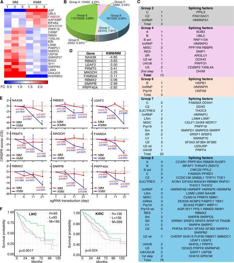
To further assess the functional significance of these splicing factors, we leveraged results from a CRISPR-Cas9 screening in KSHV-transformed (KMM) and untransformed (MM) cells (ref. 17). All cellular genes were classified into 9 groups (ref. 17). Group 1 consisted of genes that had significant increase in CRISPR score for KMM cells but significant decrease in CRISPR score for MM cells; Group 2 consisted of genes that had significant increase in CRISPR score for KMM cells but no significant change in CRISPR score for MM cells; Group 3 consisted of genes that had significant increases in CRISPR score for both KMM and MM cells; Group 4 consisted of genes that had no significant change in CRISPR score for KMM cells but had significant decrease in CRISPR score for MM cells; Group 5 consisted of genes that had no significant change in CRISPR score for both KMM and MM cells; Group 6 consisted of genes that had no significant change in CRISPR score for KMM cells but had significant increase in CRISPR score for MM cells; Group 7 consisted of genes that had significant decreases in CRISPR score for both KMM and MM cells; Group 8 consisted of genes that had significant decrease in CRISPR score for KMM cells but had no significant change in CRISPR score for MM cells; and Group 9 consisted of genes that had significant decrease in CRISPR score for KMM cells but had significant increase in CRISPR score for MM cells. This screen identified over 200 splicing-related genes, with approximately 50% being essential for KMM cell proliferation and survival, but not for MM cells (Fig. 1B) (ref. 26). Notably, these essential genes were highly enriched in Groups 7 and 8 (ref. 17), where splicing factors were nearly 10-fold overrepresented compared to other groups. Importantly, the identified splicing factors spanned all major spliceosome complexes and splicing factor families (Fig. 1C).
The top nine splicing factors in Group 8, ranked by CRISPR scores, were NAA38, RBM22, U2AF2, PRMT5, MAGOH, FAM50A, RBMX2, SNRPB, and PRPF40A (Fig. 1D). These factors all belong to core spliceosomal complexes and play key roles in pre-mRNA splicing (Fig. 1C). Notably, knockout of any of these nine genes significantly inhibited KMM cell proliferation and survival, while exerting minimal effects on MM cells (Fig. 1E). Among them, FAM50A was the only splicing factor that was upregulated in KMM cells relative to MM cells (Fig. 1A), highlighting its unique role in KSHV-driven cellular transformation.
Given the essential role of these nine splicing factors in KSHV-induced cellular transformation, we assessed their clinical relevance by analyzing their prognostic significance in cancer using TCGA survival data. High expression of any of these factors was correlated with poor survival in multiple cancer types (Fig. 1F; Fig. S1; Table S1), including FAM50A in adenoid cystic carcinoma (ACC), liver hepatocellular carcinoma (LIHC), esophageal cancer (ESCA), kidney renal clear cell carcinoma (KIRC), sarcoma (SARC), kidney chromophobe carcinoma (KICH), acute myeloid leukemia (LAML), mesothelioma (MESO), and uveal melanoma (UVM) (Fig. S1A); NAA38 in colon adenocarcinoma (COAD), LIHC, and MESO (Fig. S1B); RBM22 in KICH, LIHC, kidney renal papillary cell carcinoma (KIRP), and SARC (Fig. S1C); U2AF2 in ACC, brain lower grade glioma (LGG), LAML, MESO, LIHC, SARC, and UVM (Fig. S1D); PRMT5 in bladder urothelial carcinoma (BLCA), LIHC, head and neck squamous cell carcinoma (HNSC), and thyroid carcinoma (THCA) (Fig. S1E); MAGOH in ACC, LIHC, LGG, KIRP, MESO, and SARC (Fig. S1F); RBMX2 in ESCA, LIHC, HNSC, KIRC, KIRP, and uterine corpus endometrial carcinoma (UCEC) (Fig. S1G); SNRPB in LGG, LIHC, COAD, KIRC, KIRP, LAML, lung squamous cell carcinoma (LUSC), MESO, SARC, and UVM (Fig. S1H); and PRPF40A in ACC, LIHC, LGG, KICH, KIRP, lung adenocarcinoma (LUAD), MESO, pancreatic adenocarcinoma (PAAD), and UCEC (Fig. S1I).
These findings demonstrate that KSHV-induced cellular transformation profoundly reshapes alternative splicing regulation, highlighting the critical role of splicing factors in oncogenesis. Among the identified factors, FAM50A emerges as a key mediator of KSHV-driven splicing reprogramming, potentially contributing to the pathogenesis of KSHV-associated malignancies.
KSHV reprograms alternative pre-mRNA splicing through viral latent genes and miRNAs
To investigate how KSHV infection regulates host cell alternative splicing, we performed splicing analysis using SUPPA2 (ref. 27) and identified 131 differentially spliced transcripts between MM and KMM cells (Fig. 2A). Among these, cassette exon (SE) events accounted for 68%, while alternative first exon (AFE) events represented 19% (Fig. 2B). Gene ontology (GO) analysis of biological processes (BP) revealed that the affected transcripts were highly enriched in pathways implicated in KSHV infection and cellular transformation, including the regulation of vascular permeability (ref. 16, ref. 28, ref. 29), multiple metabolic pathways (ref. 30ref. –ref. 32), and ERK1/2 signaling cascades (ref. 33ref. –ref. 40) (Fig. 2C).
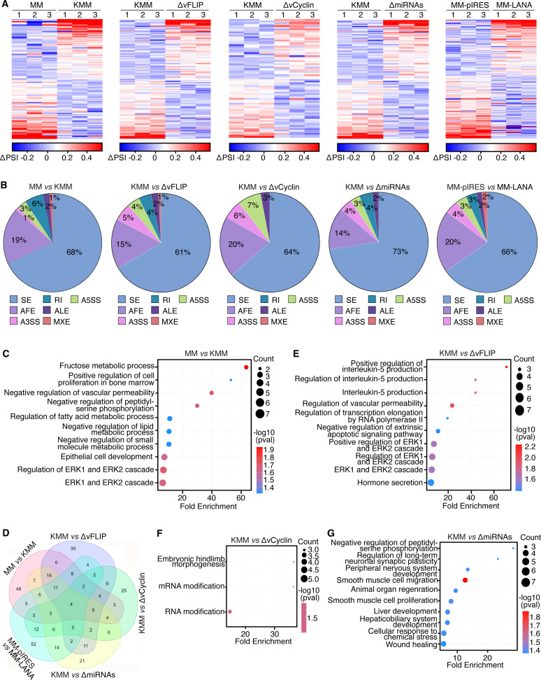
During latent infection, KSHV expresses only a limited set of viral genes, including LANA, vFLIP, vCyclin, and a cluster of viral miRNAs (ref. 14, ref. 41). To determine whether these latent genes contribute to the regulation of host alternative splicing, we performed splicing analysis in MM cells infected with KSHV mutants lacking vFLIP (ΔvFLIP), vCyclin (ΔvCyclin), or a cluster of pre-miRNAs (ΔmiRNAs) (Fig. 2A and B). Since LANA is essential for latent infection (ref. 42, ref. 43), we generated MM cells expressing LANA (MM-LANA) and compared their splicing profiles with vector control (MM-pIRES) cells. Across all conditions, SE events were the most prevalent ASEs, constituting 61%–73% of the total (Fig. 2B).
A Venn diagram analysis revealed that vFLIP and KSHV miRNAs had a more substantial impact on KSHV-induced alternative splicing reprogramming than vCyclin and LANA (Fig. 2D). Specifically, 50.4% (63/125) of differentially spliced transcripts in ΔvFLIP cells and 42.4% (55/105) in ΔmiRNAs cells overlapped with those in KMM cells. By contrast, only 37.3% (28/75) of differentially spliced transcripts in ΔvCyclin cells and 31.2% (39/125) in LANA-overexpressing cells overlapped with KMM cell transcripts (Fig. 2D).
GO analysis further confirmed that transcripts differentially spliced in ΔvFLIP cells were enriched in pathways related to vascular permeability, ERK1/2 cascades, transcription elongation by RNA polymerase II, and interleukin-5 (IL-5) signaling (Fig. 2E). Similarly, transcripts differentially spliced in ΔmiRNAs cells were enriched in pathways associated with cell proliferation, migration, wound healing, and stress response (Fig. 2G). By contrast, few (ref. 3) enriched pathways were found for differentially spliced transcripts in ΔvCyclin cells, of which two are related to RNA modification (Fig. 2F), while no significantly enriched pathways were identified in LANA-overexpressing cells (data not shown).
Together, these findings suggest that KSHV reprograms host alternative splicing primarily through vFLIP and viral miRNAs, highlighting their pivotal roles in KSHV-induced oncogenic transformation (ref. 32, ref. 44).
FAM50A is essential for KSHV-induced cellular transformation and tumorigenesis
RNA-seq analysis revealed that FAM50A is significantly upregulated in KMM cells compared to MM cells (Fig. 1A and 3A), a finding further validated by Western-blotting analysis (Fig. 3B). Similarly, elevated FAM50A expression was observed in KSHV-infected PEL cell lines (BC3 and BCP1) and KSHV-infected BJAB cells (BJAB-KSHV) compared to uninfected BJAB cells (Fig. 3C).
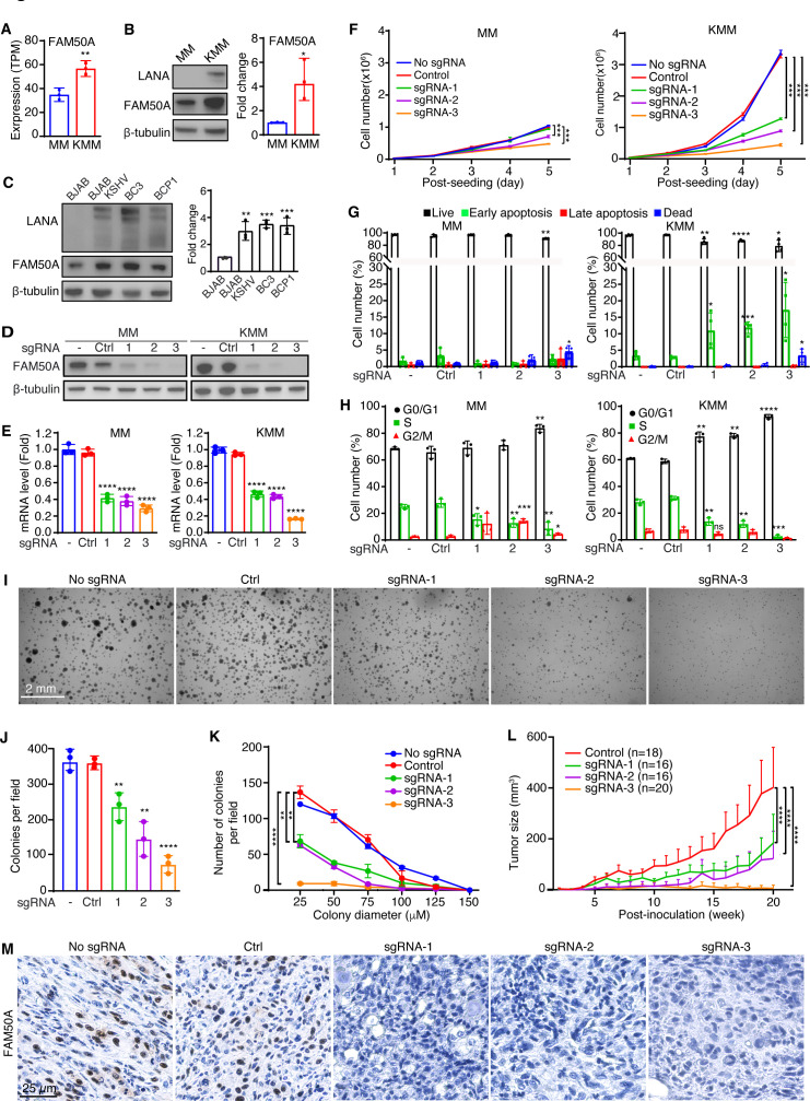
To determine the functional role of FAM50A in KSHV-induced cellular transformation, we generated FAM50A knockout cell lines in both MM and KMM cells (Fig. 3D and E). FAM50A knockout markedly reduced the proliferation of KMM cells, while only marginally affecting MM cells (Fig. 3F), suggesting FAM50A mediates KSHV-induced cell proliferation.
We then examined the impact of FAM50A depletion on KSHV gene expression. FAM50A sgRNA1 and 2 reduced the expression of 20%–30% of KSHV latent genes (LANA, vFLIP, and vCyclin), while sgRNA3 led to a more robust suppression of 70%–80% (Fig. S2A). This suppression correlated with a reduction in KSHV genome copy number per cell (Fig. S2B), implying that FAM50A may be involved in KSHV genome replication or persistence. After normalizing for viral genome copy number, the suppression effect on gene expression was no longer observed. We further evaluated KSHV lytic gene expression (RTA/ORF50, MTA/ORF57, ORF-K8, ORF-K8.1, and ORF65; Fig. S2A). Similar to latent genes, sgRNA1 and 2 downregulated most lytic genes, although sgRNA2 had no effect on MTA. By contrast, sgRNA3 increased the expression of all lytic genes by 1.6-fold to 2-fold, except for MTA, which was inhibited by over 90%. Immunofluorescence failed to detect lytic protein expression (RTA, ORF-K8.1, and ORF65), consistent with the tightly latent nature of KMM cells, suggesting that the suppression effect of FAM50A knockout is unlikely due to lytic reactivation.
Apoptosis analysis revealed that FAM50A knockout triggered early apoptosis in KMM cells while having minimal impact on MM cells (Fig. 3G). In addition, cell cycle analysis demonstrated that FAM50A knockout induced G0/G1 cell cycle arrest in both MM and KMM cells, with a more pronounced effect in KMM cells (Fig. 3H). These findings suggest that FAM50A plays a key role in promoting KSHV-induced cell cycle progression and survival.
To further assess the role of FAM50A in KSHV-driven cellular transformation, we conducted a soft agar colony formation assay. FAM50A knockout significantly impaired the ability of KMM cells to form colonies, reducing both colony number and size (Fig. 3I through K). To evaluate its role in tumorigenesis, we examined tumor growth in nude mice using FAM50A knockout cells. Loss of FAM50A markedly suppressed tumor progression in KMM-derived xenografts (Fig. 3L; Fig. S3). Immunohistochemical analysis confirmed strong FAM50A expression in tumors derived from KMM-Cas9 cells (No sgRNA) and KMM cells with scrambled sgRNAs (Control), whereas tumors from FAM50A knockout cells showed no detectable expression (Fig. 3M). Together, these results demonstrate that FAM50A plays a critical role in KSHV-induced cellular transformation and tumorigenesis.
FAM50A is essential for the proliferation of PEL cells
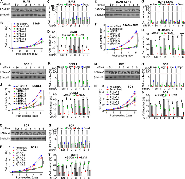
To further investigate the role of FAM50A in PEL cell proliferation, we performed siRNA-mediated knockdown of FAM50A in KSHV-infected PEL cell lines, including BCBL1, BC3, and BCP1, as well as KSHV-BJAB cells. This knockdown markedly suppressed cell proliferation in all KSHV-infected lines, whereas the effect on uninfected BJAB cells was minimal (Fig. 4A, B, E, F, I, J, M, N, Q and R). Unlike KMM cells, FAM50A depletion did not induce apoptosis in any of the tested cell lines (Fig. 4C, G, K, O and S). Instead, FAM50A knockdown led to a significant G0/G1 cell cycle arrest in KSHV-infected BCBL1, BC3, BCP1, and KSHV-BJAB cells, but not in BJAB cells (Fig. 4D, H, L, P and T). These findings demonstrate that FAM50A is a critical regulator of KSHV-driven cell proliferation and cell cycle progression in PEL cells.
FAM50A regulates alternative pre-mRNA splicing in MM and KMM cells
Given that FAM50A functions as an essential splicing factor in KMM cells, we investigated the alternative spliced transcripts regulated by FAM50A in MM and KMM cells. RNA-seq analysis of FAM50A knockout MM and KMM cells identified 34 differentially spliced transcripts in MM cells (MM vs. MM-sgFAM50A) and 28 in KMM cells (KMM vs. KMM-sgFAM50A) (Fig. 5A and B; Fig. S4). GO analysis revealed that both MM and KMM cells share common alternative spliced transcripts upon FAM50A knockout, including those related to fructose metabolism and carbohydrate phosphorylation (Fig. 5C and D). However, alternative spliced transcripts in FAM50A knockout KMM cells were also enriched in pathways crucial for KSHV-induced cellular transformation, such as apoptosis, oxidative stress, and platelet-derived growth factor (PDGF) signaling (Fig. 5D). While 16 of the 34 differentially spliced transcripts in MM FAM50A knockout cells and 12 of the 28 in KMM FAM50A knockout cells overlapped with those in KMM cells, only four were common across both cell types (Fig. 5E). This suggests that FAM50A regulates distinct ASEs in MM and KMM cells, potentially contributing to differences in biological functions.
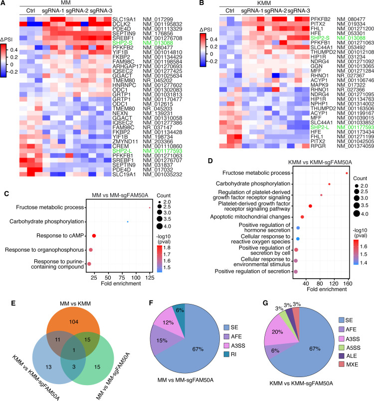
Among the various types of alternative splicing, SE events were the most predominant, accounting for 67% in both FAM50A knockout MM and KMM cells (Fig. 5F and G). Notably, alternative 3′ splice site selection (A3SS) accounted for 20% of differentially spliced transcripts in KMM FAM50A knockout cells, but only 12% in MM FAM50A knockout cells, further underscoring the differential splicing landscape shaped by FAM50A in KSHV-transformed cells.
A closer examination of the affected transcripts revealed genes with established roles in cancer development, particularly those downregulated in FAM50A-deficient KMM cells, which may be key mediators of its function in KSHV-induced oncogenesis. For instance, RHNO1, a protein that promotes homologous recombination repair and is frequently upregulated in breast, liver, and pancreatic cancers, supports tumor cell survival and chemoresistance (ref. 45), while THUMPD2 is an RNA-binding protein catalyzing N2-methylation of U6 small nuclear RNA and regulating mRNA splicing (ref. 46), and has been shown to regulate ovarian cancer progression, cell migration, and invasion (ref. 47).
A number of these genes, including ACYP1, MFF, SLC44A1, and HFE, promote cancer development by regulating metabolic pathways. ACYP1 regulates glycolytic processes and is implicated in tumor growth, invasion, and resistance to apoptosis in liver cancer (ref. 48, ref. 49), while MFF plays a central role in mitochondrial fission and is implicated in lung, colon, and breast cancers by regulating cell proliferation, apoptosis, and invasion (ref. 50). SLC44A1 is implicated in multiple cancers by regulating tumor cell proliferation, membrane biosynthesis, and potentially survival under metabolic stress through its central role in choline uptake and lipid metabolism (ref. 51), while HFE, which regulates iron metabolism, has been associated with hepatocellular carcinoma via mechanisms involving iron overload, DNA damage, and immune evasion (ref. 52).
FAM50A-regulated transcripts also include several key signaling and transcriptional regulators that influence oncogenesis. SHP2 is a pivotal mediator of RAS/MAPK, PI3K/AKT, and STAT3 signaling pathways and is broadly implicated in diverse malignancies (ref. 53). Similarly, NDRG4, known to influence PI3K/AKT and MAPK/ERK signaling, plays important roles in cell cycle regulation and the inhibition of apoptosis in breast and colorectal cancers (ref. 54, ref. 55). FHL1, a LIM-domain protein, and PITX2, a homeobox transcription factor, both function as tumor suppressors or oncogenes depending on the cellular context and are involved in regulating major signaling cascades such as MAPK, TGF-β, and WNT/β-catenin pathways (ref. 56, ref. 57). These pathways govern crucial processes like cell proliferation, epithelial-mesenchymal transition, and invasion, all of which are central to tumor initiation and progression.
Together, these findings reveal that FAM50A governs a set of alternative splicing events that significantly impact gene networks involved in DNA repair, signaling, metabolism, and transcriptional control. Its differential regulation of pre-mRNA splicing in MM and KMM cells suggests that FAM50A may act as a context-dependent modulator of oncogenic pathways, particularly in the context of KSHV-driven transformation. This underscores its potential as a key node linking RNA processing to oncogenesis in virus-associated cancers.
FAM50A enhances STAT3 activation and cell proliferation by regulating SHP2 alternative splicing
Among all identified alternative spliced transcripts, SHP2 transcripts were the most significantly affected in both MM and KMM cells following FAM50A knockout. SHP2 encodes two isoforms: a short isoform (SHP2-S) and a long isoform (SHP2-L). While FAM50A knockout did not alter the total SHP2 expression level, it upregulated SHP2-S while downregulating SHP2-L (Fig. 6A; Fig. S5A). These results were further validated by semi-quantitative RT-PCR (Fig. 6B and C; Fig. S5B).
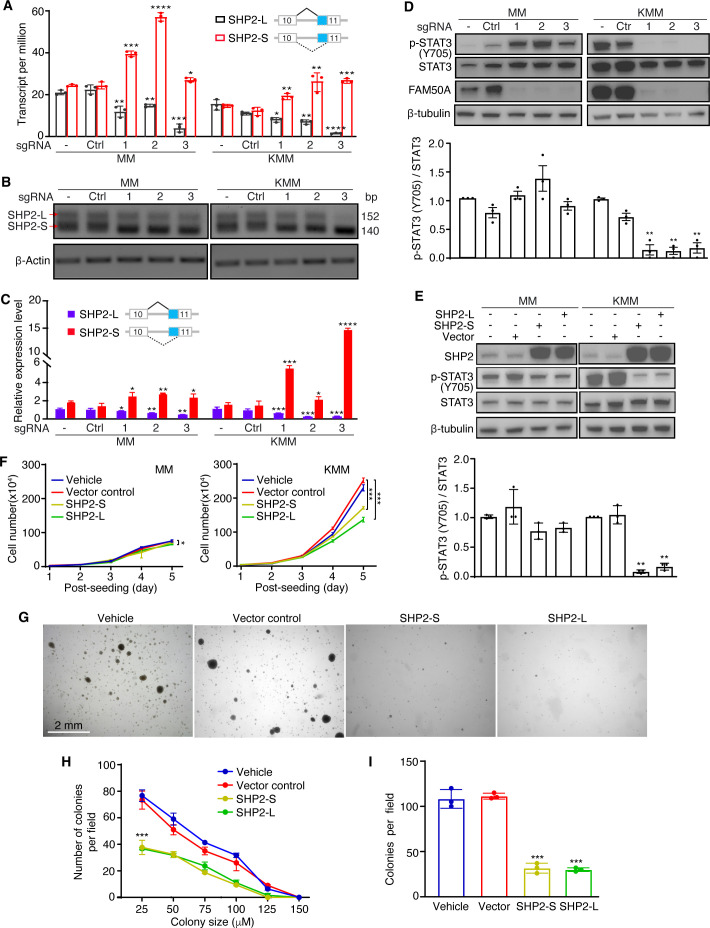
Given that SHP2-S exhibit 10-fold higher phosphatase catalytic activity than SHP2-L (ref. 58) and that FAM50A knockout leads to an increase in SHP2-S, we hypothesized that SHP2 phosphatase activity could be elevated following FAM50A knockout. To test this, we examined the phosphorylation of STAT3-Y705, a known SHP2 downstream target (ref. 59). Indeed, FAM50A knockout significantly inhibited STAT3-Y705 phosphorylation in KMM cells (Fig. 6D). Unexpectedly, STAT3-Y705 phosphorylation was increased in MM cells following FAM50A knockout, suggesting that SHP2 regulation of STAT3 activation is cell type-dependent.
To further investigate this, we overexpressed SHP2-S or SHP2-L in MM and KMM cells. Both SHP2-S and SHP2-L inhibited STAT3 activation in MM and KMM cells, but the inhibitory effect was more pronounced in KMM cells (Fig. 6E). Consistently, overexpression of SHP2-S or SHP2-L inhibited KMM cell proliferation, whereas MM cell proliferation was largely unaffected (Fig. 6F). Moreover, SHP2-S or SHP2-L overexpression suppressed colony formation of KMM cells, leading to a reduction in both colony number and size (Fig. 6G through I).
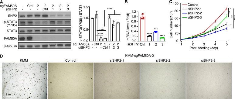
To further confirm the role of SHP2 in mediating FAM50A-dependent STAT3 regulation, we performed siRNA-mediated SHP2 knockdown in FAM50A knockout KMM cells. SHP2 depletion restored STAT3-Y705 phosphorylation (Fig. 7A and B), reinforcing the notion that SHP2 mediates STAT3 inhibition in FAM50A knockout KMM cells. However, SHP2 knockdown failed to rescue the inhibition of cell proliferation caused by FAM50A knockout (Fig. 7C). In line with this observation, SHP2 knockdown did not restore colony formation in soft agar following FAM50A depletion (Fig. 7D). These results suggest that STAT3 activation and cell proliferation are regulated by additional factors besides SHP2, implying that SHP2 is not the sole downstream target of FAM50A.
Together, our findings indicate that FAM50A plays a critical role in KSHV-induced cell proliferation and cellular transformation by regulating SHP2 alternative splicing and STAT3 activation.
DISCUSSION
Understanding how cancer cells reprogram splicing to facilitate oncogenesis is fundamental to cancer biology. In this study, we uncovered that oncogenic KSHV manipulates multiple splicing factors to promote the proliferation and survival of KSHV-transformed cells. Our findings demonstrate that KSHV systemically reprograms alternative splicing in host cells during cellular transformation and that KSHV latent genes, particularly vFLIP and KSHV miRNAs, play a crucial role in this process. While extensive research has focused on how KSHV latent genes mediate viral latent replication and transcriptional regulation, their roles in alternative splicing have remained largely unexplored (ref. 14). Here, we provide compelling evidence that KSHV infection and latent genes contribute to splicing regulation, a novel aspect of KSHV-driven oncogenesis. Further studies are warranted to determine whether these findings extend to other KSHV-latently infected cell types.
Although the precise mechanisms by which KSHV infection and latent genes regulate alternative splicing remain to be fully elucidated, our results indicate that KSHV alters the expression of multiple splicing factors in transformed cells (Fig. 1A). Beyond transcriptional control, KSHV latent proteins may directly interact with RNA-binding proteins or act as splicing regulators by associating with pre-mRNA or the spliceosome, analogous to the function of the KSHV lytic protein ORF57 (ref. 60). Notably, the alternative spliced transcripts modulated by KSHV latent genes only partially overlap with those driven by KSHV-induced cellular transformation (Fig. 2D), suggesting that additional factors contribute to splicing dysregulation. While some alternative spliced transcripts are shared, each KSHV latent gene appears to regulate a distinct subset, underscoring the complexity and specificity of host splicing alterations during KSHV-induced cellular transformation.
Aberrant splicing is a well-documented hallmark of cancer, with specific splicing factors implicated in tumorigenesis (ref. 25). Alternative splicing contributes to transcriptomic diversity, affecting key cellular processes such as proliferation, apoptosis, and immune evasion (ref. 5). However, little is known about how oncogenic viruses alter alternative splicing and splicing factors in virus-induced cancers. To address this gap, we analyzed the results of our previous genome-wide CRISPR-Cas9 screen to systematically identify splicing factors essential for KSHV-induced cellular transformation (Fig. 1C and D) (ref. 17). Among these factors, FAM50A emerged as a critical regulator, exhibiting significant upregulation in KSHV-transformed cells. Unlike well-characterized spliceosomal components, FAM50A remains largely unexplored in both cancer and viral infection. Our study has shown that FAM50A is not only upregulated in KSHV-transformed cells but also actively participates in alternative splicing regulation. These results align with prior reports describing the oncogenic roles of other spliceosomal proteins (ref. 61, ref. 62).
Importantly, we identified FAM50A as an essential factor in KSHV-induced cellular transformation, functioning in part by modulating KSHV-driven splicing reprogramming (Fig. 3). The essential role of FAM50A in KSHV-driven oncogenesis is confirmed not only in KS model KMM cells but also in PEL cells (Fig. 4). FAM50A knockout affects the alternative splicing of genes involved in diverse functions, including proliferation, survival, invasion, immune evasion, etc. (Fig. 5). Notably, FAM50A knockout selectively alters SHP2 isoform expression, shifting it toward a catalytically active short isoform in KSHV-transformed cells (Fig. 5A through C) (ref. 58). This isoform switch results in decreased phosphorylation of STAT3 at Y705 (Fig. 5D), a key driver of KSHV-induced cellular transformation (ref. 30, ref. 63, ref. 64). Consequently, FAM50A deficiency suppresses KSHV-transformed cell proliferation and anchorage-independent growth in soft agar (Fig. 6F through I). These findings implicate FAM50A as a virus-specific regulator of SHP2 catalytic activity, revealing a novel mechanism by which KSHV exploits the host splicing machinery to promote oncogenesis.
The identification of FAM50A as an essential factor for KSHV-induced transformation suggests its potential as a therapeutic target. Given that FAM50A is associated with poor prognosis in multiple cancers (Fig. 1F; Fig. S1A) (ref. 22), its inhibition could have broad therapeutic implications (ref. 65). Our findings indicate that FAM50A’s function differs between primary and KSHV-transformed cells (Fig. 6D through F), raising the possibility of selectively targeting FAM50A in virus-associated malignancies. Future research should explore potential strategies for FAM50A inhibition, including small-molecule inhibitors or RNA-based therapeutics, and evaluate possible off-target effects in non-cancerous cells.
While our study provides novel insights into FAM50A’s role in KSHV-induced cellular transformation, several limitations should be acknowledged. First, our findings are based primarily on in vitro cell culture and immunocompromised mouse models; further validation in KSHV-induced human cancers is necessary. Nevertheless, our results have confirmed the essential role of FAM50A in KSHV-driven cell proliferation of human PEL cells (Fig. 4). Second, although FAM50A has been proposed as a promising therapeutic target (ref. 22, ref. 23), its clinical potential remains unexplored, necessitating further studies to assess the safety and efficacy of its inhibition. In addition, an intriguing question remains as to whether KSHV latent proteins directly interact with FAM50A to regulate alternative splicing. Given that nuclear-localized KSHV latent proteins, such as LANA, may co-reside with spliceosomal components, they may directly interact with FAM50A and other splicing factors to mediate splicing regulation. Future studies should investigate these interactions and identify the essential ASEs driving KSHV-induced transformation.
In summary, our study demonstrates that splicing factors play fundamental roles in KSHV-induced cellular transformation. KSHV systematically reprograms host splicing, with its latent genes including vFLIP, vCyclin, LANA, and miRNAs closely involved in alternative splicing regulation. Furthermore, we identified FAM50A as a key splicing factor that promotes KSHV-induced cellular transformation by modulating alternative splicing and shifting SHP2 isoform expression, leading to decreased STAT3-Y705 phosphorylation. These findings reveal a novel mechanism by which KSHV manipulates the host splicing machinery to drive oncogenesis. Targeting FAM50A may offer new therapeutic opportunities for KSHV-associated malignancies and potentially other cancers dependent on aberrant splicing.
MATERIALS AND METHODS
Antibodies
The antibodies used for Western blotting included rabbit anti-FAM50A/XAP5 (Abcam, ab186410), rat anti-LANA (Abcam, ab4103), rabbit anti-SHP2 (Cell Signaling Technology, 3397), mouse anti-STAT3 (Cell Signaling Technology, 9139), rabbit anti-phospho-STAT3 (Tyr705) (Cell Signaling Technology, 9145), mouse anti-FLAG (Sigma-Aldrich, F1804), and mouse anti-β-tubulin (Sigma-Aldrich, 7B9). For immunohistochemistry (IHC), a rabbit anti-FAM50A antibody (Abcam, ab186410) was used.
Cell culture
MM and 293T cells were maintained in Dulbecco’s modified Eagle medium (DMEM) (Genesee, 25-500) supplemented with 10% fetal bovine serum (FBS) (Sigma-Aldrich, F2442) and 1% penicillin/streptomycin (Gibco, 15140-122) in a 5% CO₂ incubator at 37°C. KMM cells were cultured under the same conditions as MM cells but with the addition of 250 µg/mL hygromycin. MM and KMM cells with stable FAM50A knockout were maintained in their respective media. Cells with stable SHP2-S or SHP2-L expression were cultured in their respective media supplemented with 10 µg/mL Blasticidin (ThermoFisher Scientific, A1113903).
The PEL cell lines (BCBL1, BC3, and BCP1), the Burkitt lymphoma cell line (BJAB), and the KSHV-infected BJAB cell line (BJAB-KSHV) were cultured in RPMI 1640 medium (Invitrogen, Carlsbad, CA) containing 20% FBS at 37°C under 5% CO2. In addition, BJAB-KSHV cells were maintained in medium supplemented with 10 µg/mL Puromycin (Sigma-Aldrich, P8833).
All cell lines were cultured in drug-free medium for 1 week prior to experiments.
Plasmids and transfection
All plasmids were constructed using the restriction enzyme digestion and ligation method. The pBSD-FLAG-SHP2-S and pBSD-FLAG-SHP2-L plasmids were generated by inserting PCR-amplified products into the pBSD vector using the EcoRI and XbaI restriction sites. The pBSD plasmid was obtained from Addgene (Plasmid #119863).
Plasmid transfection was carried out using Lipofectamine 2000 (Invitrogen, 11668019) following the manufacturer’s protocol. Cells were transfected using a ratio of 1 µg of plasmid DNA to 3 µL of Lipofectamine 2000 and cultured for 3 days before being used for the experiments.
Generation of CRISPR/Cas9-mediated FAM50A gene knockout cell lines
FAM50A knockout KMM/MM cell lines were generated using the CRISPR/Cas9 (clustered regularly interspaced palindromic repeats/CRISPR-associated protein 9) system, following previously established protocols (ref. 17). Briefly, a single-guide RNA (sgRNA) sequence targeting the FAM50A locus (sgFAM50A sequence: 5′-TGGGCACCGGCGCACTGTTAAGG-3′) was designed on the basis of Cas-OFFinder (ref. 66) and Cas-Designer (ref. 67). The sgFAM50A plasmid was transduced into KMM/MM cells using lentivirus. The production of sgFAM50A lentivirus was performed as previously described (ref. 17).
Following transduction, cells were cultured for 48 hours before being subjected to stepwise serial dilution to isolate single-cell clones. Clones exhibiting puromycin resistance were selected and further analyzed by genomic DNA sequencing. Only clones displaying a single sequencing peak with a gap, indicative of successful gene knockout, were selected for subsequent experiments.
Small interfering RNA-mediated SHP2 knockdown
For knockdown of SHP2 in KMM cells, small interfering RNAs (siRNAs) targeting Rattus norvegicus SHP2 (SASI_Rn01_00105327, SASI_Rn01_00105328, SASI_Rn01_00105329) and the scramble control (SIC001) were purchased from Sigma-Aldrich. siRNA duplex (75 pmol) in 7.5 µL of Lipofectamine RNAiMAX Transfection Reagent (Life Technologies, 13778100) was diluted in Optimem medium (Life Technologies, 31985062) and added to the cells according to the manufacturer’s instructions. Cells were harvested 48 hours post-transfection to assess knockdown efficiency.
siRNA-mediated knockdown of human FAM50A in PEL cells and BJAB cells was similarly carried out. siRNAs targeting human FAM50A are as follows: SASI_Hs01_00137461 (si1), SASI_Hs01_00137462 (si2), SASI_Hs01_00137465 (si3), SASI_Hs01_00137467 (si4), SASI_Hs01_00137468 (si5), SASI_Hs01_00137470 (si6), and siRNA Universal Negative Control #1 (SIC001-10NMOL) (siControl or siCl) (Sigma).
RT-qPCR detection of gene expression
Total RNA was extracted from cultured cells using the TRIzol reagent (Sigma-Aldrich, T9424). First-strand cDNA synthesis was carried out with 50–100 ng of RNA per reaction using the Maxima H Minus First Strand cDNA Synthesis Kit (ThermoFisher Scientific, K1652). Quantitative real-time PCR (qPCR) was carried out using the SsoAdvanced Universal SYBR Green Supermix Kit (Bio-Rad, 172-5272) according to the manufacturer’s instructions. Gene expression levels were normalized to β-actin mRNA. The primers used for qPCR are listed in Table S2.
Validation of ASEs by semiquantitative RT-PCR
Validation of alternative spliced transcripts was performed using semiquantitative RT-PCR. Total RNA was isolated, and first-strand cDNA was synthesized as described previously. PCR amplification was carried out using the Platinum PCR SuperMix High Fidelity (ThermoFisher Scientific, 12532016). Reaction products were separated on 2% agarose gels and visualized by ethidium bromide staining. The relative abundance of each splicing isoform was quantified using ImageJ software. Primers used for splicing assays are listed in Table S2.
Purification and quantification of viral DNA by qPCR
Total DNAs in KMM, KMM-Cas9 control, and FAM50A knockout KMM cells (sgFAM50A-1, 2, and 3) were collected and extracted by the Total DNA Isolation Kit (ThermoFisher Scientific, K0512). KSHV DNA was measured using SsoAdvanced Universal SYBR Green Supermix (BioRad, 172-5272) based qPCR assay with ORF71-73 primers along with the cellular actin gene primers in triplicate. The average values were used to determine the viral copies. The cell-associated KSHV DNA values were converted to copies per million cells using a cell quantitation assay based on the viral gene with an assay sensitivity of 10 copies/106 cells.
Cell proliferation assay
Cells were seeded into 12-well plates at a density of 30,000 cells per well. Three biological replicates were performed for each condition. At the indicated time points, cells were harvested and counted using a hemocytometer.
Cell cycle and apoptosis assays
Cells were seeded into six-well plates and cultured overnight. For cell cycle analysis, 5-bromo-2′-deoxyuridine (BrdU; 10 µM; Sigma-Aldrich, B5002) was added to the culture medium at 10 µM for 2 hours to label replicating DNA. Cells were then fixed with 70% ethanol, permeabilized with 2 M hydrochloric acid, and stained with an anti-BrdU monoclonal antibody (ThermoFisher Scientific, B35129). Cells were further stained with propidium iodide (PI), and flow cytometry was performed using a FACS Canto II system (BD Biosciences). Apoptotic cells were detected by flow cytometry using Fixable Viability Dye eFluor 660 (Invitrogen, 65-0864) and Annexin V Apoptosis Detection Kits (Invitrogen, 88-8103-74) according to the manufacturer’s instructions. Cells treated with 100 µM Menadione (Sigma-Aldrich, M5625) served as positive controls for apoptosis induction. All experiments were performed in three biological replicates, and data were analyzed using FlowJo software (BD Biosciences).
Soft agar assay
The soft agar assay was performed as previously described (ref. 16). Briefly, 5 × 10⁴ cells were suspended in 1 mL of 0.3% top agar (Sigma-Aldrich, A5431) and plated onto a solidified 0.5% base agar layer in six-well plates. The wells were then overlaid with culture medium to maintain cell viability. After 2 weeks of incubation, colonies were visualized using a microscope with a 2 × objective lens, and colonies with a diameter greater than 50 µm were counted.
Animal studies
Six-week-old male Hsd:Athymic Nude-Foxn1nu mice (Envigo, Inotiv) were maintained under standardized pathogen-free conditions. FAM50A knockout KMM cells (sgFAM50A-1, 2, and 3), KMM-Cas9 cells, and KMM cells with scrambled sgRNAs (Control) were subcutaneously (IC) injected into both flanks of the mice at a density of 5 × 10⁶ cells in 0.1 mL of PBS per site using a 25-gauge needle. Tumor size was monitored twice weekly. The mice were euthanized at 20 weeks post-injection, and the tumors were excised, weighed, and processed for immunohistochemistry.
Immunohistochemistry staining
Formalin-fixed, paraffin-embedded (FFPE) tissue sections were rehydrated through a series of xylene and ethanol washes (100%, 95%, and 75%), followed by rinsing in water. Antigen retrieval was performed by pressure cooking the sections in citrate buffer at 110°C for 20 minutes. After washing to remove citrate buffer, endogenous peroxidase activity was quenched with 3% hydrogen peroxide in methanol for 30 minutes at room temperature. Sections were blocked with 5% BSA in 1× TBST for 1 hour at 37°C, followed by overnight incubation with the primary antibody (1:100 dilution in 2.5% BSA in 1× TBST) in a humidity-controlled chamber. The secondary antibody was applied at a 1:100 dilution in 2.5% BSA in 1× TBST and incubated for 60 minutes at 37°C. Slides were developed using the ImmPACT DAB Substrate Kit, Peroxidase (Vector, SK-4105), according to the manufacturer’s instructions, and counterstained with hematoxylin QS (Vector, H-3404-100). Finally, sections were dehydrated through graded ethanol (75%, 95%, and 100%) and xylene washes before being mounted with xylene-based mounting media (Epredia Shandon-Mount, 1900333).
Analysis of splicing factors identified by CRISPR-Cas9 screening
Our prior CRISPR-Cas9 screening of MM and KMM cells identified essential genes for cell proliferation and survival (ref. 17). From this data set, we mapped over 200 splicing-related factors (ref. 26) into nine functional groups based on the CRISPR-Cas9 screening results. The percentage of enrichment for each group was calculated, and splicing factors were further categorized based on their roles within the spliceosome.
RNA-seq
Total RNA was extracted from cells using TRI Reagent (Sigma-Aldrich, T9424) following the manufacturer’s instructions. Approximately 10 µg of RNA from each sample was used for mRNA library preparation. The TruSeq Stranded mRNA Library Prep Kit (Illumina) was used to construct next-generation sequencing libraries according to the manufacturer’s protocol. Paired-end sequencing (150 base pairs) was performed on an Illumina HiSeq 4000 platform (Illumina).
RNA-seq data analysis
RNA-seq data analysis was performed by first processing raw reads in fastq format to obtain clean reads, which were then aligned to the rat genome mRatBN7.2 using STAR (ref. 68). Transcripts were assembled, and raw gene counts were estimated using featureCounts (ref. 69) and presented in transcripts per million (TPM). Differentially expressed genes (DEGs) were identified using DESeq2 (ref. 70) with a significance threshold of P-adjusted value < 0.05 and fold change ≥ 2. ASEs were analyzed with SUPPA2 (ref. 27), and results were filtered based on significance (P-adjusted value < 0.05), percent spliced-in (PSI) changes (|ΔPSI| > 0.1), and SUM (TPMs of three replicates) ≥2.5. Gene ontology (GO) analysis for functional annotation of candidate alternative spliced transcripts was conducted using the online database for annotation, visualization, and integrated discovery (ref. 71).
Statistical analysis
Statistical analyses were conducted using GraphPad Prism software for experimental data, while data from TCGA and GEO repositories were analyzed using R software. Comparative analyses between experimental groups and their respective controls were performed using Student’s t-test, log-rank test, Wilcoxon matched-pairs signed-rank test, Mann-Whitney U test, and one-way analysis of variance (ANOVA), as appropriate. Data are presented as mean ± SD, and statistical significance was defined as P < 0.05.
References
- The discovery of split genes and RNA splicing.. Trends Biochem Sci, 2005. [DOI | PubMed]
- RNA splicing by the spliceosome.. Annu Rev Biochem, 2020. [DOI | PubMed]
- Alternative isoform regulation in human tissue transcriptomes.. Nature, 2008. [DOI | PubMed]
- RNA splicing: a new paradigm in host-pathogen interactions.. J Mol Biol, 2019. [DOI | PubMed]
- Alternative splicing and cancer: a systematic review.. Sig Transduct Target Ther, 2021. [DOI]
- RBM5, 6, and 10 differentially regulate NUMB alternative splicing to control cancer cell proliferation.. Mol Cell, 2013. [DOI | PubMed]
- The RNA-binding protein AKAP8 suppresses tumor metastasis by antagonizing EMT-associated alternative splicing.. Nat Commun, 2020. [DOI | PubMed]
- TGF-β-induced alternative splicing of TAK1 promotes EMT and drug resistance.. Oncogene, 2019. [DOI | PubMed]
- Kaposi’s sarcoma-associated herpesvirus-like DNA sequences in AIDS-related body-cavity-based lymphomas.. N Engl J Med, 1995. [DOI | PubMed]
- Identification of herpesvirus-like DNA sequences in AIDS-associated Kaposi’s sarcoma.. Science, 1994. [DOI | PubMed]
- Kaposi’s sarcoma-associated herpesvirus-like DNA sequences in multicentric Castleman’s disease.. Blood, 1995. [PubMed]
- An interleukin-6-related systemic inflammatory syndrome in patients co-infected with Kaposi sarcoma-associated herpesvirus and HIV but without multicentric Castleman disease.. Clin Infect Dis, 2010. [DOI | PubMed]
- Kaposi sarcoma.. Nat Rev Dis Primers, 2019. [DOI | PubMed]
- Regulation of KSHV latency and lytic reactivation.. Viruses, 2020. [DOI | PubMed]
- 15 Schulz TF, Chang Y. 2007. KSHV gene expression and regulation. In Arvin A, Campadelli-Fiume G, Mocarski E, Moore PS, Roizman B, Whitley R, Yamanishi K (ed), Human herpesviruses: biology, therapy, and immunoprophylaxis. Cambridge.
- Direct and efficient cellular transformation of primary rat mesenchymal precursor cells by KSHV.. J Clin Invest, 2012. [DOI | PubMed]
- CRISPR-Cas9 screening of Kaposi’s sarcoma-associated herpesvirus-transformed cells identifies XPO1 as a vulnerable target of cancer cells.. mBio, 2019. [DOI | PubMed]
- Differentially regulated splice variants and systems biology analysis of Kaposi’s sarcoma-associated herpesvirus-infected lymphatic endothelial cells.. Nucleic Acids Res, 2011. [DOI | PubMed]
- Regulation of 3’ splice site selection after step 1 of splicing by spliceosomal C* proteins.. Sci Adv, 2023. [DOI | PubMed]
- Combinatorial CRISPR screen identifies fitness effects of gene paralogues.. Nat Commun, 2021. [DOI | PubMed]
- Mutations in FAM50A suggest that Armfield XLID syndrome is a spliceosomopathy.. Nat Commun, 2020. [DOI | PubMed]
- Upregulation of FAM50A promotes cancer development.. Med Oncol, 2023. [DOI | PubMed]
- Proto-oncogene FAM50A can regulate the immune microenvironment and development of hepatocellular carcinoma In Vitro and In Vivo.. Int J Mol Sci, 2023. [DOI | PubMed]
- Co-regulated gene expression of splicing factors as drivers of cancer progression.. Sci Rep, 2019. [DOI | PubMed]
- RNA splicing dysregulation and the hallmarks of cancer.. Nat Rev Cancer, 2023. [DOI | PubMed]
- The spliceosome: design principles of a dynamic RNP machine.. Cell, 2009. [DOI | PubMed]
- SUPPA2: fast, accurate, and uncertainty-aware differential splicing analysis across multiple conditions.. Genome Biol, 2018. [DOI | PubMed]
- Latent KSHV infection increases the vascular permeability of human endothelial cells.. Blood, 2011. [DOI | PubMed]
- Kaposi’s sarcoma-associated herpesvirus disrupts adherens junctions and increases endothelial permeability by inducing degradation of VE-cadherin.. J Virol, 2008. [DOI | PubMed]
- Oncogenic Kaposi’s sarcoma-associated herpesvirus upregulates argininosuccinate synthase 1, a rate-limiting enzyme of the citrulline-nitric oxide cycle, to activate the STAT3 pathway and promote growth transformation.. J Virol, 2019. [DOI | PubMed]
- A citical role of glutamine and asparagine γ-nitrogen in nucleotide biosynthesis in cancer cells hijacked by an oncogenic virus.. mBio, 2017. [DOI | PubMed]
- An oncogenic virus promotes cell survival and cellular transformation by suppressing glycolysis.. PLoS Pathog, 2016. [DOI | PubMed]
- Mechanism of sustained activation of ribosomal S6 kinase (RSK) AND ERK by kaposi sarcoma-associated herpesvirus ORF45: multiprotein complexes retain active phosphorylated ERK AND RSK AND protect them from dephosphorylation.. J Biol Chem, 2009. [DOI | PubMed]
- Pseudomonas aeruginosa stimulates inflammation and enhances kaposi’s sarcoma herpesvirus-induced cell proliferation and cellular transformation through both lipopolysaccharide and flagellin.. mBio, 2020. [DOI | PubMed]
- Kaposi’s sarcoma-associated herpesvirus induces the phosphatidylinositol 3-kinase-PKC-zeta-MEK-ERK signaling pathway in target cells early during infection: implications for infectivity.. J Virol, 2003. [DOI | PubMed]
- Modulation of Kaposi’s sarcoma-associated herpesvirus infection and replication by MEK/ERK, JNK, and p38 multiple mitogen-activated protein kinase pathways during primary infection.. J Virol, 2006. [DOI | PubMed]
- Sangivamycin induces apoptosis by suppressing Erk signaling in primary effusion lymphoma cells.. Biochem Biophys Res Commun, 2014. [DOI | PubMed]
- Reactivation of Kaposi’s sarcoma-associated herpesvirus from latency requires MEK/ERK, JNK and p38 multiple mitogen-activated protein kinase pathways.. Virology (Auckl), 2008. [DOI]
- Kaposi’s sarcoma-associated herpesvirus induction of AP-1 and interleukin 6 during primary infection mediated by multiple mitogen-activated protein kinase pathways.. J Virol, 2005. [DOI | PubMed]
- Kaposi’s sarcoma-associated herpesvirus promotes angiogenesis by inducing angiopoietin-2 expression via AP-1 and Ets1.. J Virol, 2007. [DOI | PubMed]
- Mechanisms of Kaposi’s sarcoma-associated herpesvirus latency and reactivation.. Adv Virol, 2011. [DOI | PubMed]
- Genetic disruption of KSHV major latent nuclear antigen LANA enhances viral lytic transcriptional program.. Virology (Auckl), 2008. [DOI]
- Disruption of Kaposi’s sarcoma-associated herpesvirus latent nuclear antigen leads to abortive episome persistence.. J Virol, 2004. [DOI | PubMed]
- KSHV microRNAs mediate cellular transformation and tumorigenesis by redundantly targeting cell growth and survival pathways.. PLoS Pathog, 2013. [DOI | PubMed]
- RHNO1: at the crossroads of DNA replication stress, DNA repair, and cancer.. Oncogene, 2024. [DOI | PubMed]
- N 2-methylguanosine modifications on human tRNAs and snRNA U6 are important for cell proliferation, protein translation and pre-mRNA splicing.. Nucleic Acids Res, 2023. [DOI | PubMed]
- RNA-binding protein THUMPD2 inhibits proliferation and promotes metastasis in epithelial ovarian cancer.. Heliyon, 2024. [DOI | PubMed]
- Development and validation of a carbohydrate metabolism-related model for predicting prognosis and immune landscape in hepatocellular carcinoma patients.. Curr Med Sci, 2024. [DOI | PubMed]
- Targeting ACYP1-mediated glycolysis reverses lenvatinib resistance and restricts hepatocellular carcinoma progression.. Drug Resist Updat, 2023. [DOI | PubMed]
- Mitochondrial fission in human diseases.. Handb Exp Pharmacol, 2017. [DOI | PubMed]
- Choline transporter-like proteins CTLs/SLC44 family as a novel molecular target for cancer therapy.. Biopharm Drug Dispos, 2014. [DOI | PubMed]
- Hepatic iron overload and hepatocellular carcinoma: new insights into pathophysiological mechanisms and therapeutic approaches.. Cancers (Basel), 2025. [DOI | PubMed]
- Targeting SHP1 and SHP2 to suppress tumors and enhance immunosurveillance.. Trends Cell Biol:, 2024. [DOI]
- DNA methylation-based diagnostic, prognostic, and predictive biomarkers in colorectal cancer.. Biochim Biophys Acta Rev Cancer, 2022. [DOI | PubMed]
- NDRGs in breast cancer: a review and In silico analysis.. Cancers (Basel), 2024. [DOI | PubMed]
- Pitx genes in development and disease.. Cell Mol Life Sci, 2021. [DOI | PubMed]
- The role of FHL1 in tumors.. Gene, 2024. [DOI | PubMed]
- RNA splicing regulates the activity of a SH2 domain-containing protein tyrosine phosphatase.. J Biol Chem, 1994. [PubMed]
- Targeting SHP2 as a promising strategy for cancer immunotherapy.. Pharmacol Res, 2020. [DOI | PubMed]
- Genome-wide regulation of KSHV RNA splicing by viral RNA-binding protein ORF57.. PLoS Pathog, 2022. [DOI | PubMed]
- Aberrant RNA splicing in cancer; expression changes and driver mutations of splicing factor genes.. Oncogene, 2016. [DOI | PubMed]
- Splicing factor mutations and cancer.. Wiley Interdiscip Rev RNA, 2014. [DOI | PubMed]
- TLR4-mediated inflammation promotes KSHV-induced cellular transformation and tumorigenesis by activating the STAT3 pathway.. Cancer Res, 2017. [DOI | PubMed]
- Exploitation of the complement system by oncogenic Kaposi’s sarcoma-associated herpesvirus for cell survival and persistent infection.. PLoS Pathog, 2014. [DOI | PubMed]
- Roles and mechanisms of alternative splicing in cancer – implications for care.. Nat Rev Clin Oncol, 2020. [DOI | PubMed]
- Cas-OFFinder: a fast and versatile algorithm that searches for potential off-target sites of Cas9 RNA-guided endonucleases.. Bioinformatics, 2014. [DOI | PubMed]
- Cas-Designer: a web-based tool for choice of CRISPR-Cas9 target sites.. Bioinformatics, 2015. [DOI | PubMed]
- STAR: ultrafast universal RNA-seq aligner.. Bioinformatics, 2013. [DOI | PubMed]
- featureCounts: an efficient general purpose program for assigning sequence reads to genomic features.. Bioinformatics, 2014. [DOI | PubMed]
- Moderated estimation of fold change and dispersion for RNA-seq data with DESeq2.. Genome Biol, 2014. [DOI | PubMed]
- Gene ontology analysis for RNA-seq: accounting for selection bias.. Genome Biol, 2010. [DOI]