
Antigonococcal Activity and Cytotoxicity Screening of Three Ziziphus Species Indigenous to South Africa
Abstract
Despite the extensive use of Ziziphus species in South African traditional medicine to treat various ailments, they remain poorly studied. Therefore, this study aimed to scientifically validate the ethnomedicinal use of Z. mucronata Willd., Z. rivularis Codd, and Z. zeyheriana Sond. in the treatment of gonorrhea. Their roots, stem bark, and leaves were sequentially extracted using five different solvents. The extracts were evaluated against Neisseria gonorrhoeae using the broth microdilution assay and cytotoxicity in rat skeletal (L6) myoblast cell lines. Of the 48 successive extracts evaluated, 12.5% exhibited high antigonococcal activity (MIC < 1 mg/mL), 33.3% showed moderate activity (MIC 1–6.25 mg/mL), and 54.1% were inactive (MIC > 6.25 mg/mL). Furthermore, the extracts displayed low cytotoxicity profiles, with 81% exhibiting no cytotoxicity (CC50 > 0.05 mg/mL) and the remaining 19% cytotoxic (CC50 < 0.05 mg/mL). Phytoconstituents tentatively identified and putatively attributed to the observed antigonococcal activity were n‐hexadecanoic acid, phytol, and betulin. These have not been previously studied against N. gonorrhoeae; further bioactivity screening in both in vivo and in vitro models is still needed to confirm their antigonococcal activity. Future research should focus on isolating phytoconstituents and testing the antigonococcal activity in vitro and in vivo.
Article type: Research Article
Keywords: antigonococcal activity, cytotoxicity, South Africa
Affiliations: Department of Plant and Soil Sciences University of Pretoria Pretoria South Africa; Department of Life and Consumer Sciences University of South Africa Florida South Africa; Swiss Tropical and Public Health Institute Allschwil Switzerland; Department of Zoology and Entomology University of Pretoria Hatfield South Africa
License: © 2026 The Author(s). Chemistry & Biodiversity published by Wiley‐VHCA AG. CC BY 4.0 This is an open access article under the terms of the http://creativecommons.org/licenses/by/4.0/ License, which permits use, distribution and reproduction in any medium, provided the original work is properly cited.
Article links: DOI: 10.1002/cbdv.71477 | PubMed: 42407016 | PMC: PMC13336808
Relevance: Moderate: mentioned 3+ times in text
Full text: PDF (1.0 MB)
Introduction
Neisseria gonorrhoeae remains a significant and evolving bacterial threat, disproportionately affecting the African continent. A high number of cases have been reported among sexually active populations between the ages of 15 and 49 years [ref. 1]. Globally, approximately 82 million new gonorrhea infections are reported annually, with a significant increase in infections among men who have sex with men [ref. 2]. This trend is particularly pronounced in sub‐Saharan Africa, where South Africa bears the largest share of the burden [ref. 3, ref. 4]. Consequently, the gonorrhea burden is further exacerbated by the syndemic relationship between gonorrhea and human immunodeficiency virus (HIV), which complicates the clinical management of gonorrhea [ref. 5]. This places additional strain on already constrained healthcare systems and treatment strategies for better managing these diseases. Although effective gonorrhea chemotherapeutic regimens are available, access, especially in low– and middle– income regions in Africa, is uneven [ref. 6, ref. 7]. This unequal access is compounded by reliance on a limited number of antimicrobial drugs, which exerts selective pressure on N. gonorrhoeae populations. There is currently no approved vaccine to treat gonorrhea, making antibiotics the primary treatment option. This highlights the necessity of discovering novel drug leads with different chemical structures and varying modes of action [ref. 8, ref. 9, ref. 10, ref. 11].
The emergence of widespread antimicrobial resistance (AMR) to prescribed antibiotic regimens threatens the efficacy of currently available drugs, leaving a few treatment alternatives in the antigonococcal therapeutic pool [ref. 12]. While AMR was initially associated with mono‐chemotherapeutics, recent findings suggest that N. gonorrhoeae has also developed resistance to dual‐combination treatments, thereby worsening the gonorrhea burden [ref. 13, ref. 14, ref. 15]. In response to this, new effective antibiotics with different modes of action and chemical scaffolds need to be developed [ref. 16, ref. 17]. It is against this backdrop that medicinal plants are resorted to as potential sources of new chemotherapeutic agents. These have the potential to provide chemical compounds for the development of novel antibiotics [ref. 18]. Although no commercial antibiotics have been directly derived from plants, the extensive chemical constituents found in them are promising candidates for antibiotic discovery and further development [ref. 19]. Examining medicinal plants may lead to the development of next‐generation antibiotics with diverse chemical scaffolds that effectively target N. gonorrhoeae.
Examples of plants with known antimicrobial properties include species of the cosmopolitan genus Ziziphus. These hold great significance in South African traditional medicine, with Z. mucronata Willd., Z. rivularis Codd, and Z. zeyheriana Sond. having been used for generations. The ethnomedicinal significance of these species has been observed in their ability to treat fevers, diabetes, malaria, swollen glands, respiratory ailments, dysentery, septic wounds, and STIs, including gonorrhea [ref. 20, ref. 21, ref. 22, ref. 23, ref. 24, ref. 25, ref. 26]. Despite their traditional use to treat various STIs, they have not been extensively evaluated for their antigonococcal activity or cytotoxicity. Furthermore, information on their chemical profiling and isolated phytoconstituents has not been investigated in depth. The aim of this study was therefore to investigate the in vitro antigonococcal activity and cytotoxicity of Z. mucronata, Z. rivularis, and Z. zeyheriana. In addition, it sought to tentatively identify antigonococcal chemical classes and phytoconstituents using 1H NMR‐based metabolomics and GC–MS, respectively.
Results and Discussion
The cytotoxicity screening of all 48 successive extracts revealed that 27% were non‐cytotoxic, while 54% exhibited moderate cytotoxicity, and 19% were highly cytotoxic to L6 rat skeletal cells (Table 1). A comparative analysis of the different plant parts highlighted that the cytotoxicity observed in the leaf extracts was very low, while the roots were moderately cytotoxic, and the stem bark extracts were highly cytotoxic. Notably, as previously reported, the leaf extracts of Z. rivularis demonstrated the most favorable cytotoxicity profile, achieving CC50> 0.05 mg/mL [ref. 27]. Hence, Ziziphus species are generally regarded as non‐cytotoxic to various human cell lines, and the findings of this study support this observation [ref. 28]. Although there was no established correlation between solvent polarity and cytotoxicity, it was noted that cytotoxicity was limited to extracts that were associated with the use of dichloromethane, ethyl acetate, or a combination of ethyl acetate and methanol (1:1; v/v) as extraction solvents. The decoctions showed limited cytotoxicity on rat myoblast cell lines, except Z. rivularis leaf and Z. zeyheriana root extracts, which reported CC50 values below 0.05 mg/mL. Typically, samples with low cytotoxicity are favored for further exploration in drug discovery and development. However, it’s crucial to recognize that in vitro tests alone cannot predict the clinical safety of any crude extract, and thus in vivo evaluations are necessary to determine the clinical safety of these plant extracts [ref. 29, ref. 30].
TABLE 1: The minimum inhibitory concentration (MIC) values of South African Ziziphus species against N. gonorrhoeae (ATCC 49226) and mammalian L‐6 cells (CC50). The indicated MIC and CC50 values represent the means of three independent assays conducted in triplicate (n = 3).
| Plant (parts) | Sample ID (solvents) | Yield (%) | MIC (mg/mL)cbdv71477-tbl1-note-0003 | CC50 (mg/mL)cbdv71477-tbl1-note-0004 | Selectivity index (SI) |
|---|---|---|---|---|---|
| Z. mucronata (leaf) | ZML1 (n‐hexane) | 1.22 | 12.5 ± 2.1 | >0.1 | ND |
| ZML2 (DCM) | 3.38 | 3.13 ± 1.1 | 0.059 ± 2.4 | 0.02 | |
| ZML3 (EtOAc) | 2.49 | 3.13 ± 0.2 | 0.069 ± 0.7 | 0.02 | |
| ZML4 (EtOAc:MeOH (1:1)) | 11.97 | 1.56 ± 3.2 | >100 | ND | |
| ZML5 (MeOH) | 22.10 | 12.5 ± 0.7 | 0.064 ± 4.1 | 0.01 | |
| ZML6 (H2O)cbdv71477-tbl1-note-0005 | 3.40 | >12.5 | 0.080 ± 1.1 | ND | |
| Z. mucronata (stem bark) | ZMS1 (n‐hexane) | 0.16 | 1.56 ± 0.6 | 0.022 ± 0.9 | 0.01 |
| ZMS2 (DCM) | 0.54 | >12.5 | 0.016 ± 0.1 | ND | |
| ZMS3 (EtOAc) | 0.13 | 12.5 ± 0.4 | 0.020 ± 2.5 | 0.001 | |
| ZMS4 (EtOAc:MeOH (1:1)) | 7.33 | 3.13 ± 2.2 | 0.054 ± 3.7 | 0.02 | |
| ZMS5 (MeOH) | 6.03 | 6.25 ± 0.7 | 0.051 ± 2.1 | 0.01 | |
| ZMS6 (H2O)cbdv71477-tbl1-note-0005 | 7.17 | 12.5 ± 0.9 | 0.066 ± 17.9 | 0.01 | |
| Z. mucronata (root bark) | ZMR1 (n‐hexane) | 1.20 | 1.56 ± 2.6 | 0.055 ± 6.6 | 0.04 |
| ZMR2 (DCM) | 0.84 | 6.25 ± 0.8 | 0.024 ± 4.1 | 0.004 | |
| ZMR3 (EtOAc) | 0.89 | 6.25 ± 2.1 | 0.07 ± 17.6 | 0.01 | |
| ZMR4 (EtOAc:MeOH (1:1)) | 4.83 | 6.25 ± 1.1 | 0.050 ± 3.5 | 0.01 | |
| ZMR5 (MeOH) | 10.69 | >12.5 | 0.050 ± 5.1 | ND | |
| ZMR6 (H2O)cbdv71477-tbl1-note-0005 | 0.98 | >12.5 | 0.053 ± 0.1 | ND | |
| Z. rivularis (leaf) | ZRL1 (n‐hexane) | 1.05 | 0.39 ± 0.2 | >0.1 | ND |
| ZRL2 (DCM) | 5.31 | 0.2 ± 04 | >0.1 | ND | |
| ZRL3 (EtOAc) | 0.79 | 0.2 ± 0.2 | >0.1 | ND | |
| ZRL4 (EtOAc:MeOH (1:1)) | 27.94 | 0.39 ± 0.9 | 0.050 ± 1.8 | 0.13 | |
| ZRL5 (MeOH) | 8.88 | 0.78 ± 0.3 | 0.050 ± 2.4 | 0.06 | |
| ZRL6 (H2O)cbdv71477-tbl1-note-0005 | 0.21 | >12.5 | 0.046 ± 3.1 | ND | |
| Z. rivularis (stem bark) | ZRS1 (n‐hexane) | 0.19 | 6.25 ± 1.5 | >0.1 | ND |
| ZRS2 (DCM) | 0.40 | 6.25 ± 2.2 | >0.1 | ND | |
| ZRS3 (EtOAc)* | 0.17 | 0.39 ± 0.1 | 0.062 ± 11.3 | 0.16 | |
| ZRS4 (EtOAc:MeOH (1:1)) | 0.37 | 3.13 ± 1.1 | 0.073 ± 21.8 | 0.02 | |
| ZRS5 (MeOH) | 0.36 | 3.13 ± 2.9 | 0.053 ± 7.1 | 0.02 | |
| ZRS6 (H2O)cbdv71477-tbl1-note-0005 | 2.31 | 6.25 ± 0.2 | >0.1 | ND | |
| Z. rivularis (root bark) | ZRR1 (n‐hexane) | 0.05 | 6.25 ± 0.9 | >0.1 | ND |
| ZRR2 (DCM) | 0.06 | 6.25 ± 2.1 | >0.1 | ND | |
| ZRR3 (EtOAc) | 0.32 | 6.25 ± 4.5 | >100 | ND | |
| ZRR4 (EtOAc:MeOH (1:1)) | 6.98 | 6.25 ± 0.6 | 0.052 ± 2.5 | 0.01 | |
| ZRR5 (MeOH) | 7.37 | 3.13 ± 1.8 | 0.068 ± 26.8 | 0.02 | |
| ZRR6 (H2O)cbdv71477-tbl1-note-0005 | 4.83 | 6.25 ± 3.8 | >0.1 | ND | |
| Z. zeyheriana (stem) | ZZS1 (n‐hexane) | 0.16 | 6.25 ± 2.3 | 0.057 ± 10.4 | 0.01 |
| ZZS2 (DCM) | 0.47 | 6.25 ± 4.3 | 0.058 ± 1.3 | 0.01 | |
| ZZS3 (EtOAc) | 0.21 | 3.13 ± 0.2 | 0.067 ± 8.3 | 0.02 | |
| ZZS4 (EtOAc:MeOH (1:1)) | 2.95 | 1.56 ± 0.7 | 0.047 ± 0.4 | 0.03 | |
| ZZS5 (MeOH) | 1.80 | 3.13 ± 0.4 | 0.062 ± 17.4 | 0.02 | |
| ZZS6 (H2O)cbdv71477-tbl1-note-0005 | 1.04 | 12.5 ± 1.2 | >0.1 | ND | |
| Z. zeyheriana (root) | ZZR1 (n‐hexane) | 0.19 | 6.25 ± 2.9 | 0.055 ± 6.2 | 0.01 |
| ZZR2 (DCM) | 0.78 | 1.56 ± 4.2 | 0.019 ± 0.1 | 0.01 | |
| ZZR3 (EtOAc) | 0.43 | 0.2 ± 0.3 | 0.016 ± 0.4 | 0.08 | |
| ZZR4 (EtOAc:MeOH (1:1)) | 5.97 | 1.56 ± 0.6 | 0.048 ± 0.9 | 0.03 | |
| ZZR5 (methanol) | 5.74 | 1.56 ± 1.6 | 0.066 ± 0.2 | 0.04 | |
| ZZR6 (H2O)cbdv71477-tbl1-note-0005 | 1.21 | 3.13 ± 2.6 | 0.046 ± 0.5 | 0.01 | |
| 10% DMSOcbdv71477-tbl1-note-0006 | 12.5 | ||||
| Ciprofloxacincbdv71477-tbl1-note-0007 | 0.09 | ||||
| Podophyllotoxincbdv71477-tbl1-note-0008 | 0.008 |
Note: Samples with high antigonococcal activity (bold).
Abbreviations: ND, not determined; DCM, dichloromethane.
N. gonorrhoeae.
Rat skeletal myoblast human cell lines (L‐6 cell lines).
Decoction.
Microdilution assay negative control.
Microdilution assay positive control.
Cytotoxicity assay control.
Screening of the extracts against N. gonorrhoeae (ATCC 49226) showed a range of antigonococcal activity (Table 1). Of the 48 extracts tested, 12.5% showed high activity (MIC < 1 mg/mL), 33.3% were moderately active (1 mg/mL ≤ MIC ≤ 6.25 mg/mL), and the majority (54.1%) were inactive (MIC > 6.25 mg/mL). Although a smaller proportion of the extracts exhibited limited antigonococcal activity, it is a common occurrence, especially among crude plant extracts. Their complex metabolome can mask or interfere with the activity of bioactive compounds [ref. 16, ref. 17]. When comparing the observed antigonococcal activity across the different plant parts, the leaf extracts, particularly those from Z. rivularis, exhibited the highest antigonococcal activity. The hexane (ZRL1), dichloromethane (ZRL2), ethyl acetate (ZRL3), ethyl acetate:methanol (1:1; v/v) (ZRL4), and methanol (ZRL5) displayed the strongest inhibitory effects, with MIC values of 0.39, 0.2, 0.2, 0.39, and 0.78 mg/mL, respectively. This consistent antigonococcal bioactivity across extracts of different polarities may suggest a broad spectrum of chemical constituents that span a wide polarity range in the leaf extracts. In contrast, the root extracts were relatively poorly active, except for the ethyl acetate root extract of Z. zeyheriana (ZZR3), which showed high activity (MIC = 0.2 mg/mL). This observation indicates that although the roots may contain some bioactive constituents, their distribution and relative concentrations differ from those in the leaves. The higher bioactivity observed in the leaves can be attributed to the accumulation of defensive secondary metabolites in the plant’s aerial parts, which are mostly exposed to environmental stresses compared to the roots and stem bark.
Furthermore, the current findings revealed that non‐polar and semi‐polar extracts demonstrate enriched antigonococcal bioactivity compared to polar extracts. This observed trend may suggest that the larger proportion of the bioactive constituents is likely lipophilic in nature, potentially including chemical classes like terpenoids and some alkaloids known to elicit antimicrobial effects [ref. 31, ref. 32]. The low antigonococcal activity observed in most polar extracts further supports the idea that highly polar extracts do not elicit the desired antigonococal activity [ref. 33]. This is corroborated by the majority of the tested decoctions, which were inactive, except for the root extract of Z. zeyheriana, which demonstrated activity with an MIC value of 3.13 mg/mL. Consequently, these findings contrast with previous observations by Erasmus et al., where antigonococcal activity was observed but at an MIC value of 1.56 mg/mL for Z. mucronata across different boiling times [ref. 34]. The decreased activity observed among the decoctions in this study may be attributed to the prolonged boiling duration, which might have affected thermolabile phytoconstituents [ref. 35, ref. 36]. Additionally, different geographical locations and harvesting seasons could also contribute to the observed discrepancy in the observed antigonococcal activity across the different plants tested [ref. 37, ref. 38]. While the microdilution assay (incorporating INT as an indicator reagent) used to determine the MICs provides a reliable endpoint MIC determination, it does not account for the continuous quantitative data needed to generate dose‐response curves and IC50 computation. Future studies will incorporate microplate reader‐based assays for more detailed pharmacodynamic analysis.
There was no correlation observed between the percentage yield and observed antigonococcal activity. In some instances, high‐yield extracts like methanol and the decoctions (e.g., ZML5, 22.10%, MIC = 12.5 mg/mL) displayed poor antigonococcal activity, stressing the idea that yield doesn’t directly translate to increased bioactivity [ref. 40]. Conversely, some of the extracts with low yield exhibited the highest antigonococcal activity, such as the dichloromethane and ethyl acetate extracts of Z. rivularis and Z. zeyheriana (e.g., ZRL2, 5.31%, MIC = 0.2 mg/mL; ZZR3, 0.43%, MIC = 0.2 mg/mL). These observations suggest that the observed antigonococcal activity is primarily driven by the presence of specific metabolites as compared to the overall extract yield obtained. Despite the promising antigonococcal activity observed and low in vitro cytotoxicity profiles across most extracts, the selectivity indices fall short of the desirable threshold (SI<10). The low SI observed suggests poor selectivity of the extracts to N. gonorrhoeae, suggesting potential cytotoxicity to mammalian cells [ref. 41]. An ideal drug candidate ought to demonstrate high selectivity (SI>10), indicating preferential toxicity to N. gonorrhoeae over the host cells. The low SI thus limits the therapeutic potential of these crude extracts. However, poor selectivity is common, especially at the crude extract level, and improvements are often observed with further bioassay‐guided fractionation and isolation of bioactive compounds [ref. 42]. Furthermore, the inactive extracts should not be interpreted as evidence of the absence of antigonococcal activity. Crude extracts are made up of complex metabolite mixtures in which synergistic and antagonistic interactions can mask or enhance the efficacy of the tested extracts [ref. 43]. As such, extracts that are inactive at the crude‐extract stage may later yield active fractions upon bioactivity‐guided fractionation and should not be prematurely excluded from further analysis. While the findings present significant antigonococcal bioactivity in vitro, further in vivo studies are needed to validate these under physiological conditions.
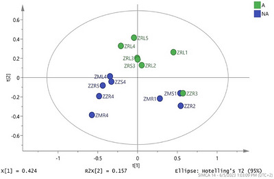
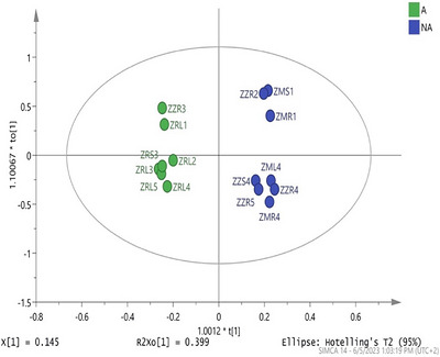
To further gain insight into the relationship between phytoconstituent composition and antigonococcal bioactivity, multivariate statistical analysis was conducted using principal component analysis (PCA). To minimize data skewing and improve the model’s accuracy and robustness, extracts with MIC < 1 mg/mL were classified as active, while those with MIC values ranging from 1 to 3.13 mg/mL were deemed inactive. Extracts exhibiting an MIC > 3.13 mg/mL were excluded from analysis, resulting in a dataset of 15 samples that were subjected to 1H NMR‐based metabolomics profiling. The PCA scores plot displayed a general clustering pattern that distinguished between active and inactive samples of the training set (Figure 1). However, the ethyl acetate root extract from Z. zeyheriana (ZZR3; MIC = 0.2 mg/mL) clustered with the inactive group. The model produced R 2 and Q 2 values of 0.67 and 0.47, respectively. While the R 2 value indicates that the data fit perfectly in the PCA model, the moderate Q 2 value suggests limited capability for predicting antigonococcal bioactivity, especially when applied to unknown samples. Such limitations are suggestive of inherent variability within the metabolite profile and other limitations of an unsupervised PCA classification. To improve the discriminatory capabilities of the PCA model, the supervised orthogonal projections to latent structures‐discriminant analysis (OPLS‐DA) was applied. This model demonstrated a clear separation between active and inactive samples (Figure 2) and exhibited improved performance (R 2 = 0.69 and Q 2 = 0.77). The higher Q 2 value indicates improved prediction capabilities and model robustness, which was further validated by ANOVA cross‐validation (p = 0.05). The previously misclassified ZZR3 extract was correctly clustered with active samples, demonstrating the advantage of a supervised OPLS‐DA model in the resolution of sample overlapping in the PCA scores plot. Despite the improved classification observed in the OPLS‐DA model, there is some degree of in‐group variation observed, particularly among the inactive extracts. Such variability is attributed to the inherent chemical diversity within plant extracts, even in species of the same genus. When the 1H NMR spectra were processed, solvent peaks, water signals, and background noise were excluded for model clarity. Nevertheless, small phytochemical variations, especially among crude extracts, remain a significant challenge as related species often share major similarities in their metabolic profiles, further complicating discrimination solely based on spectral data [ref. 39].
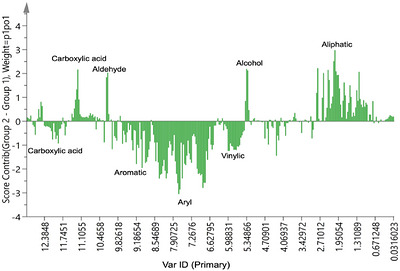
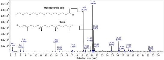
Subsequent to the observed clear discrimination of the training set, a contribution plot was generated (Figure 3) to identify the specific chemical classes that may be associated with the observed antigonococcal activity. The classes tentatively linked to the observed antigonococcal activity include aliphatic compounds (δ 0–1.9 ppm), alcohols (δ 5.3–6.6 ppm), aldehydes (δ 9.8–10.4 ppm), and carboxylic acids (δ 11–12 ppm) (Figure 3). Conversely, certain chemical shifts in the carboxylic acid region suggested the presence of classes not contributing to the observed antigonococcal bioactivity. These results emphasize the complexity of plant metabolomes, particularly where specific morphological features within a chemical class may determine biological activity. To further understand the chemical basis of the observed antigonococcal bioactivity, selected extracts were subjected to GC–MS analysis. The dichloromethane leaf extract of Z. rivularis revealed the occurrence of hexadecanoic acid (Rt = 19.38 min) and phytol (Rt = 21.30 min) (Figure 4). Similarly, hexadecanoic acid (Rt = 19.37 min) and phytol (Rt = 21.20 min) were identified in the ethyl acetate:methanol (1:1; v/v) leaf extract of Z. rivularis (Figure 5), while hexadecanoic acid (Rt = 20.41 min) and betulin (Rt = 30.41 min) were tentatively identified in the ethyl acetate root extract (Figure 6). These peak sizes correlate to the relative extract concentration of 1 mg/mL, where larger peaks indicate a higher availability.
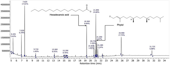
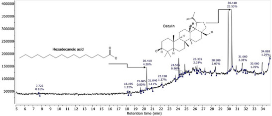
Although the identified phytoconstituents were not previously tested against N. gonorrhoeae, they have demonstrated antimicrobial biological activity against some clinical Gram‐negative bacteria. Hexadecanoic acid, for instance, displayed moderate activity at a maximum concentration of 50 mg/mL against E. coli and Klebsiella pneumoniae, exhibiting inhibition zones of 11.1 and 11.93 mm, respectively [ref. 44]. Phytol demonstrated significant antimicrobial activity against E. coli, Salmonella, and H. pylori, revealing inhibition zones of 10 – 20 mm. Further molecular docking of phytol on DNA gyrase revealed a good binding affinity, tentatively confirming its potential as an antigonococcal lead constituent [ref. 45]. Betulin, previously isolated from Z. mucronata root bark, displayed reduced antibacterial effects against E. coli; however, its derivatives exhibited increased activity from 24.0 to 0.015 mM [ref. 46]. The occurrence of these phytoconstituents in active extracts may suggest that they contribute either individually or synergistically to the observed antigonococcal activity. These phytoconstituents have demonstrated antimicrobial activity against Gram‐negative bacteria, similar to N. gonorrhoeae; nevertheless, additional testing, especially on N. gonorrhoeae, is necessary to fully assess their antigonococcal potential. Overall, these findings highlight that Z. rivularis and Z. zeyheriana, particularly the non‐polar and semi‐polar extracts, have significant antigonococcal potential. However, the low SI and the outstanding in vivo validation emphasize the need for further investigation, including bioassay‐guided fractionation, constituent isolation, and cytotoxicity evaluation. The integration of multivariate analysis and metabolomics provided valuable insights into the chemical basis of antigonococcal activity among Ziziphus species indigenous to South Africa. The application of a supervised OPLS‐DA was effective at discriminating between active and inactive samples, while the contribution plot and GC–MS analysis tentatively revealed key chemical classes as well as compounds behind the observed antigonococcal activity. Collectively, these findings offer a solid foundation for further bioassay‐guided isolation and subsequent inclusion into drug discovery initiatives.
Conclusions
This study has successfully validated the traditional medicinal use of Z. mucronata, Z. rivularis, and Z. zeyheriana in treating gonorrhea, with Z. rivularis and Z. zeyheriana showing antigonococcal activity for the first time. The low cytotoxicity observed in most of the extracts further reinforces the traditional applications of these plants in South African indigenous communities. It demonstrated the effectiveness of 1H NMR‐based metabolomics in accelerating the identification of potential bioactive chemical classes within Ziziphus species, using a robust OPLS‐DA model. The contribution plot generated from the OPLS‐DA model revealed chemical classes associated with the observed antigonococcal activity, setting the stage for future isolation and detailed phytoconstituent characterization. Notable compounds, including hexadecanoic acid, phytol, and betulin, were identified in Z. rivularis and Z. zeyheriana using GC–MS. Although previously reported to possess antimicrobial properties against some Gram‐negative bacteria, no studies have specifically tested their efficacy against N. gonorrhoeae, highlighting an area ripe for in‐depth research. Future research should focus on the bioassay‐guided isolation of active phytoconstituents to confirm their contribution to the observed antigonococcal activity in the crude extracts. Further structure elucidation and constituent quantification should be conducted to shed insights into the structure‐activity relationship. Given the limitations of in vitro assays, in vivo studies need to be conducted to establish the therapeutic efficacy of these isolated constituents in animal models.
Experimental Section
Plant Collection
Ziziphus mucronata plant samples were collected on January 5, 2022, from Limpopo Province in the Vhembe district Municipality (22°00’S; 29°12’E), while Z. rivularis and Z. zeyheriana samples were obtained on May 22, 2022 from the Manie Van Der Schijff Botanical Garden (25°45′ S; 28°13′ E) and the University of Pretoria Experimental Farm (25°00′ S; 28°00′ E), respectively. Ms Magda Nel performed all taxonomic identifications of the three Ziziphus plant species. Voucher specimens of the respective collected Ziziphus species were prepared and deposited in the HGWJ Schweickerdt Herbarium (PRU) under the following accession numbers: Z. mucronata (130883), Z. rivularis (130882), and Z. zeyheriana (0125140). The root, stem bark, and leaf materials were collected for Z. mucronata and Z. rivularis. For Z. zeyheriana, only the root and stem bark samples were obtained. Seasonal senescence prevented the collection of the leaves as they were not available. In total, eight plant samples were collected from the three species, air‐dried, and powdered using an Ultracfugal Mill (Retsch, Germany). The samples were then stored at room temperature (20°C–25°C) until further extraction experiments.
Plant Extraction
The dried and powdered plant material (40 g per sample) was extracted sequentially using a series of solvents with increasing polarity. The extraction solvents used were: (1) n‐hexane, (2) dichloromethane, (3) ethyl acetate, (4) ethyl acetate:methanol (1:1; v/v), and (5) methanol. For each extraction step, 400 mL of analytical‐grade solvent was added to the plant material, followed by homogenization in a blender (Philips, Pretoria) for 5–10 min. Subsequently, the texture was sonicated in an ultrasonic water bath (Labotec, Midrand) for 10 min and filtered through a Whatman No.1 filter paper (Merck, Germany). Each respective solvent extraction was performed in triplicate (three successive cycles) on the same plant material to ensure an exhaustive extraction before proceeding to the next solvent. The final combined filtrates for each respective solvent were concentrated under reduced pressure utilizing a rotary evaporator (Buchi, R‐200, Switzerland) and then transferred into pre‐weighed glass vials. In addition to the organic solvent extractions, aqueous decoctions were also prepared to mimic traditional preparation methods [ref. 47]. For this, 40 g of each plant sample was boiled in 500 mL of tap water at 100°C for 45 min [ref. 48]. The resulting mixture was cooled in an ice‐water bath, frozen at −80°C overnight, and subsequently freeze‐dried using a manifold freeze dryer (Virtis, New Zealand) to obtain dried extracts. In total, 48 successive extracts were obtained from the combined sequential extraction and decoction procedures.
Bacterial Culturing
N. gonorrhoeae (ATCC 49226) was maintained on chocolate agar (Sigma‐Aldrich, Missouri, USA) and cultured in Mueller‐Hinton (MH) broth (Merck, New Jersey, USA) for a 24‐h incubation period following the procedure described by Wiegand et al. and Klančnik et al. [ref. 49, ref. 50]. For inoculum preparation, a single colony was transferred into 50 mL MH broth and further incubated for 20 h at 37°C in a shaking incubator (Labotec, South Africa). To provide a CO2‐enriched atmosphere required by the N. gonorrhoeae to grow optimally, the bacterial cultures were incubated in an airtight container containing CO2‐generating sachets to create an anaerobic environment (Thermo Scientific, South Africa). After the incubation, the bacterial suspension was standardized to an optical density (OD600) of 0.2 using sterile MH broth and a spectrophotometer (Labotec, South Africa). To avoid contamination and maintain optimal sterility, all procedures were conducted in a sterile biosafety cabinet (ESCO, Singapore). All experiments were conducted as two independent assays, with each assay conducted in triplicate (n = 3). The results are expressed as the mean of the combined replicates.
Minimum Inhibitory Concentration (MIC) Assay
The minimum inhibitory concentration (MIC) of all 48 successive extracts was determined using the broth microdilution assay, a cost‐effective, reliable, and reproducible method. This approach is best suited, especially when assessing the microbial effects of plant extracts [ref. 49, ref. 50]. Briefly, 50 mg of each extract was dissolved in 1 mL of 10% dimethyl sulfoxide (DMSO) to obtain a 50 mg/mL stock solution. Ciprofloxacin (Merck, Modderfontein) served as the positive control, while the negative control consisted of 10% DMSO and broth (1:1, v/v). Twofold serial dilutions of each extract were prepared to yield final concentrations ranging from 12.5 to 0.01 mg/mL. Two independent assays were conducted, with each extract tested in triplicate (n = 3) in 96‐well ELISA microtiter plates (Sigma‐Aldrich, Missouri, USA). After incubation, bacterial proliferation was assessed by adding 40 µL of p‐iodonitrotetrazolium (INT) solution (Sigma, Missouri, USA) followed by a further 2‐h incubation. Development of a pink color indicated bacterial proliferation, whereas the absence of color change indicated inhibition of bacterial growth. The MIC was defined as the lowest concentration at which no visible color change was observed in the microtiter plates. The antigonococcal activity was classified according to MIC values, with extracts exhibiting MIC < 1 mg/mL considered highly active, values between 1 and 6.25 mg/mL were moderately active, and values above 6.25 were considered inactive [ref. 51].
Cytotoxicity Assay
The cytotoxicity of the 48 successive extracts was tested for cytotoxicity in vitro using rat skeletal myoblast (L6) cells (CVCL_0385), procured from Banco de Células do Rio de Janeiro (BCRJ, Brazil; Cat. no. 0141). Podophyllotoxin (Sigma‐Aldrich, P440) was used as the positive control. The cells were seeded in 96‐well ELISA microtiter plates at a density of 4 × 10³ cells per well in 100 µL of RPMI 1640 medium supplemented with 10% fetal bovine serum (FBS) and 1% L‐glutamine and further incubated under standard cell culture conditions. Eleven threefold serial dilutions of the respective extracts were prepared to yield final concentrations ranging from 100 to 0.002 µg/mL. This was followed by a 70‐h incubation period, cell morphology, and confluence examination using an inverted microscope. Cell viability was then assessed by the addition of resazurin solution to a final concentration of 10 µg/mL, followed by another 2‐h incubation. Fluorescence was measured at excitation and emission wavelengths of 536 nm and 589 nm, respectively, using a SpectraMax Gemini XS microplate fluorometer (Molecular Devices, California, USA). The required concentration to inhibit 50% of viability (CC50) was determined by non‐linear regression of sigmoidal curves. Cytotoxicity was classified as cytotoxic (CC50< 0,05 mg/mL), moderately cytotoxic (CC50 between 0.05 and 0.1 mg/mL), and non‐cytotoxic (CC50> 0.1 mg/mL) [ref. 52].
Nuclear Magnetic Resonance and Multivariate Data Analysis
All 48 extracts were subjected to 1H NMR analysis to compare the chemical profiles of the extracts based on their chemical profiles. Each extract was dissolved in 700 mL of deuterated dimethyl sulfoxide (DMSO‐d6 to a final concentration of 15 mg/mL. The solutions were then vortexed to dissolve all particulates and then transferred into 5 mm NMR tubes (Sigma‐Aldrich, Missouri, USA). 1H NMR spectra were recorded at 25°C on a 400 MHz Varian NMR spectrometer. For each sample, spectra were acquired over a spectral width of 0–14 ppm using 64 scans with standard acquisition parameters. Before the data acquisition, the magnetic field homogeneity was optimized by manual shimming to ensure consistent spectral resolution across all samples. The spectra were processed using MestReNova software (Version 14.20, Mestrelab). Free induction decays (FIDs) were Fourier transformed, and the resulting spectra were then manually phase‐corrected and automatically baseline corrected using the Whittaker smoothing algorithm [ref. 53, ref. 54]. Chemical shifts were referenced to the residual solvent signal of DMSO‐d6 (δ 2.50 ppm). For multivariate data analysis, all processed spectra were uniformly binned into regions of 0.04 ppm over the δ 0.00–14.00 ppm range. Regions on the spectra that correspond to the residual solvent signal (δ 2.40–2.60 ppm) and residual water signal (δ 3.20–3.40 ppm) were excluded before further analysis. The binned data were subsequently exported as ASCII files and then imported into SIMCA‐P software (version 14.1, Umetrics, Umeå, Sweden) for multivariate statistical analysis. 1H NMR data acquisition, processing, and multivariate analysis were performed according to established NMR‐based metabolomics protocols for plant extracts, including spectral preprocessing (phase and baseline correction), binning, and statistical analysis using SIMCA‐P [ref. 54].
Gas Chromatography–Mass Spectrometry (GC–MS) Analysis
Three extracts exhibiting the best antigonococcal activity (MIC < 1 mg/mL) were selected for GC–MS analysis to tentatively identify compounds attributed to the observed bioactivity in Z. rivularis and Z. zeyheriana. The successive extracts analyzed included the dichloromethane leaf extract of Z. rivularis (ZRL2), the ethyl acetate:methanol (1:1) leaf extract of Z. rivularis (ZRL4), as well as the ethyl acetate root extract of Z. zeyheriana (ZZR3). Each extract was resuspended in the respective extraction solvent before the analysis. Derivatization was performed using N,O‐bis(trimethylsilyl)trifluoroacetamide (BSTFA) at room temperature for 5 h to produce trimethylsilyl (TMS) derivatives, thus enhancing the volatility and thermal stability of the polar metabolites. The GC–MS analysis was conducted as previously described by Mabuza et al. with minor modifications [ref. 27]. Briefly, 1 µL of each derivatized extract was injected in a split mode at an injector temperature of 250°C into a Shimadzu GC–MS (QP2010 SE, Shimadzu, Kyoto, Japan) equipped with an Rxi‐5MS capillary column (30 m × 0.25 mm, 0.25 µm film thickness; Restek, Bellefonte, PA, USA) composed of 5% diphenyl/95% dimethylpolysiloxane stationary phase. The oven temperature was set to an initial temperature of 60°C (held for 3 min), followed by an increase to 250°C. The oven was programmed to 60°C, held for 3 min, and then increased to a final temperature of 280°C at a rate of 15°C per minute. While the ion source temperature was set at 250°C, the interface temperature was also set at 250°C, at 15°C/min. The ion source and interface temperatures were maintained at 250°C. Mass spectra were acquired in electron ionization (EI) mode at 70 eV over a mass range of m/z 30–550 with a 2000 scan speed. The tentative identifications of the phytoconstituents were conducted by comparing the obtained mass spectral fragmentation patterns with those in the NIST 11 and Wiley 09 mass spectral libraries.
Author Contributions
Mcebisi Junior Mabuza conceptualized the study, conducted formal analysis, methodology, and visualization, and wrote the original draft. Marcel Kaiser assisted with the biological activity screening and cytotoxicity. Mahwahwatse Johanna Bapela provided supervision, resources, and proofreading of the manuscript. Thilivhali Emanuel Tshikalange provided resources and supervision and assisted with reading the original draft manuscript. Abdullahi Ahmed Yusuf assisted with GC–MS analysis and proofreading the manuscript.
Conflicts of Interest
The authors declare no conflicts of interest.
References
- 1 World Health Organization (WHO) , Sexually Transmitted Infections (STIs), https://www.who.int/news‐room/fact‐sheets/detail/sexually‐transmitted‐infections‐(stis) (Accessed March 5, 2025).
- Sexually Transmitted Infection Prevalence and Neisseria gonorrhoeae Antimicrobial Resistance Patterns in Men Who Have Sex With Men With or Without Urethral Discharge Syndrome in Johannesburg, South Africa, 2024,”. PLOS Global Public Health, 2026. [DOI | PubMed]
- Prevalence and Incidence of Sexually Transmitted Infections Among South African Women Initiating Injectable and Long‐Acting Contraceptives,”. PLoS ONE, 2023. [DOI | PubMed]
- Prevalence of Chlamydia, Gonorrhoea, and Trichomoniasis Among Male and Female General Populations in Sub‐Saharan Africa From 2000‐2024: a Systematic Review and Meta‐Regression Analysis,”. medRxiv, 2025. [DOI]
- Gonorrhoea (Neisseria gonorrhoeae Infection),”. 2024
- Mitigating Inequitable Access to Appropriate Antibiotics in Low‐ and Middle‐Income Countries,”. JAC‐Antimicrobial Resistance, 2025. [DOI]
- Gonorrhea Management in High‐, Limited‐ and No‐Resource Settings: Implications in the Context of Antimicrobial Resistance,”. International Journal of Dermatology, 2025. [DOI]
- Future Prospects for Neisseria gonorrhoeae Treatment,”. Antibiotics, 2018. [DOI | PubMed]
- The Continuing Evolution of Antibiotic Resistance in Neisseria gonorrhoeae: Past, Present, and Future Threats to Effective Treatment,”. Journal of Antimicrobial Chemotherapy, 2025. [DOI | PubMed]
- Antibiotic Resistance in Neisseria gonorrhoeae: Challenges in Research and Treatment,”. Microorganisms, 2022. [DOI | PubMed]
- Sexually Transmitted Neisseria gonorrhoeae Infections—Update on Drug Treatment and Vaccine Development,”. Medicines, 2021. [DOI | PubMed]
- Antimicrobial Resistance in Neisseria gonorrhoeae in Nine Sentinel Countries Within the World Health Organization Enhanced Gonococcal Antimicrobial Surveillance Programme (EGASP), 2023: a Retrospective Observational Study,”. Lancet Reg Health West Pac, 2025. [DOI]
- Drug Combinations Targeting Antibiotic Resistance,”. Antimicrobials and Resistance, 2024. [DOI | PubMed]
- Chemical Basis of Combination Therapy to Combat Antibiotic Resistance,”. Journal of the American Chemical Society Au, 2023. [DOI | PubMed]
- Antimicrobial Resistance of Neisseria Gonorrhoeae in Sub‐Saharan Populations,”. Bacteria, 2022. [DOI]
- Antimicrobial Resistance: a Concise Update,”. Lancet Microbe, 2025. [DOI | PubMed]
- Multidrug‐resistant Gonorrhea: a Research and Development Roadmap to Discover New Medicines,”. Plos Medicine, 2017. [DOI | PubMed]
- Back to Nature: Medicinal Plants as Promising Sources for Antibacterial Drugs in the Post‐antibiotic Era,”. Plants, 2023. [DOI | PubMed]
- Established Antibacterial Drugs From Plants,”. Advances in Botanical Research, 2023. [DOI]
- Ethnomedicinal Plants Used by Traditional Healers in the Management of HIV/AIDS Opportunistic Diseases,”. South African Journal of Botany, 2019. [DOI]
- Hierarchies of Knowledge: Ethnobotanical Knowledge, Practices and Beliefs of the Vhavenda in South Africa for Biodiversity Conservation,”. Journal of Ethnobiology and Ethnomedicine, 2018. [DOI | PubMed]
- Anti‐inflammatory, Anticholinesterase and Antioxidant Activity of Leaf Extracts of Twelve Plants Used Traditionally to Alleviate Pain and Inflammation in South Africa,”. Journal of Ethnopharmacology, 2015. [DOI | PubMed]
- 23 A. Hutchings and A. H. Scott , Zulu Medicinal Plants: An Inventory (University of Natal Press, 1996).
- Ethnoveterinary Medicine Practices among Tsonga‐speaking People of South Africa,”. Onderstepoort Journal of Veterinary Research, 2006. [DOI | PubMed]
- 25 E. Palmer and N. Pitman , Trees of Southern Africa: Covering All Known Indigenous Species in the Republic of South Africa, South‐West Africa, Botswana, Lesotho & Swaziland, Balkema, Cape Town, (1972).
- The Ethnobotany and Antimicrobial Activity of Selected Medicinal Plants From Ga‐Mashashane,”. South African Journal of Botany, 2022. [DOI]
- In Vitro Antiplasmodial Activity and Cytotoxicity of Three Ziziphus (Rhamnaceae) Species From South Africa,”. Journal of Ethnopharmacology, 2025. [DOI | PubMed]
- Phytochemical Diversity and Pharmacological Effects of Triterpenes From Genus Ziziphus: a Comprehensive Review,”. Phytochemistry Reviews, 2022. [DOI]
- A Review of the Toxicity and Phytochemistry of Medicinal Plant Species Used by Herbalists in Treating People Living With HIV/AIDS in Uganda,”. Frontiers in Pharmacology, 2021. [DOI | PubMed]
- The In‐Vitro Pharmacokinetics of Medicinal Plants: a Review,”. Pharmaceuticals, 2025. [DOI | PubMed]
- Terpene Derivatives as a Potential Agent against Antimicrobial Resistance (AMR) Pathogens,”. Molecules, 2019. [DOI | PubMed]
- Assessment of Lipophilicity Parameters of Antimicrobial and Immunosuppressive Compounds,”. Molecules, 2023. [DOI | PubMed]
- Comparison of Techniques and Solvents on the Antimicrobial and Antioxidant Potential of Extracts From Acacia dealbata and Olea europaea ,”. Antibiotics, 2020. [DOI | PubMed]
- 34 L. J. C. Erasmus , M. J. Potgieter , and T. E. Tshikalange , Impact of Various Boiling Intervals on the Antimicrobial Efficacy and Phytochemical Profile of Selected Crude Aqueous Plant Extracts, Used by Bapedi Traditional Healers in the Treatment of Sexually Transmitted Infections (University of Limpopo, 2014).
- The Effects of Varying Ingredients Combination and Boiling Time on Total Phenolic Content, Antioxidant Activity, and Antimicrobial Properties of Lemongrass‐Ginger Tea,”. Heliyon, 2024. [DOI | PubMed]
- The Stability and Degradation Products of Polyhydroxy Flavonols in Boiling Water,”. Current Research in Food Science, 2023. [DOI | PubMed]
- Effect of Different Climatic Regions and Seasonal Variation on the Antibacterial and Antifungal Activity, and Chemical Profile of Helichrysum Aureonitens Sch. Bip,”. Metabolites, 2022. [DOI | PubMed]
- Geographical Variation in the Chemical Profile and Antimicrobial Activity of Solidago Gigantea Essential Oils,”. Frontiers in Microbiology, 2026. [DOI | PubMed]
- Exploring the Known Chemical Space of the Plant Kingdom: Insights Into Taxonomic Patterns, Knowledge Gaps, and Bioactive Regions,”. Journal of Cheminformatics, 2023. [DOI | PubMed]
- Impact of Extraction Techniques on Phytochemical Composition and Bioactivity of Natural Product Mixtures,”. Frontiers in Pharmacology. [DOI]
- Clinical Potential of Essential Oils: Cytotoxicity, Selectivity Index, and Efficacy for Combating Gram‐Positive ESKAPE Pathogens,”. Molecules, 2025. [DOI | PubMed]
- Bioassay‐Guided Fractionation of Pittosporum angustifolium and Terminalia Ferdinandiana With Liquid Chromatography Mass Spectroscopy and Gas Chromatography Mass Spectroscopy Exploratory Study,”. Plants, 2024. [DOI | PubMed]
- Quantifying Synergy in the Bioassay‐guided Fractionation of Natural Product Extracts,”. PLoS ONE, 2020. [DOI | PubMed]
- Structural Characterization of n‐Hexadecanoic Acid From the Leaves of Ipomoea Eriocarpa and Its Antioxidant and Antibacterial Activities,”. Biomass Conversion and Biorefinery, 2022. [DOI]
- Exploring the Bioactive Compounds of Carica Papaya Leaves: Phytol’s Role in Combatting Antibiotic‐resistant Bacteria,”. Frontiers in Cellular and Infection Microbiology, 2025. [DOI | PubMed]
- Analysis of the Antibiotic‐Potentiating Activity, Absorption, Distribution, Metabolism, and Excretion (ADME) and the Molecular Docking Properties of Phytol against Multi‐Drug‐Resistant (MDR) Strains,”. Antibiotics, 2024. [DOI | PubMed]
- S‐Alkylated Sulfonium Betulin Derivatives: Synthesis, Antibacterial Activities, and Wound Healing Applications,”. Bioorganic Chemistry, 2025. [DOI | PubMed]
- Influence of Direct and Sequential Extraction Methodology on Metabolic Profiling,”. Journal of Chromatography B, 2018. [DOI]
- Agar and Broth Dilution Methods to Determine the Minimal Inhibitory Concentration (MIC) of Antimicrobial Substances,”. Nature Protocols, 2008. [DOI]
- Evaluation of Diffusion and Dilution Methods to Determine the Antibacterial Activity of Plant Extracts,”. Microbiology Methods, 2010. [DOI]
- Potential of Cameroonian Plants and Derived Products against Microbial Infections,”. Planta Medica, 2010. [DOI | PubMed]
- Vitro Cytotoxic Activity of African Plants: a Review,”. Molecules, 2022. [DOI | PubMed]
- NMR‐based Metabolomic Analysis of Plants,”. Nature Protocols, 2010. [DOI | PubMed]
- A Perfect Smoother,”. Analytical Chemistry, 2003. [DOI | PubMed]