
Polymorphism in Cannabinol Piperazine Cocrystals: Structural, Morphological, and Energetic Perspectives
Abstract
Polymorphism remains a major challenge in the development of pharmaceutical solid products as even small changes in the crystal arrangement can influence key properties such as stability and solubility. In this study, we mechanochemically prepared a novel cannabinol piperazine cocrystal, which exists in three polymorphic forms. The formation of these polymorphs was systematically investigated by varying the solvents, temperature, and milling time during the ball mill experiments. Interestingly, the phenomenon of disappearing polymorphs was observed under repeated milling. To get insights into the polymorphic behavior and assess the relative stability of the forms, we analyzed their crystal structures, morphologies, and hydrogen bond motifs and performed particle energy calculations using density functional theory. The theoretical results show good correlation with the experimental data and provide valuable and deep insights into the polymorphic behavior and the disappearance of the metastable form. Overall, this work highlights the importance of integrating structural analysis with energetic evaluations to rationalize and predict polymorph stability and transformations.
Affiliations: † Department of Chemical Engineering, 534467University of Chemistry and Technology in Prague, Technická 3, 16628 Prague 6, Czech Republic; ‡ Institute of Physics of the Czech Academy of Sciences, Na Slovance 2, 182 00 Prague 8, Czech Republic; § 537141Zentiva k.s., U Kabelovny 130, 10237 Prague 10, Czech Republic
License: © 2025 The Authors. Published by American Chemical Society CC BY 4.0 This article is licensed under CC-BY 4.0
Article links: DOI: 10.1021/acs.cgd.5c01390 | PubMed: 41427022 | PMC: PMC12715752
Relevance: Moderate: mentioned 3+ times in text
Full text: PDF (6.4 MB)
Introduction
Polymorphism, which is defined as the ability of a compound to crystallize in more than one crystal structure, is a common phenomenon in solid-state chemistry. In pharmaceutical development, polymorphism plays a crucial role because it directly affects key physicochemical properties of solid forms, including stability, solubility, and thermal properties.ref1−ref2ref3ref4ref5ref6 While multiple polymorphic forms are often observed, their experimental availability is not always straightforward. In some cases, a previously obtained polymorphic form cannot be reproduced under identical or similar crystallization conditions. These cases, widely referred to as “disappearing polymorphs”,ref7−ref8ref9 highlight the sensitivity of the crystallization processes to factors such as impurities, seeding effect, or competitive nucleation. From a pharmaceutical point of view, disappearing polymorphs present a significant challenge, as they complicate reproducibility and control of the desired polymorphic form.ref6,ref9 Nevertheless, in the literature, we can also find examples, where these disappeared polymorphic forms have been successfully reproduced under controlled conditions, such as adjusting particle size or using virgin glassware.ref9−ref10ref11ref12 Thus, understanding and controlling polymorphism is essential for manufacturability of pharmaceutical compounds and also for achieving consistent therapeutic effects.
The polymorphism is not limited only to the single-component solids, but it has also been observed in multicomponent systems.ref13−ref14ref15ref16ref17 Cocrystals, a well-known system in the pharmaceutical area, consist of an active pharmaceutical ingredient (API) and one or more pharmaceutically acceptable coformers in a defined stoichiometric ratio.ref18−ref19ref20 They are usually stabilized through noncovalent interactions, such as hydrogen bonding, π–π stacking, or van der Waals forces. The formation of cocrystals enables a fine-tuning of the physicochemical properties, including solubility, thermal stability, mechanical properties, etc.ref19,ref21 However, polymorphic behavior in cocrystals remains less studied compared to single-component systems, partly due to the limitations of conventional solution-based screening strategies, especially when the API and coformer solubilities differ significantly.ref. ref22
As an alternative way, mechanochemistry has recently emerged with considerable attention as a sustainable, efficient, and green approach for the synthesis of novel solid forms.ref23,ref24 Among these methods, ball milling is the most commonly used and allows preparation under either neat grinding (NG) or liquid-assisted grinding (LAG) conditions.ref25−ref26ref27 Contrary to the traditional solution-based methods,ref. ref13 ball milling operates under nonequilibrium conditions, which facilitate the formation of kinetically metastable phases.ref25,ref28,ref29 The formation of the metastable forms is highly dependent on many parameters, such as the presence of additives,ref2,ref30 used frequency,ref31,ref32 temperatureref25,ref33 or the material of the milling jars.ref34−ref35ref36
Cannabinol (CBN) is a nonpsychoactive phytocannabinoid, naturally found in Cannabis sativa, alongside more widely studied cannabinoids such as tetrahydrocannabinol (THC) and cannabidiol (CBD).ref37,ref38 CBN has attracted attention due to its potential therapeutic properties, including anticancer,ref. ref39 anti-inflammatory,ref. ref40 and analgesic effects.ref. ref41 Similar to CBD, cannabinol suffers from poor aqueous solubility and limited stability, which do not enable its direct pharmaceutical applications.ref. ref38 The patent literatureref42,ref43 has already described four CBN cocrystals, namely, betaine, d-proline, l-proline, and tetramethylpyrazine, which highlights the potential of cocrystallization strategies to optimize CBN physicochemical properties.
In this study, we present a novel unexplored CBN cocrystal with piperazine (CBN-PI), which exhibits rich polymorphic behavior. Three polymorphic forms were obtained through ball milling, and their formations were systematically investigated using a combination of experimental and computational approaches. We employed a combination of experimental and computational approaches to understand their relative stability, crystallographic features, and transformation pathways. In particular, density functional theory (DFT)-based particle energy calculations were used to evaluate the influence of lattice energy, conformational strain, and surface attachment energies on the observed polymorphic landscape. The results provide new insights into the stability of the CBN-PI cocrystal polymorphs and offer a rational explanation for the experimentally observed disappearance of a specific polymorphic form.
Materials and Methods
Materials
Cannabinol was purchased from Pharmabinoid B.V. (Uden, Netherlands). Piperazine was purchased from Sigma-Aldrich (St. Louis, USA) and was used as received with a purity higher than 98%. Solvents such as acetone (AE), butyl acetate (BA), dichloromethane (DCM), heptane (HP), methanol (MeOH), and tetrahydrofuran (THF) were purchased from PENTA (Prague, Czech Republic).
Milling Experiments
All grinding experiments were carried out with a Retsch MM500 mixer mill. Approximately 50 mg of CBN and piperazine were mixed in a 2:1 molar ratio in a 2 mL polypropylene milling jar with two 5 mm stainless steel balls. All experiments were carried out at a frequency of 25 Hz. Initial screening experiments were carried out for 20 min using heptane as a LAG additive in an amount of 10 μL. Subsequent experiments focused on studying different milling conditions. Various liquid additives (AE, BA, DCM, HP, MeOH, THF, water) were tested in an amount of 10 μL. The milling times varied from 5 to 60 min at room temperature, and in a separate set of experiments, the milling time was fixed at 20 min, while the temperature was adjusted to 5, 25, and 35 °C.
Single-Crystal Preparation
The single crystals of CBN-PI Form I and Form III were prepared by combining CBN and piperazine in a cyclohexane solution in a 2:1 ratio. The mixtures were heated to 35 °C to dissolve the powder and then allowed to cool, followed by slow evaporation until the single crystals were formed. The resulting single crystals were subsequently analyzed according to the conditions that are described below. The preparation of both single crystals was straightforward, in contrast to Form II. Since Form II is not the most stable polymorph, we conducted several seeding experiments from different solvents (cyclohexane, HP, hexane); however, these experiments resulted in the formation of Form III. For this reason, we employed XRPD to solve the crystal structure from powder data.
Characterization
Powder X-ray Diffraction (XRPD)
X-ray diffraction patterns were obtained for both the raw powders and the prepared materials using a powder diffractometer X’Pert3 Powder (PANanalytical, Holland) equipped with Cu anode Kα (λ = 1.542 Å) with a tube voltage of 40 kV and a tube current of 30 mA. The data were collected from 4 to 40° 2θ with 0.026° 2θ step size and 56.87 s per step.
Single-Crystal and Powder X-ray Diffraction for the Structure Solution
Single-crystal X-ray diffraction (SCXRD) measurement was performed using two four-circle CCD Rigaku diffractometers: Supernova, equipped with a microfocus Mo X-ray tube and a CCD detector Atlas S2 (Form I, Pc), and XtaLAB Synergy R, equipped with a rotating anode Cu X-ray tube and a HyPix-Arc 150 detector (Form III, P21/n). Both diffractometers use a Cryojet chiller.
The data reduction and absorption correction were done with CrysAlisPro software.ref. ref44 The structures were solved by charge flipping methods using Superflip software and refined by full matrix least-squares on squared value using Crystalsref. ref45 and Jana2020 software.ref. ref46 MCE softwareref. ref47 was used for the visualization of residual electron density maps. All H atoms were placed from the residual electron density map, and the C–H atoms were constrained to ideal geometries. The structure of Form I is slightly disordered, with one of the CBD side chains being in two positions with occupancies of 0.828 and 0.172. The sample CBN-PI Form II was ground and placed in a 0.5 mm borosilicate-glass capillary. Powder diffraction data were collected using the Debye–Scherrer transmission configuration on the powder diffractometer Empyrean of PANalytical (λCu,Kα = 1.54184 Å) that was equipped with a focusing mirror, a capillary holder, and a PIXcel3D detector. The data was collected from 3° to 80° 2θ, with a step of 0.013° and with an overall 20 h measurement time.
The structures were deposited into the Cambridge Structural Database under numbers 2483132 (CBN-PI Form I), 2483133 (CBN-PI Form II), and 2483134 (CBN-PI Form III). Details of crystal structure solutions are in the Supporting Information in Section SI 1.
Thermal Analysis
Thermal properties were analyzed using differential scanning calorimetry DSC 3+ (Mettler Toledo, Switzerland) and thermogravimetric analysis (TGA). For DSC measurements, approximately 2 mg of the sample was placed into an aluminum pan, which was sealed and pierced to allow possible solvent vapor to escape and prevent an explosion. The temperature range for DSC experiments was from 20 °C to the specific degradation temperature of each sample with a heating rate of 10 °C/min. For TGA experiments, the pan was filled with approximately 5 mg of the sample and heated from 30 to 300 °C at a rate of 5 °C/min. All measurements were carried out under an inert nitrogen atmosphere.
Solution and Solid-State Nuclear Magnetic Resonance (NMR)
Solution NMR was employed to determine the stoichiometry and purity of the prepared materials. Each sample was dissolved in d 6-DMSO, and the 1H NMR spectra were measured with an Avance III 500 MHz NMR spectrometer (Bruker, USA) equipped with a Prodigy probe and with a repetition delay of 10 s.
Solid-state NMR was utilized to confirm cocrystal formation and purity of the obtained material. The 13C NMR spectra were measured by an Avance III 400 MHz NMR spectrometer (Bruker, USA) equipped with a 4 mm probe and with 13 kHz spinning.
Computational Procedures
Interaction Energy Calculations and Energy Frameworks
Interaction energies and energy frameworks were calculated using the software CrystalExplorer17 (version 17.5, revision f4e298a).ref. ref48 Molecular wave functions were generated using the built-in Tonto utility in the “accurate” mode using the B3LYP/6-31G(d,p) level of theory. Visualizations of the energy frameworks were created by using the same software.
Surface and Morphology Analysis
The morphology of each polymorph surface was calculated using the CSD-Particle tool,ref49,ref50 which is a part of the Mercury software (2025.1.1, build 448738).ref. ref51 The Dreinding II force field with a limiting radius of 30 Å was used for the calculation of the lattice energies and crystal shape with the VisualHabit model, where the lattice energy value can also be found. The surface morphology and topology were calculated by using the Surface Analysis tool. The analysis was focused on the largest predicted facet for each form. The surface offset was automatically selected by the software to be the smoothest one. The examined descriptors included hydrogen bond (HB) acceptors, HB donors, and aromatic bonds.
Calculation of Hydrogen Bonding Propensity and Full Interaction Maps
The hydrogen bonding propensity (HBP) and hydrogen bond coordination (HBC) analysesref. ref52 were performed using the CSD-Material tool,ref. ref53 a part of Mercury software (4.3.0, build 270015). For the analyses, the default donor/acceptor definitions (O–H and N–H donors and N and O acceptors) and a 0.25 Å heavy-atom distance tolerance were used. For each polymorph, the top-scoring hydrogen bond networks were enumerated and plotted on the HBP/HBC landscape, and the experimentally observed network for each form was overlaid. Full interaction maps (FIMs) were generated with default NH and OH donor probes and N/O acceptor probes for the isolated component molecules. The experimental packings were then inspected against the predicted hotspots. No user retraining or reweighting of the statistical model was applied.
Calculation of Conformational Strain Energy
Conformational strain energies were calculated using DFT in Gaussian 16, applying the B3LYP functional with a D3BJ dispersion correction and the 6-311G(d,p) basis set. For each polymorph, a single molecule of CBN was extracted from the crystal structure using Mercury. A constrained geometry optimization was performed, where torsion angles were frozen to retain the crystal conformation, while bond lengths and angles were allowed to relax. A fully relaxed gas-phase optimization of the same molecule (with no constraints) was then performed using the same level of theory.ref. ref54 The conformational strain energy was calculated using eq eq1 :
where the first term corresponds to the crystal energy of the constrained structure and the second term is the fully optimized gas-phase structure. Energies were obtained in Hartree and converted to kJ mol–1 using the factor 1 hartree = 2625.5 kJ mol–1.
Particle Energy Calculations
The particle energy was calculated using eq eq2 :ref. ref10
The first contribution in the equation corresponds to the lattice energy, which describes the strength of intermolecular interactions that hold the molecules together in the bulk. The second term is the conformational energy penalty, representing the energy cost of a molecule to adopt the specific conformation required within the crystal lattice from the lowest energy reference state. The last term is the surface energy penalty, describing how surface termination influences particle stability. It includes the fractional surface area of each crystal face, with the corresponding attachment energy values.
Results and Discussion
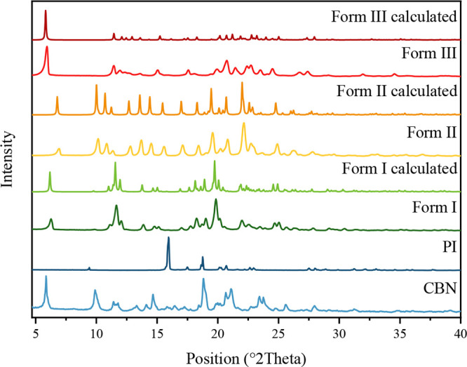
Mechanochemical preparation of pharmaceutical cocrystals is known to be sensitive to both kinetic and thermodynamic factors, often giving rise to polymorphic diversity. In this study, initial screening milling experiments using heptane as a LAG additive resulted in the formation of a CBN-PI cocrystal, in a 2:1 ratio, referred to as Form I. However, subsequent repetitions of the same milling conditions unexpectedly resulted in the appearance of a new polymorph, Form II, followed by the emergence of a third form, Form III, under identical milling parameters, some months later. All polymorphs were initially obtained in their pure forms. Notably, after the appearance of Form III, Form I could not be obtained anymore despite repeating the experiments under the same previously successful conditions. The XRPD patterns of all three polymorphs are shown in Figure , confirming distinct phases.
Characterization of the Obtained CBN-PI Polymorphs
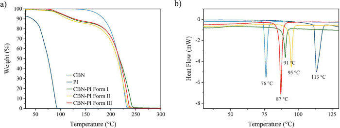
To evaluate the thermal behavior and stability of the obtained polymorphs, their thermodynamic properties were investigated using TGA and DSC. The TGA curves (Figure a) showed comparable behavior among all polymorphs. All of them show a weight loss of around 13.5% near 150 °C, which is in accordance with the calculated values of 12.2% of the weight loss in a 2:1 ratio, which is connected to the sublimation of the piperazine within the cocrystal lattice.ref. ref55 The DSC thermographs (Figure b) show a single sharp endothermic peak for each polymorph, which indicates their phase purity. The melting points of the polymorphs lie between those of the starting materials: CBN shows a melting peak at 76 °C (T onset = 74 °C) and a pure piperazine peak at 113 °C (T onset = 111 °C). The CBN-PI Form I melts at 91 °C (T onset = 88 °C), Form II melts at 95 °C (T onset = 91 °C), and Form III melts at 87 °C (T onset = 84 °C). Notably, Form I, which was initially obtained but could not be reproduced in later experiments, does not exhibit the lowest melting point among the three polymorphs, as might be expected.ref11,ref56 However, the stability of the polymorph is determined by more than just its melting point, encompassing parameters such as lattice energy, symmetry, hydrogen bonding network, etc.ref55,ref57
The 1H NMR spectroscopy confirmed a 2:1 molar ratio in the prepared polymorphic forms. Solid-state NMR was used to confirm the cocrystal formation in the CBN-PI samples and to reveal distinct chemical shift differences, which confirms that each sample represents a unique polymorph. The ssNMR spectra are shown in Figure SI 2.
To better understand and control the formation of each polymorph, a systematic screening was conducted. This included evaluating the effect of different solvents, investigating the influence of milling time, and finally investigating the effect of the milling temperature.
Influence of Different Experimental Conditions on the Polymorphic Form Obtained
Influence of the Solvent
In these experiments, seven different solvents were selected along with NG as a solvent-free condition. The chosen solvents represent a range of commonly used liquid additives in mechanochemistry and also include a variety of functional groups with different polarities. The results are shown in Table . The corresponding XRPD patterns are presented in Figure SI 3. Milling in the presence of BA, DCM, and HP resulted in the complete formation of Form III. In contrast, milling experiments with AE and water resulted in partial conversion of the cocrystal in Form III, alongside still present unreacted starting materials. However, the presence of MeOH caused the appearance of a mixture of Forms II and III. Finally, using neat grinding conditions led to partial conversion to Form II in the presence of starting materials. Interestingly, the use of THF did not lead to any cocrystal transformation, and the XRPD pattern corresponds to the physical mixture of pure CBN and PI. Overall, the results reveal a correlation between the solvent polarity and the outcome of the milling experiments. Weakly polar or nonpolar solvents (BA, DCM, HP) do not form any hydrogen bonds with the starting components and lead directly to the interaction between CBN and PI, thus promoting rapid nucleation and growth of the most stable Form III. In contrast, polar liquid additives (AE, MeOH, THF, water) can compete for the hydrogen bonds with the reactants, forming solvated layers or sticky pastes, or even dissolve part of the reactants. These effects reduce or inhibit the efficiency of the nucleation and might stabilize a mixture with a metastable form.ref. ref58
1: Polymorphic Forms Obtained at Different Temperatures Using Various Solvents and Neat Grinding Conditions
| solvent | polymorphic form |
|---|---|
| AE | CBN + PI + Form III |
| BA | Form III |
| DCM | Form III |
| HP | Form III |
| MeOH | Form II + Form III |
| H2O | CBN + PI + Form III |
| THF | CBN + PI |
| NG | CBN + PI + Form II |
In conclusion, the results show that the choice of solvents influences the efficiency of cocrystal formation based on the solvent polarity. In most cases, Form III tends to be the dominant polymorph. Form II was obtained only under solvent-free conditions, together with the starting materials, and as a mixture with Form III, when milling with MeOH. Based on these findings, we have selected HP, MeOH, and NG for subsequent experiments, as they provide different results and influence the polymorphic forms. Moreover, they represent a diverse set of solvents, including both a nonpolar and a polar solvent and a solvent-free approach.
Influence of Temperature
Another factor that was systematically studied was the influence of milling temperature. Experiments were performed at 5, 25, and 35 °C to simulate varying conditions that might affect polymorphic formation. The results are reported in Table , and XRPD patterns are shown in Figure SI 4. Milling in the presence of methanol and heptane led to the formation of pure Form III across all three temperatures except at room temperature with MeOH, where a mixture of Forms II and III was obtained. Interestingly, under NG conditions, incomplete conversion of Form II was observed only at room temperature, while at 5 and 35 °C, the incomplete formation of Form III was detected. These results indicate that temperature has no significant effect on the polymorphic formation under these solvent conditions and highlight the dominance of Form III.
2: Polymorphic Forms Obtained at Different Temperatures Using Methanol, Heptane, and Neat Grinding Conditions
| 5 °C | 25 °C | 35 °C | |
|---|---|---|---|
| HP | Form III | Form III | Form III |
| MeOH | Form III | Form II + III | Form III |
| NG | CBN + PI + Form III | CBN + PI + Form II | CBN + PI + Form III |
Influence of Time
Lastly, the effect of milling time on polymorphic formation was investigated. The experiments were carried out using heptane, methanol, and under NG conditions with milling durations ranging from 5 to 60 min. The obtained polymorphic forms are summarized in Table , and the corresponding XRPD patterns are presented in Figure SI 5. When heptane was used, the pure polymorphic form III was obtained across all time points. In contrast, milling with methanol led to the consistent formation of a mixture of Forms II and III throughout the entire time. Under NG conditions, no full cocrystal conversion of Form II was observed at any time point. These findings show that the use of a liquid additive facilitates full conversion of the cocrystal. Furthermore, the type of the solvent plays a key role in the resulting polymorphic form, favoring specific Form III or stabilizing a mixture of Forms II and III.
3: Polymorphic Forms Obtained at Different Time Points Using Methanol, Heptane, and Neat Grinding Conditions
| 5 min | 10 min | 15 min | 20 min | 30 min | 40 min | 60 min | |
|---|---|---|---|---|---|---|---|
| HP | Form III | Form III | Form III | Form III | Form III | Form III | Form III |
| MeOH | Form II + III | Form II + III | Form II + III | Form II + III | Form II + III | Form II + III | Form II + III |
| NG | CBN + PI + Form II | CBN + PI + Form II | CBN + PI + Form II | CBN + PI + Form II | CBN + PI + Form II | CBN + PI + Form II | CBN + PI + Form II |
In conclusion, despite a systematic investigation of various experimental parameters, including solvent, temperature, and milling time, it was not possible to selectively obtain each polymorphic form under specific conditions. Moreover, the Form I could not be reproduced under any of the tested conditions, which suggests the occurrence of the well-known “disappearing polymorph” phenomenon.ref7,ref8,ref11 Form II was obtained only as a minor phase, either in mixtures with Form III or along with incompletely converted starting materials. Interestingly, in most cases, Form III was the dominant polymorphic form, indicating its strong preference as the thermodynamically most stable form despite having a slightly lower melting point compared to that of Forms I and II.
Crystal Structure Characterization
To further understand the observed polymorphic behavior of the CBN-PI cocrystal polymorphs, we determined the crystal structures of all prepared polymorphs. Crystallographic data and details of the structure refinement are listed in Section S1. Furthermore, we calculated the lattice energies and intermolecular interaction energies to evaluate the packing efficiency and stability of each polymorph.
CBN
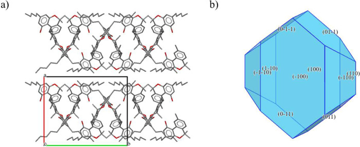
The crystal structure of pure CBN has been known since 1977.ref. ref59 It crystallizes in the monoclinic space group P 21/c. The asymmetric unit (Figure SI 6a) consists of two molecules of CBN, while in the unit cell (Figure a), there are eight molecules of CBN. The two unique molecules in the asymmetric unit are connected by a hydrogen bond via oxygen atoms (Figure SI 6b). The molecules of CBN form infinite chains linked by hydrogen bonds within each chain. The calculated crystal shape (Figure b) shows that the largest facets are (100) and (−100), which are equal and parallel, and together account in total 44.75% of the crystal surface area. The CBN chains create channels in which the slip planes are oriented and align with the direction of the largest facets of the crystal. The calculated lattice energy for this crystal is −180.0 kJ mol–1. The strongest calculated interaction (−48.5 kJ mol–1) is between two unique CBN molecules (Figure SI 6c), which correspond to the described hydrogen bonding motif.
CBN-PI Form I
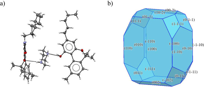
CBN-PI Form I crystallizes in the monoclinic system with space group Pc. In the asymmetric unit (Figure a), there are two unique molecules of CBN and one piperazine molecule, with one CBN molecule exhibiting disorder in its aliphatic chain. The unit cell (Figure SI 7a) comprises eight CBN and four PI molecules. The hydrogen bonding network (Figure SI 7b) is relatively complex: one PI molecule connects to three molecules of CBN. Both N atoms in piperazine each accept a hydrogen bond from the hydroxyl group of two neighboring CBN molecules. There is a third hydrogen bond, where N50 also acts as a donor, forming a hydrogen bond to a third molecule of CBN through an NH···O (ether) interaction. These molecular subunits are further arranged in the crystal packing to form a zigzag chain running along axis c. The calculated crystal shape (Figure b) shows a noncentrosymmetric crystal shape with two different parallel largest facets, (100) and (−100), which together comprise 42.15% of the total crystal surface area. The calculated lattice energy is −119.7 kJ mol–1. The strongest interaction energies (Figure SI 7c) align with the hydrogen bonding direction, with values of approximately −58.7 and −56.5 kJ mol–1.
CBN-PI Form II
The polymorphic Form II of the CBN-PI cocrystal crystallizes in the orthorhombic system with space group P b c n. The asymmetric unit (Figure a) comprises one CBN and a half PI molecule. The unit cell (Figure SI 8a) contains eight CBN and four PI molecules. Each piperazine N accepts a hydrogen bond (Figure SI 8b) from a hydroxyl group of a neighboring CBN, creating a centrosymmetric trimer. Weak C–H···O hydrogen bonds (C2a–H2C2a···O2 with C···O ∼ 3.60 Å and H···O ∼ 2.70 Å) link these trimers into columns extended along axis c. The calculated crystal shape (Figure b) displays a crystal shape dominated by two identical largest facets, (200) and (−200), with an area of 51.64% of the crystal surface. The lattice energy was calculated to be −118.2 kJ mol–1. Interestingly, the strongest calculated interaction (−30.1 kJ mol–1) occurs between two CBN molecules, which creates the chain motif, while the PI molecule is bonded (Figure SI 8c) in the hydrogen bond direction with an energy of −29.1 kJ mol–1.
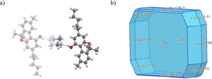
CBN-PI Form III
CBN-PI Form III has a monoclinic system with space group P 21/n. In its asymmetric unit (Figure a), it has two molecules of CBN and one molecule of PI. The unit cell (Figure SI 9a) consists of eight CBN and four PI molecules. Each piperazine N accepts a hydrogen bond (Figure SI 9b) from a hydroxyl group of a neighboring CBN, creating a pseudocentrosymmetric trimer.
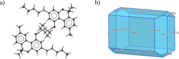
Weak C–H···O hydrogen bonds (C49–H491···O24 and C52–H521···O1 with C···O < 3.52 Å and H···O < 2.67 Å) link the trimers into layers parallel to the ab plane. The calculated crystal shape (Figure b) shows that the largest facets are (002) and (00–2), covering 41.58% of the crystal surface. The lattice energy was determined to be −122.9 kJ mol–1. The strongest calculated interactions (Figure SI 9c) occur between the CBN and PI molecules (approximately −69.4 kJ mol–1) in the hydrogen bond direction. Furthermore, the CBN molecules between the layers within the chain are slightly weaker (−58.6 kJ mol–1).
In summary, Form I exhibits a distinct packing pattern compared to Forms II and III, which probably contributes to its bulkier crystal morphology compared to the plate-like shapes of Forms II and III. In Forms II and III, the hydrogen bond motif is consistent with subunits being arranged into long chains, which create natural spaces corresponding to the slip planes. In contrast, the hydrogen bond system of Form I is arranged as an infinite chain, with one PI molecule connecting three CBN molecules. Interestingly, the Form III exhibits the highest lattice energy (−122.9 kJ mol–1) among all polymorphs. Nevertheless, all three forms showed lower lattice energy values than pure CBN (−180.0 kJ mol–1). The observed trend in lattice energy is in accordance with the calculated interaction energies, where Form III again shows the highest interaction energy (−69.4 kJ mol–1) among all polymorphs, even though it has the lowest melting point (87 °C). Furthermore, its interaction energy is higher than that of pure CBN, which indicates that the thermal properties alone are not the sole determining factor for polymorph stability.ref55,ref57 Molecular packing efficiency, crystal symmetry, interaction energies, etc. also contribute to the stability difference among the solid-state forms. This is further supported by the unit cell volumes at room temperature (Table SI 5), which revealed that Form III possesses the smallest volume, corresponding to the highest crystal density and closest packing, consistent with its experimentally observed high stability.
Polymorph Comparison
Surface Morphology Analysis
To investigate morphological factors influencing the polymorphic behavior of CBN–PI, we performed surface analyses using CSD-Particle tools. The comparison of the functional groups displayed on the largest facets is shown in Figure a, and they are visualized in Figure . The functional group density analysis revealed that HB acceptors were present on the surface of Form I (0.014 counts/Å2) and Form II (0.012 counts/Å2), while HB donors were only found on the surface of Form I, and they are located at the same position as HB acceptors, which correspond to the −OH groups on the CBN molecule. The Form III does not exhibit with any HB donors and acceptors on the studied surfaces. The aromatic bonds were identified on the surfaces of all polymorphs with a higher density than HB acceptors and donors. This might be caused by the presence of two aromatic rings in each CBN molecule, which, when they are oriented parallel to the surface plane, terminate at the crystal surface.
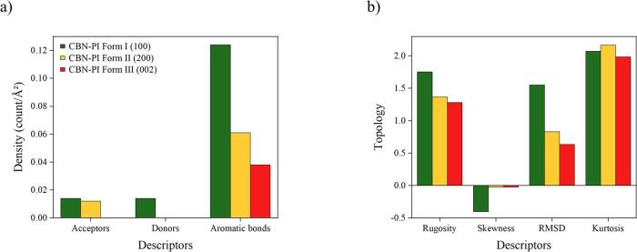
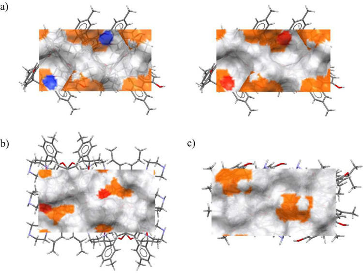
The topography descriptors, presented in Figure b and visualized in Figure SI 10, indicate that Form I exhibits the highest rugosity (1.752), which reflects a rougher surface. Forms II and III exhibit a smoother surface, with rugosity 1.366 and 1.281, respectively. The RMSD (root mean square difference) values follow the same trend observed for rugosity. The skewness value was negative for all forms, which indicates that the height distribution is found below the mean plane. However, the skewness value for Forms II and III was close to zero, meaning the surface is flatter. The kurtosis value was below 3 for all polymorphs, which means the absence of significant hills or valleys.ref. ref49
Based on these obtained results, Form I appears to have the roughest and most chemically interactive surface due to the presence of both HB donors and acceptors, which may enhance its reactivity and facilitate transformation under milling. In contrast, Form III exhibits a smoother and less interactive surface, making it the most stable polymorph from a morphological perspective.
Hydrogen Bond Propensity and Interaction Mapping
To complement morphological insights, hydrogen bond propensity (HBP) calculations and full interaction maps (FIMs) were used to evaluate the statistical favorability of the hydrogen bonding networks inside the crystal structure of each polymorph. The hydrogen bond propensity landscape (Figure ) plots the mean hydrogen bond propensity on the horizontal axis against the mean hydrogen bond coordination on the vertical axis. Each point corresponds to one of the top-scoring predicted hydrogen bond networks (smaller triangles) or an experimentally observed polymorph structure (large symbol). Networks clustering toward the lower right corner are predicted to be the most favorable in the solid state. The experimentally observed forms are highlighted.
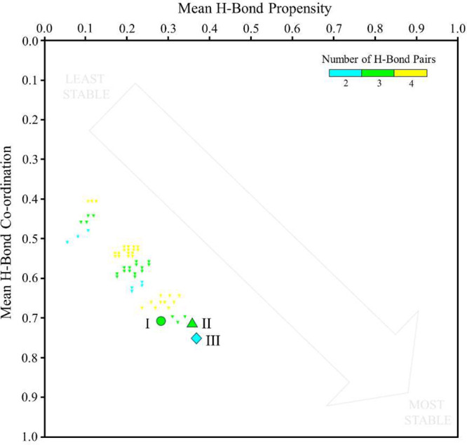
Across the three polymorphs, Form III has the highest values on both axes (HBP = 0.363, HBC = 0.737), suggesting the best use of available donor–acceptor groups within the crystal structure. Form II (0.350, 0.714) and Form I (0.275, 0.710) rank lower.
Form III contains two intermolecular hydrogen bond pairs, whereas Forms I and II each present three. The greater stability of Form III arises from the quality rather than the number of contacts. In Form III, both CBN phenolic O–H donors address piperazine N acceptors, which is consistent with high likelihood usage for these functional groups. By contrast, Form I introduces a piperazine N–H···O (cycloether) contact, which is statistically less probable and geometrically less favorable. Form II is structurally closer to Form III, but its donor–acceptor vectors are less well aligned. FIM analysis (Figure SI 11) further supports these observations. In Form III, donor and acceptor hotspots align well with actual interactions. In Form I, cycloether O lies in a region of weak acceptor probability, while in Form II, displaced acceptor hotspots lead to geometrical compromises that reduce network quality.
Overall, Form III achieves a “network-economical” structure that leverages strong, accessible hydrogen bonding motifs without introducing weak or frustrated interactions. This likely explains why Form III becomes the dominant product after repeated milling, while Form I disappears. Notably, the most probable NH···N (piperazine) homosynthon is absent in all polymorphs, explaining the modest absolute HBP values but reinforcing the relative advantage of Form III.
In conclusion, comparative analysis of the morphology and hydrogen bonding networks reveals distinct characteristics across the three CBN-PI polymorphs. Form I appears to have the most chemically reactive surface and exhibits the highest surface rugosity and a unique NH···O (ether) interaction, which is statistically weak. This higher reactivity may influence its tendency to disappear upon repeated milling. Form II shows intermediate characteristics, with a smoother surface than Form I but some geometric misalignment in its hydrogen bond contacts, which may limit its stability and allow it to be stable only in a specific mixture, e.g., with Form III. In contrast, Form III exhibits the smoothest surface morphology with no HB donors and acceptors available on the largest surface and the simplest, most favorable hydrogen bonding network. These structural and interaction advantages contribute to its stability and probably explain why Form III becomes the dominant polymorph under the studied conditions.
Particle Energy Calculation
As mentioned above, the stability of the polymorphs is a complex phenomenon, which depends on multiple factors, such as interaction energy, lattice energy, melting points, arrangement in the crystal lattice, etc. To further explore these contributions, we performed DFT-based calculations of the particle energy of the CBN-PI polymorphs. This approach incorporates three key energetic components: lattice energy, conformational energy penalty, and surface energy of the particle. This calculation approach estimates the total energy of a finite crystal particle, thereby providing deeper insights into the solid form stability and enabling the assessment of which contribution plays a dominant role in stabilizing each polymorph. This value was then scaled by the number of formula units per unit cell (Z) to obtain the particle energy per unit cell. The calculated values of each contribution, together with the resulting total particle energy for all polymorphs, are shown in Table .
4: Calculated Energetic Contributions for the CBN-PI Polymorph Particle Energy
| Eparticle (kJ mol–1) | Eparticle per unit cell (kJ mol–1) | Elatt (kJ mol–1) | ΔE conf (kJ mol–1) | 0.5∑x (hkl) × E (hkl) (kJ mol–1) | |
|---|---|---|---|---|---|
| Form I | –88.8 | –355.3 | –119.7 | 2.4 | –28.5 |
| Form II | –99.5 | –796.2 | –118.2 | 12.8 | –6.5 |
| Form III | –113.0 | –903.8 | –122.9 | 4.3 | –5.7 |
The lattice energy contribution has already been discussed above (Section sec3.3 ), and it was found that the lattice energy differs only in a few kJ mol–1 and From III (−122.9 kJ mol–1) showed the highest one. The conformational energy penalty was highest for Form II (12.8 kJ mol–1), which means that the molecules must undergo significant distortion from their lowest energy conformation (under vacuum) in order to accommodate the crystal lattice. Conversely, Form I exhibits the lowest conformational energy penalty (2.4 kJ mol–1), suggesting the molecular conformation within the crystal is close to the relaxed state in vacuum. Concerning the surface energy penalty, Form I shows the highest value (−28.5 kJ mol–1), which means that the surfaces grow faster, producing smaller facets, which are more reactive and result in bulkier crystals,ref. ref60 which is in accordance with the calculated crystal shape (Figure b). In contrast, Form II (−6.5 kJ mol–1) and Form III (−5.7 kJ mol–1) exhibit much lower attachment energy, corresponding to slower facet growth, with less reactive facets and resulting usually in the plate-like morphologiesref. ref60 (Figures b and fig6b). Finally, the total energy of the particles correlates well with the observed stability of the polymorphs. Form I, with the lowest total energy (−88.8 kJ mol–1), was found to disappear completely under the experimental conditions. Form II, with a total particle energy (−99.5 kJ mol–1), appeared only under specific conditions along with Form III or with unconverted starting materials. Form III, with the highest total particle energy (−113.0 kJ mol–1), proves to be the most stable polymorph. These findings complement the structural and morphological analyses and confirm that Form III is the most stable under the studied conditions.
Conclusions
In this study, we successfully prepared and characterized a novel cannabinol (CBN) piperazine cocrystal, which exists in three distinct polymorphic forms. By using ball milling, we systematically investigated the influence of various parameters such as the choice of solvents, temperature, and milling time on selective polymorph formation. However, repeated milling experiments revealed the phenomenon of disappearing polymorphs. While Form I was observed at the starting experiments, later on Form III emerged as a dominant phase under nearly all tested conditions. To understand the phenomenon, we further investigated the stability relationship between the polymorphs by performing detailed structural analysis, including crystal packing, hydrogen bond motifs, and morphology. Form I showed a bulkier morphology, with a more reactive surface, whereas Forms II and III exhibited a plate-like morphology with a less reactive surface. Among them, Form III exhibited the highest values of interaction energies among all polymorphs, indicating its superior stability. Furthermore, hydrogen bond propensity and full interaction maps (FIMs) identified that Form III displayed the most favorable, directional hydrogen bonds between CBN hydroxyl groups and piperazine nitrogen atoms. In contrast, Form I contained a statistically weak NH···O (ether) interaction, contributing to the lower network quality. By calculating particle energies, we observed that Form I, despite its strong lattice energy comparable to Forms II and III, exhibited low overall energy (−88.8 kJ mol–1), consistent with its disappearance. Form II showed an intermediate energy value (−99.5 kJ mol–1), reflecting its stability only under specific conditions and a combination with Form III or the starting material. Form III had the highest overall energy (−113.0 kJ mol–1), confirming its dominant stability under the experimental conditions despite its lowest melting point among the polymorphs. This proves that the melting point is not the sole determining factor of the polymorph stability, and other parameters such as crystalline lattice energy, symmetry, and hydrogen bonding system have to be also considered. Together, all of these findings can explain why Form I disappeared and why Form III prevailed. This study provides valuable insights into the polymorphic behavior of cannabinol piperazine cocrystals and highlights the importance of combining computational and experimental approaches in polymorph research. It also emphasizes the importance of a multifaceted approach in the development of a new solid-state form for pharmaceutical applications.
Supplementary Materials
References
- A. J. Cruz-Cabeza, J. Bernstein. Conformational Polymorphism. Chem. Rev., 2014. [DOI | PubMed]
- A. Chatziadi, E. Skořepová, J. Rohlíček, M. Dušek, L. Ridvan, M. Šoóš. Mechanochemically Induced Polymorphic Transformations of Sofosbuvir. Cryst. Growth Des., 2020. [DOI]
- E. Skořepová, J. Rohlíček, A. Chatziadi, V. Zvoníček, J. Jirát, J. Čejka, L. Ridvan, M. Šoóš. Low-temperature polymorphs of lacosamide. J. Cryst. Growth, 2021. [DOI]
- J.-P. Brog, C.-L. Chanez, A. Crochet, K. M. Fromm. Polymorphism, what it is and how to identify it: a systematic review. RSC Adv., 2013. [DOI]
- E. Skořepová, D. Bím, M. Hušák, J. Klimeš, A. Chatziadi, L. Ridvan, T. Boleslavská, J. Beránek, P. Šebek, L. Rulíšek. Increase in Solubility of Poorly-Ionizable Pharmaceuticals by Salt Formation: A Case of Agomelatine Sulfonates. Cryst. Growth Des., 2017. [DOI]
- D. Singhal, W. Curatolo. Drug polymorphism and dosage form design: a practical perspective. Adv. Drug Delivery Rev., 2004. [DOI]
- J. Bauer, S. Spanton, R. Henry, J. Quick, W. Dziki, W. Porter, J. Morris. Ritonavir: An Extraordinary Example of Conformational Polymorphism. Pharm. Res., 2001. [DOI | PubMed]
- T. Madan, A. P. Kakkar. Preparation and Characterization of Ranitidine-HC1 Crystals. Drug Dev. Ind. Pharm., 1994. [DOI]
- D.-K. Bučar, R. W. Lancaster, J. Bernstein. Disappearing Polymorphs Revisited. Angew. Chem., Int. Ed., 2015. [DOI]
- P. Sacchi, S. E. Wright, P. Neoptolemou, G. I. Lampronti, A. K. Rajagopalan, W. Kras, C. L. Evans, P. Hodgkinson, A. J. Cruz-Cabeza. Crystal size, shape, and conformational changes drive both the disappearance and reappearance of ritonavir polymorphs in the mill. Proc. Natl. Acad. Sci. U. S. A., 2024. [DOI | PubMed]
- I. Barsky, J. Bernstein, P. W. Stephens, K. H. Stone, E. Cheung, M. B. Hickey, J.-O. Henck. Disappearing and Reappearing Polymorphism in p-Methylchalcone. Cryst. Growth Des., 2008. [DOI]
- J.-O. Henck, J. Bernstein, A. Ellern, R. Boese. Disappearing and Reappearing Polymorphs. The Benzocaine:Picric Acid System. J. Am. Chem. Soc., 2001. [DOI | PubMed]
- Y. Yan, X.-L. Dai, J.-L. Jia, X.-H. Zhao, Z.-W. Li, T.-B. Lu, J.-M. Chen. Crystal Structures, Stability, and Solubility Evaluation of Two Polymorphs of a 2:1 Melatonin–Piperazine Cocrystal. Cryst. Growth Des., 2020. [DOI]
- H. Kulla, C. Becker, A. A. L. Michalchuk, K. Linberg, B. Paulus, F. Emmerling. Tuning the Apparent Stability of Polymorphic Cocrystals through Mechanochemistry. Cryst. Growth Des., 2019. [DOI]
- R. Prohens, R. Barbas, A. Portell, M. Font-Bardia, X. Alcobé, C. Puigjaner. Polymorphism of Cocrystals: The Promiscuous Behavior of Agomelatine. Cryst. Growth Des., 2016. [DOI]
- F. Fischer, A. Heidrich, S. Greiser, S. Benemann, K. Rademann, F. Emmerling. Polymorphism of Mechanochemically Synthesized Cocrystals: A Case Study. Cryst. Growth Des., 2016. [DOI]
- S. Aitipamula, P. S. Chow, R. B. H. Tan. Polymorphism in cocrystals: a review and assessment of its significance. CrystEngComm, 2014. [DOI]
- N. Schultheiss, A. Newman. Pharmaceutical Cocrystals and Their Physicochemical Properties. Cryst. Growth Des., 2009. [DOI]
- M. Guo, X. Sun, J. Chen, T. Cai. Pharmaceutical cocrystals: A review of preparations, physicochemical properties and applications. Acta Pharm. Sin. B, 2021. [DOI | PubMed]
- N. Qiao, M. Li, W. Schlindwein, N. Malek, A. Davies, G. Trappitt. Pharmaceutical cocrystals: An overview. Int. J. Pharm., 2011. [DOI | PubMed]
- N. Shan, M. J. Zaworotko. The role of cocrystals in pharmaceutical science. Drug Discovery Today, 2008. [DOI | PubMed]
- M. D. Eddleston, S. Sivachelvam, W. Jones. Screening for polymorphs of cocrystals: a case study. CrystEngComm, 2013. [DOI]
- K. Nagapudi, E. Y. Umanzor, C. Masui. High-throughput screening and scale-up of cocrystals using resonant acoustic mixing. Int. J. Pharm., 2017. [DOI | PubMed]
- S. Aher, R. Dhumal, K. Mahadik, A. Paradkar, P. York. Ultrasound assisted cocrystallization from solution (USSC) containing a non-congruently soluble cocrystal component pair: Caffeine/maleic acid. Eur. J. Pharm. Sci., 2010. [DOI | PubMed]
- K. Linberg, B. Röder, D. Al-Sabbagh, F. Emmerling, A. A. L. Michalchuk. Controlling polymorphism in molecular cocrystals by variable temperature ball milling. Faraday Discuss., 2023. [DOI | PubMed]
- A. M. Belenguer, G. I. Lampronti, A. A. L. Michalchuk, F. Emmerling, J. K. M. Sanders. Quantitative reversible one pot interconversion of three crystalline polymorphs by ball mill grinding. CrystEngComm, 2022. [DOI]
- N. Kamali, K. Gniado, P. McArdle, A. Erxleben. Application of Ball Milling for Highly Selective Mechanochemical Polymorph Transformations. Org. Process Res. Dev., 2018. [DOI]
- A. M. Belenguer, T. Friščić, G. M. Day, J. K. M. Sanders. Solid-state dynamic combinatorial chemistry: reversibility and thermodynamic product selection in covalent mechanosynthesis. Chem. Sci., 2011. [DOI]
- H. Kulla, S. Greiser, S. Benemann, K. Rademann, F. Emmerling. Knowing When To StopTrapping Metastable Polymorphs in Mechanochemical Reactions. Cryst. Growth Des., 2017. [DOI]
- F. Fischer, S. Greiser, D. Pfeifer, C. Jäger, K. Rademann, F. Emmerling. Mechanochemically Induced Conversion of Crystalline Benzamide Polymorphs by Seeding. Angew. Chem., Int. Ed., 2016. [DOI]
- P. A. Julien, I. Malvestiti, T. Friščić. The effect of milling frequency on a mechanochemical organic reaction monitored by in situ Raman spectroscopy. Beilstein J. Org. Chem., 2017. [DOI | PubMed]
- R. Schmidt, H. Martin Scholze, A. Stolle. Temperature progression in a mixer ball mill. Int. J. Ind. Chem., 2016. [DOI]
- K. Linberg, P. C. Sander, F. Emmerling, A. A. L. Michalchuk. In situ investigation of controlled polymorphism in mechanochemistry at elevated temperature. RSC Mechanochem., 2024. [DOI]
- E. Losev, S. Arkhipov, D. Kolybalov, A. Mineev, A. Ogienko, E. Boldyreva, V. Boldyrev. Substituting steel for a polymer in a jar for ball milling does matter. CrystEngComm, 2022. [DOI]
- L. S. Germann, M. Arhangelskis, M. Etter, R. E. Dinnebier, T. Friščić. Challenging the Ostwald rule of stages in mechanochemical cocrystallisation. Chem. Sci., 2020. [DOI | PubMed]
- A. Chatziadi, E. Skořepová, M. Kohout, L. Ridvan, M. Šoóš. Exploring the polymorphism of sofosbuvir via mechanochemistry: effect of milling jar geometry and material. CrystEngComm, 2022. [DOI]
- K. Cohen, A. Weizman, A. Weinstein. Positive and Negative Effects of Cannabis and Cannabinoids on Health. Clin. Pharmacol. Ther., 2019. [DOI | PubMed]
- A. Khouchlaa, S. Khouri, A. Hajib, I. Zeouk, S. Amalich, S. Msairi, N. El Menyiy, C. Rais, M. Lahyaoui, A. Khalid. Health benefits, pharmacological properties, and metabolism of cannabinol: A comprehensive review. Ind. Crops Prod., 2024. [DOI]
- N. Zhong, D. Li, B. Wang, O. Kovalchuk, I. Kovalchuk. Cannabinol inhibits cell growth and triggers cell cycle arrest and apoptosis in cancer cells. Biocatal. Agric. Biotechnol., 2023. [DOI]
- M. Zaiachuk, S. V. Suryavanshi, N. Pryimak, I. Kovalchuk, O. Kovalchuk. The Anti-Inflammatory Effects of Cannabis sativa Extracts on LPS-Induced Cytokines Release in Human Macrophages. Molecules, 2023. [DOI | PubMed]
- H. Wong, B. E. Cairns. Cannabidiol, cannabinol and their combinations act as peripheral analgesics in a rat model of myofascial pain. Arch. Oral Biol., 2019. [DOI | PubMed]
- Solid compositions of cocrystals of cannabinoids. Spain. 2021
- Cocrystals of cannabinoids. US. 2023
- Agilent CrysAlis PRO; Agilent Technologies Ltd.: Yarnton, England, 2014 (accessed).
- P. W. Betteridge, J. R. Carruthers, R. I. Cooper, K. Prout, D. J. Watkin. CRYSTALS version 12: software for guided crystal structure analysis. J. Appl. Crystallogr., 2003. [DOI]
- V. Petříček, L. Palatinus, J. Plášil, M. Dušek. Jana2020 – a new version of the crystallographic computing system Jana. Z. Kristallogr. Cryst. Mater., 2023. [DOI]
- J. Rohlíček, M. Hušák. MCE2005–a new version of a program for fast interactive visualization of electron and similar density maps optimized for small molecules. J. Appl. Crystallogr., 2007. [DOI]
- C. F. Mackenzie, P. R. Spackman, D. Jayatilaka, M. A. Spackman. CrystalExplorer model energies and energy frameworks: extension to metal coordination compounds, organic salts, solvates and open-shell systems. IUCrJ., 2017. [DOI | PubMed]
- A. A. Moldovan, A. G. P. Maloney. Surface AnalysisFrom Crystal Structures to Particle Properties. Cryst. Growth Des., 2024. [DOI]
- E. Prandini, E. Calì, A. G. P. Maloney, E. Parisi, E. Simone. Predicting particle quality attributes of organic crystalline materials using Particle Informatics. Powder Technol., 2024. [DOI]
- C. F. Macrae, I. J. Bruno, J. A. Chisholm, P. R. Edgington, P. McCabe, E. Pidcock, L. Rodriguez-Monge, R. Taylor, J. van de Streek, P. A. Wood. Mercury CSD 2.0 – new features for the visualization and investigation of crystal structures. J. Appl. Crystallogr., 2008. [DOI]
- K. Kopczyńska, C. J. Kingsbury, E. Pidcock, A. A. Moldovan, I. D. Madura. A Case of Unusual Cocrystal Polymorphs of Caffeine and Phenylboronic Acid: Cambridge Structural Database Tools in Action. Cryst. Growth Des., 2024. [DOI]
- C. F. Macrae, I. Sovago, S. J. Cottrell, P. T. A. Galek, P. McCabe, E. Pidcock, M. Platings, G. P. Shields, J. S. Stevens, M. Towler. Mercury 4.0: from visualization to analysis, design and prediction. J. Appl. Crystallogr., 2020. [DOI | PubMed]
- R. M. Bhardwaj, J. A. McMahon, J. Nyman, L. S. Price, S. Konar, I. D. H. Oswald, C. R. Pulham, S. L. Price, S. M. Reutzel-Edens. A Prolific Solvate Former, Galunisertib, under the Pressure of Crystal Structure Prediction, Produces Ten Diverse Polymorphs. J. Am. Chem. Soc., 2019. [DOI | PubMed]
- A. Koryt́áková, A. Chatziadi, J. Rohlíček, E. Zmeškalová, J. Beránek, M. Šoóš. Stability study and structural insights into cannabidiol cocrystals. CrystEngComm, 2025. [DOI]
- S. Aitipamula, P. S. Chow, R. B. H. Tan. Dimorphs of a 1:1 cocrystal of ethenzamide and saccharin: solid-state grinding methods result in metastable polymorph. CrystEngComm, 2009. [DOI]
- A. Chatziadi, E. Skorepova, J. Jirat, J. Rohlíček, M. Šoóš. Characterization and Insights into the Formation of New Multicomponent Solid Forms of Sofosbuvir. Cryst. Growth Des., 2022. [DOI]
- S. Pagola. Outstanding Advantages, Current Drawbacks, and Significant Recent Developments in Mechanochemistry: A Perspective View. In Crystals, 2023. [DOI]
- T. Ottersen, E. Rosenqvist, C. Turner, F. El-Feraly. The crystal and molecular structure of cannabinol. Acta Chem. Scand., 1977. [DOI]
- T. T. H. Nguyen, I. Rosbottom, I. Marziano, R. B. Hammond, K. J. Roberts. Crystal Morphology and Interfacial Stability of RS-Ibuprofen in Relation to Its Molecular and Synthonic Structure. Cryst. Growth Des., 2017. [DOI]