
Cannabis Use and Glutamate across the Psychosis Spectrum: In Vivo Evidence from 7T Proton Magnetic Resonance Spectroscopy
Abstract
Cannabis use is linked to elevated psychosis risk, yet the neurobiological mechanisms that couple use to symptom expression remain unclear. Because glutamatergic dysregulation has been implicated in both cannabis effects and psychosis vulnerability, we examined whether brain glutamate relates to dimensional symptoms as a function of cannabis use across the psychosis spectrum. Seventy-nine participants—typically developing controls, clinical high-risk individuals, and patients with psychosis—completed dimensional clinical assessments, detailed cannabis surveys, urine toxicology, and ultra-high-field 7T 1HMRS quantification of anterior cingulate cortex (ACC) glutamate levels. Linear models assessed the main and interactive effects of ACC glutamate and cannabis use on positive and negative symptoms. Self-reported cannabis use showed strong concordance with urine toxicology. Cannabis use was associated with higher positive and negative symptoms. Independently, higher ACC glutamate predicted greater positive and negative symptoms. Notably, lower glutamate levels were associated with higher positive symptoms in cannabis users. Exploratory analyses suggested interactions for depressive and manic symptoms, indicating that glutamatergic abnormalities may amplify the overall severity of cannabis-related symptoms. Sensitivity analyses revealed lower ACC glutamate in psychosis patients—especially cannabis users—highlighting diagnostic group differences and reinforcing the link between cannabis exposure and glutamatergic dysfunction. These findings implicate ACC glutamatergic dysfunction as a transdiagnostic correlate of symptom burden, particularly in those with psychosis who are cannabis users. Glutamate-targeted interventions and longitudinal designs will be needed to examine causal pathways linking cannabis exposure to psychosis-relevant outcomes.
Affiliations: University of Pennsylvania; Perelman School of Medicine; Center for Metabolic Imaging in Precision Medicine (CAMIPM), Perelman School of Medicine at the University of Pennsylvania
License: CC BY 4.0 This work is licensed under a Creative Commons Attribution 4.0 International License, which allows reusers to distribute, remix, adapt, and build upon the material in any medium or format, so long as attribution is given to the creator. The license allows for commercial use.
Article links: DOI: 10.21203/rs.3.rs-7802376/v1 | PubMed: 41333424 | PMC: PMC12668139
Relevance: Moderate: mentioned 3+ times in text
Full text: PDF (1.4 MB)
Introduction
Recent changes in cannabis laws and the rise in nonmedical cannabis use have intensified concerns about the long-term effects of cannabis on mental health, particularly among youth. While cannabis use has been implicated in a range of psychiatric outcomes1, accumulating evidence highlights its association with dimensional psychopathology, including positive and negative psychosis-spectrum symptoms2 and mood disturbances3. Epidemiological studies show that frequent use—especially of high-potency cannabis—and earlier age of initiation are linked to greater risk for adverse outcomes4–7, with a dose–response relationship, particularly in psychosis8, 9. Cannabis use during adolescence has been associated not only with the onset of psychotic disorders in early adulthood but also with elevated subclinical symptoms and poorer functional outcomes across multiple domains10, 11. In clinical high-risk for psychosis (CHR) samples12–14, cannabis use is associated with worsened subthreshold symptoms, and among those with established schizophrenia, it may exacerbate symptom severity and increase relapse risk15–17. These effects may be partly mediated by cannabis-induced disruption of the glutamatergic system18, 19, a key neurochemical pathway implicated in mood, psychosis, and neurodevelopmental disorders. Both cannabis use18, 19 and psychiatric disorders20–24 have been independently associated with glutamate dysregulation, yet the mechanistic interplay remains poorly understood. Here we begin to address this gap by integrating detailed self-reported cannabis use, urine drug screening, and ultra–high field 7T proton magnetic resonance spectroscopy (¹HMRS) of the ACC to examine glutamate (Glu) alterations associated with cannabis use across a transdiagnostic sample.
The primary psychoactive component in cannabis, Δ9-tetrahydrocannabinol (THC), modulates neurometabolic and neurotransmitter systems—most notably dopamine and glutamate19, 25, both central to psychosis.
Sustained cannabis use reduces glutamate signaling via Cannabinoid receptor 1 (CB1R) activation, and CB1R agonists induce psychosis-like behaviors26. Glutamate hypofunction20, 21 contributes to dopamine dysregulation in schizophrenia, and recent studies have documented glutamatergic metabolite alterations in individuals with or at risk for schizophrenia20, 22–24 and depression27. However, systematic in vivo investigations of glutamatergic alterations associated with cannabis use in humans using ultra-high field MRI are limited, hindering efforts to clarify how cannabis may heighten psychopathological vulnerability and symptoms.
Glutamatergic alterations are central to the pathophysiology of psychosis. Historically, psychosis has been associated with dopaminergic dysfunction due to its link to positive symptoms28, but accumulating evidence emphasize the relevance of glutamate dysregulation in psychosis29. A growing literature implicates glutamate dysfunction in psychosis, especially in schizophrenia30, 31, including data from animal models32–36, postmortem studies37–40, genetic analyses41–45, peripheral biomarkers46, and in vivo 1HMRS31. While early 1HMRS meta-analyses at 3T reported higher Glu metabolites in cases vs. controls47, subsequent mega-analyses20, 7T GluCEST investigations22, 24, and 7T 1HMRS23 studies have demonstrated regionally lower Glu in individuals with psychosis, but see48. Region-specific effects include lower Glu in mPFC, frontal white matter, medial temporal lobe, and thalamus, with elevated Glu in cerebellum and basal ganglia.47 Taken together, 1HMRS studies reveal regionally heterogeneous findings in glutamatergic levels in CHR patients to those with threshold psychosis disorder,30, 49–54 suggesting that glutamate alterations may be part of illness progression providing significant rationale for studying glutamate across the psychosis spectrum.
Few studies have directly examined cannabis–glutamate associations across clinical dimensions of the psychosis spectrum, and existing work has been limited to 3T MRI. Early 1HMRS work found that patients with early psychosis who used cannabis exhibited lower prefrontal Glu concentrations than both non-using psychosis patients and healthy controls, along with a steeper age-related Glu decline, suggesting that cannabis may exacerbate glutamatergic dysfunction and influence disease progression55. Another 3T 1HMRS study in early psychosis patients observed limited differences in Glutamate+Glutamine (Glx) between cannabis users and nonusers but identified associations between regional gray matter volume (caudate, hippocampus, cortex) and caudate Glx in cannabis users, implying that heavy cannabis use may alter structure–neurochemical connectivity in CB1R-rich regions56. In healthy individuals, acute THC induced greater Glx surges in those who developed transient psychotic-like symptoms, particularly among participants with lower baseline Glx and greater prior cannabis exposure57. These findings collectively suggest that baseline glutamatergic state and cannabis responsiveness may interact to influence psychosis risk and expression. However, prior studies have been limited to 3T 1HMRS, where sensitivity to glutamate is lower, and they have only measured cannabis–glutamate associations in categorical clinical groupings.
In summary, converging epidemiological, preclinical, and neuroimaging evidence implicates glutamate as a key modulator of the cannabis–psychosis relationship. By integrating detailed cannabis use phenotyping, high–field 7T 1HMRS measures, and dimensional clinical assessments across individuals who are typically developing, CHR or experience psychosis (PSY), our study aims to elucidate how cannabis use is associated with glutamatergic levels in those with and at risk for psychosis. In line with recent RDoC and NIMH initiatives, we assess clinical symptoms dimensionally rather than relying solely on categorical diagnoses. Dimensional measurement provides greater sensitivity to subthreshold variation58, 59, allowing us to capture the full spectrum of psychopathology and potential associations with cannabis use, including effects that may not meet diagnostic thresholds. This approach also facilitates transdiagnostic comparisons and improves power to detect brain–behavior associations by reducing heterogeneity within broad diagnostic categories. Understanding this interplay may identify neurobiological and clinical markers of risk and ultimately inform targeted prevention and intervention strategies for this high-risk population.
Methods
Participants To capture a broad range of psychopathology, we studied both healthy individuals and those with subthreshold and threshold psychiatric symptoms. The study sample (N = 79, ages 14–32, 35 female) included a typically-developing (TD) group (no Axis I diagnoses or subthreshold psychosis symptomatology, no history of psychotropic medication use; N = 31) and a clinical group composed of individuals classified by experienced clinician scientists (CK, MEC) as having psychosis spectrum disorders (N = 48), including 27 with CHR and 21 with schizophrenia spectrum and other psychotic disorders (PSY; N = 21). Although our primary analyses treat psychopathology as a continuous, transdiagnostic construct, demographic and clinical characteristics are displayed by categorical diagnosis for clarity and interpretability. This allows for evaluation of the representativeness of the sample and provides context for the dimensional analyses presented below. Complete demographic and clinical information are presented in Table 1, stratified by cannabis user status and clinical diagnosis. In addition, sensitivity analyses are conducted by diagnostic group (Supplemental Results). Brief cognitive (Montreal Cognitive Assessment (MoCA60)) and functional assessments (Global Assessment of functioning (GAF61)) were also administered.
Table 1: Participant demographics and clinical characteristics.
| DxGroup | N | Age (years) | Percent Female | SIPS Positive | SIPS Negative | SIPS Disorganzied | SIPS Genralized | GAF | MoCA |
|---|---|---|---|---|---|---|---|---|---|
| TD | |||||||||
| Cannabis User | |||||||||
| No | 16 | 20.7 ± 2.7 | 63% | 1.3 ± 1.8 | 1.1 ± 1.0 | 0.4 ± 0.6 | 0.7 ± 0.9 | 87.4 ± 4.5 | 26.5 ± 2.8 |
| Yes | 15 | 22.8 ± 3.0 | 27% | 1.7 ± 1.8 | 1.5 ± 1.8 | 0.3 ± 0.6 | 1.5 ± 2.0 | 81.2 ± 13.7 | 27.7 ± 1.6 |
| CHR | |||||||||
| Cannabis User | |||||||||
| No | 14 | 20.8 ± 3.6 | 50% | 8.1 ± 4.7 | 7.6 ± 6.0 | 4.1 ± 2.5 | 3.7 ± 3.4 | 56.9 ± 8.0 | 24.8 ± 2.9 |
| Yes | 13 | 22.6 ± 2.8 | 31% | 6.8 ± 3.1 | 7.6 ± 5.5 | 4.0 ± 3.0 | 3.2 ± 2.5 | 61.0 ± 12.2 | 27.0 ± 2.7 |
| PSY | |||||||||
| Cannabis User | |||||||||
| No | 5 | 26.8 ± 4.1 | 60% | 14.0 ± 6.1 | 9.4 ± 5.6 | 5.4 ± 1.1 | 5.6 ± 3.6 | 47.4 ± 4.0 | 22.0 ± 3.9 |
| Yes | 16 | 24.8 ± 3.5 | 44% | 19.7 ± 6.0 | 12.0 ± 2.7 | 7.6 ± 4.2 | 8.5 ± 6.0 | 42.4 ± 9.0 | 25.6 ± 2.4 |
This table summarizes demographic and clinical characteristics of the study sample. Demographic variables include age, sex distribution, and relevant clinical status or group designation. Mean (+/− s.d.) positive, negative, disorganized and generalized SIPS, GAF and MoCA scores are shown.
Clinical Assessment All participants received detailed clinical assessments, including a semi-structured diagnostic interview to assess a broad spectrum of psychosis-relevant experiences, psychopathology, treatment history, and medications. Participants diagnostic assessments by highly-trained assessors using a computerized version of a modified and adapted Schedule for Affective Disorders and SZ for School Age Children – Present and Lifetime Version (K-SADS-4.0)62, which includes semi-structured assessments of mood, ADHD and substance use disorders. Psychosis spectrum symptoms were assessed using the Structured Interview for Prodromal Syndromes (SIPS, v. 4.0), in which selected modules of the Structured Clinical Interview for DSM-IV (SCID-IV)63 are integrated to facilitate differential diagnosis. The Scale of Prodromal Symptoms (SOPS)64, embedded within the SIPS65, describes and dimensionally rates the severity of positive, negative, disorganized, and general symptoms occurring within the past 6 months. To facilitate efficient data collection, the instruments were integrated into one administrative tool, the Computer-Assisted Psychopathology Assessment (CAPA)62. Interviewers underwent formal training including lectures and mock interviews, followed by supervised administration and performance evaluation until competency was established62. For minors, collateral information was provided by an independent interview with a parent or guardian. All information was integrated to yield consensus diagnoses and clinical symptom ratings at a clinical case conference led by at least two doctoral-level clinicians. CHR was defined as the presence of at least one positive symptom rated 3–5, or at least two negative and/or disorganized symptoms rated 3–6. Psychosis (PSY) patients are individuals with a DSM-5 schizophrenia spectrum disorder diagnosis (e.g., SZ). Clinical scores are shown in Table 1.
Estimation of Correlated Traits Clinical Factor scores. Clinical factor scores were derived using a framework that incorporated longitudinal computerized assessments (e.g. GOASSESS66, 67 and CAPA68) used in our laboratory group since the landmark Philadelphia Neurodevelopmental Cohort study.69–71 We used a bank of all clinically available data from our laboratory to inform a dimensional factor analysis (e.g. GOASSESS66, 67 and CAPA68) and generate clinical factor scores for participants enrolled in the current study.
Because the same instruments were not used across historical time points, historical data were harmonized across longitudinal visits by using only items that overlapped between GOASSESS and CAPA interviews. These included the screen items from the depression, mania, and ADHD sections of the GOASSESS/CAPA, as well as the PRIME72, 73 screen and a subset of SIPS items. Using this harmonized framework, dimensional clinical scores (correlated trait factor scores) were generated for each participant in the present study based on the CAPA data collected (described in the Clinical Assessment section). Dimensional clinical factors scores are shown, by diagnostic group, in Supplemental Table 2.
Cannabis Use Cannabis use was measured using a computerized version of the Minnesota Center for Twin and Family Research self-report substance use assessment74, which was privately self-administered on a computer. The measure assessed lifetime use of several substances; for cannabis and alcohol, additional questions queried age at first use and frequency of past year use. To obtain more detailed data regarding patterns of cannabis use, several questions from the DFAQ-CU (Daily Sessions, Frequency, Age of Onset, and Quantity of Cannabis Use Inventory)75 were also administered. This measure provided data for individual methods of cannabis use, with visual depictions to indicate quantities of cannabis across forms (e.g., joint, flower). Summary measures were generated for frequency, age of onset, and quantity of cannabis use across methods of use, including increasingly popular methods (e.g., edibles, concentrates). The DFAQ-CU shows convergent, predictive, and discriminant validity, including high expected correlations (> 0.7) with other frequency measures (e.g., timeline follow-back), modulates correlations with cannabis use disorder symptoms and cannabis-related problems, and low correlations with alcohol use problems75, 76. Consistent with prior research77, 78, we classified individuals as cannabis users if they reported use more than once per month over the past year; for exploratory analyses we also classified individuals as frequent cannabis users if they reported using cannabis on a daily or nearly daily basis for the past 12 months. We also administered sections of the Cannabis Experiences Questionnaire (CEQ) that assess motivations for cannabis use and positive and negative experiences associated with use, consistent with prior studies in early psychosis79.
Urine Drug Screen for Cannabis In line with expert consensus groups recommendations80, we also used a urine drug test to identify presence/absence of cannabis use. All urine samples (90ml) were collected using Redwood Toxicology iCUPs™, which detect one or more of the primary metabolites of THC. The immunoassay is binary (positive or negative). If the sample is below threshold, it is reported as negative; if above, it is reported as positive. Detection threshold for presence/absence is 50 ng/mL of THC-COOH. Cannabis metabolites are typically detected for up to 3 days after a single use in occasional users, while frequent users typically test positive for a much longer period—sometimes up to several weeks—due to the slow release of stored THC from fatty tissues and repeated, high-level exposure. Body fat percentage, metabolism, hydration level, and dosage all affect how long THC-COOH remains detectable in urine. Urine drug screening occurred prior to the 7T MRI session. If a participant reported cannabis use on the day of the MRI, or if acute intoxication was suspected, the study visit was rescheduled.
7T1HMRS Data Acquisition All 1HMRS data were acquired on a whole-body 7 Tesla Terra MRI system (Siemens, Erlangen, Germany) equipped with a single-channel volume transmit and 32-channel receive proton phased array head radiofrequency coil (Nova Medical, Wilmington, MA, USA). Prior to spectroscopy, high-resolution structural images were obtained using a magnetization-prepared rapid acquisition gradient echo (MP2RAGE) sequence (TE = 2.52 ms, TR = 5000 ms, TI1 = 700 ms, TI2 = 2500 ms, flip angle 1 = 7°, flip angle 2 = 5°, voxel size = 0.8 mm3). These anatomical images served both for voxel placement and for later tissue-segmentation corrections.
Voxel Localization and Shimming1HMRS voxels (10 × 40 × 15 mm³) were manually placed in anterior cingulate cortex, using the mid-sagittal anatomical image as reference. Care was taken to align all voxel faces orthogonal to the cortical surface and to minimize inclusion of cerebrospinal fluid (CSF)-rich sulcal regions. First- and second-order B0 shimming was performed manually over each voxel to achieve a full-width at half-maximum (FWHM) linewidth of ≤ 20 Hz for the unsuppressed water peak within the voxel.
Spectroscopy Sequence Spectra were acquired using a point-resolved spectroscopy (PRESS) sequence optimized for 7T. Sequence parameters were as follows: TE = 23 ms, TR = 3,000 ms, number of averages (NA) = 64 (total acquisition time ≈ 3 min 24 s per voxel). Unsuppressed water reference scan preceded this acquisition for reference and calibration to quantify and correct for eddy-currents. Sequence parameters of the unsuppressed scan (TE = 23 ms, TR = 3,000 ms, NA = 8) used identical localization and shimming settings.
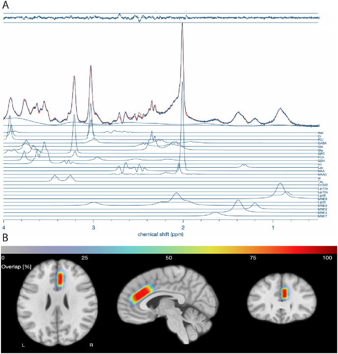
Data Processing and Quantification All spectroscopy data were exported in RDA format, which were coil-combined and averaged at the scanner. All spectra were processed, modeled, and quantified using Osprey (v2.9.0; Osprey Project, Johns Hopkins University)81 running in MATLAB (R2023b; MathWorks, Natick, MA, USA). Spectral fitting was performed using the LCModel package (v6.3–1N) implemented in Osprey software. Prior to fitting, imported raw spectra were processed through the ‘OspreyProcess’ module, which includes eddy-current correction, frequency and phase alignment, water removal, frequency referencing, and initial phasing. Default parameters were utilized for modeling and quantification in ‘OspreyFit’, including a metabolite fit range of 0.5 to 4.0 ppm, a water fit range of 2.0 to 7.4 ppm, and a knot spacing of 0.4 ppm. The basis set provided for LCModel included simulated metabolite spectra for N-acetylaspartate (NAA), creatine (Cr), choline (Cho), glutamate (Glu), glutamine (Gln), myo-inositol (mI), γ-aminobutyric acid (GABA), and others, generated with density-matrix simulations at 7T incorporating sequence-specific TE and TM. In addition to the basis set, default macromolecular and lipid components provided by Osprey were fitted to each spectrum. Each participant’s 1HMRS voxel was then co-registered to a T1-weighted MP2RAGE provided for each individual. Co-registration in ‘OspreyCoReg’ also produces a voxel mask that is subsequently used in ‘OspreySeg’ to segment the voxel into gray matter, white matter, and CSF using SPM12. Example spectra and voxel overlap across participants are shown in Fig. 1. In this module, fractional tissue volumes are also determined. Tissue- and relaxation-corrected metabolite molal concentrations were then estimated in ‘OspreyQuantify’ according to the Gasparovic method82. Only tissue-corrected metabolite estimates passing quality control (Cramér–Rao lower-bound (CRLB) ≤ 20%) were included in subsequent analyses.
Statistical Analysis Group differences in cannabis use rates were assessed using independent-samples t tests and chi-squared tests, as appropriate. To examine the effects of 1HMRS glutamate, cannabis use, and their interaction on clinical symptom dimensions, we conducted a series of linear regression models. The primary outcome measures of interest were those associated with psychosis: positive and negative symptoms. Each of the measures was modeled as a continuous outcome. All linear models controlled for sex and age at scan. Cannabis use was coded dichotomously (0 = non-user, 1 = user), and glutamate was entered as a continuous variable. Interaction terms between glutamate and cannabis user status were included to assess modulation effects. Significance was assessed at p < 0.05, with trend-level effects noted at p < 0.10. Residuals were visually inspected for normality and homoscedasticity. Results are reported as F-statistics with associated degrees of freedom and p-values. Exploratory analyses in other symptom domains (depression, mania, and ADHD) were also conducted. As a sensitivity analysis, these analyses were repeated with categorical diagnostic group (TD, CHR, PSY; See Supplemental Results). Demographic data were compared across diagnostic groups (e.g., diagnosis, cannabis user status) using t-tests and chi-square as appropriate. Rates of cannabis use and urine drug screen data were compared across all three diagnostic groups using an omnibus Chi-square test, with planned post-hoc pairwise comparisons. Post-hoc comparisons were completed using simple slopes (‘sim_slopes’) and least squares means approach (‘lsmeans’) using the ‘interactions’83 and ‘lsmeans’84 libraries in R (V4.3.3), respectively.
Results
Self-reported cannabis use rates converge with results of urine toxicology screening
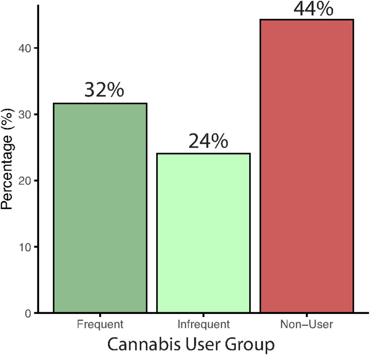
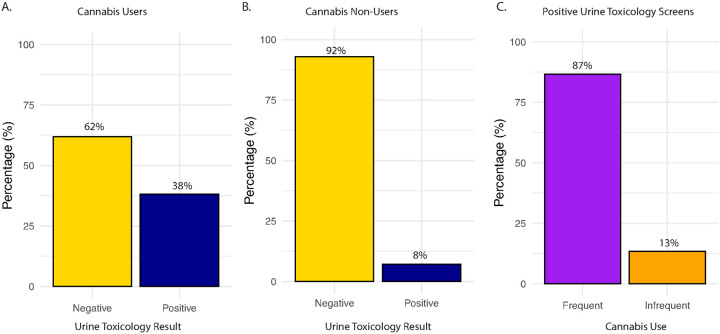
Across the sample, 56% (n = 44) self-reported lifetime cannabis use, including 32% (n = 25) frequent users and 24% (n = 19) infrequent users, while 44% (n = 35) were cannabis non-users (Fig. 2). To assess the validity of self-reported cannabis use, we compared responses on cannabis use self-report assessments to results from urine toxicology across all individuals. 89% (70/79) of individuals had a valid urine toxicology screen. A chi-square test revealed a significant association between self-reported use and toxicology results, χ2(1) = 7.62, p < 0.01. The vast majority (92%) of cannabis non-users tested negative on the urine toxicology screen (Fig. 3A), while self-reported cannabis users were more likely to test positive on the urine toxicology screen (Fig. 3B), but overall rates of positive screens were low (38%), which is not unexpected given the typical window of cannabis metabolite detection. Indeed, self-reported frequent users (87%) were more likely to test positive on urine toxicology than infrequent users (13%; Fig. 3C). Overall, these results indicate good concordance between self-report and biological verification of cannabis use. Note that two individuals who denied cannabis use tested positive on urine toxicology; these individuals were considered infrequent cannabis users in subsequent analyses.
Cannabis use is higher in psychosis patients
76% of PSY participants, 48% of CHR participants, and 45% of TD participants reported cannabis use (Supplemental Fig. 1). A chi-square test of independence comparing all three groups was not significant (χ2(2) = 5.51, p = 0.06), but planned pairwise comparisons indicated that cannabis use was more prevalent in the PSY group compared to the TD group, χ2(1) = 3.78, p = 0.05. The rate of use between PSY and CHR groups showed nominally more cannabis use in PSY (χ2(1) = 2.80, p = 0.09), but CHR and TD groups did not differ in cannabis use, χ2(1) < 0.01, p = 0.99. Notably, analysis of frequency of cannabis use (Supplemental Fig. 2) in CHR (33%) and PSY (32%) showed similar rates of frequent use (3–4 times a week or daily).
Glutamate and cannabis use are associated with dimensional clinical symptoms
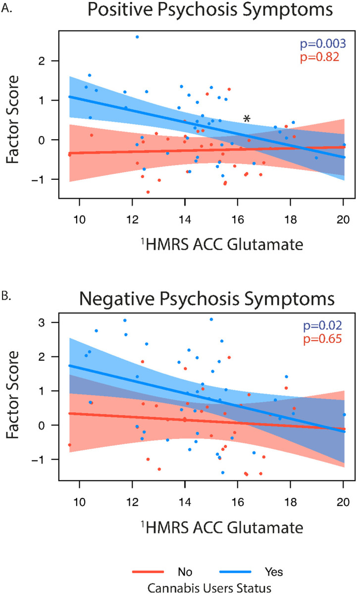
We examined the relationship of 1HMRS glutamate levels, cannabis use, and their interaction with dimensional clinical symptoms across the entire sample. A significant Glu × cannabis interaction was observed for positive psychosis symptoms (F(1,68) = 4.33, p = 0.041), indicating that the relationship between glutamate and positive symptom severity differed by cannabis use status (Fig. 4A). Using a simple slopes follow-up analysis, in cannabis users, lower glutamate was significantly associated with greater positive symptom severity (β = − 0.15, SE = 0.05, t(68) = − 3.11, p = 0.003), but this association was not significant in non-users (β = 0.01, SE = 0.06, t(68) = 0.23, p = 0.82). These findings suggest that glutamate levels are differentially linked to positive psychosis symptoms depending on cannabis use history, with effects most pronounced among cannabis users. Additionally, both glutamate (p = 0.012) and cannabis use (p < 0.001) independently predicted greater positive symptoms. For negative psychosis symptoms (Fig. 4B), glutamate (p = 0.01) and cannabis use (p = 0.02) were also significant predictors, but there was no interaction, suggesting additive effects of elevated glutamate and cannabis exposure. Follow-up sensitivity analysis comparing glutamate levels by psychosis diagnostic group showed similar results, including when stratified by cannabis use status (Supplemental Results).
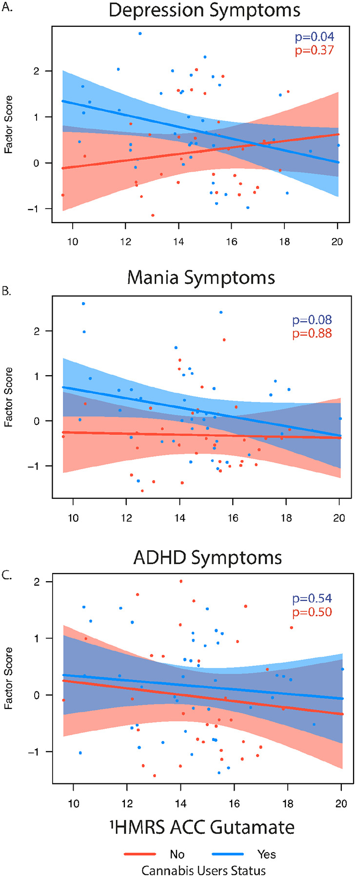
In the exploratory analysis of other dimensional symptoms, cannabis use was associated with greater depressive symptoms severity (p = 0.011), and a trend-level interaction with glutamate (p = 0.05), which suggested a potential cannabis-dependent relationship between glutamate and depressive symptoms (Fig. 5A). There was no main effect of glutamate (p = 0.22). A similar pattern emerged for manic symptoms, where cannabis use again predicted greater symptom burden (p = 0.01), while glutamate showed a marginal association (p = 0.09), but with no significant interaction (Fig. 5B). In contrast, ADHD symptoms were not significantly associated with either glutamate or cannabis use (Fig. 5C). Higher glutamate levels were also associated with overall functioning as measured by the GAF (F(1,66) = 4.50. p = 0.03), with cannabis non-users showing trend-level (p = 0.06) higher GAF scores than cannabis users. Within the cannabis user group, there were no associations between glutamate level and age of onset, length of use, or paranoia scores on the CEQ. There was a trend level association for cannabis-associated euphoria, with lower levels of glutamate associated with higher reported CEQ euphoric scores (p = 0.07). Analyses stratified by clinical diagnosis showed similar trends (Supplemental Results).
Discussion
We combined high-resolution 7T 1HMRS with detailed cannabis use assessments to examine glutamatergic alterations in a transdiagnostic sample of patients with psychosis, clinical high risk for psychosis, and typically developing youth. Cannabis use was common overall but most prevalent among individuals with psychosis. Self-reported use showed strong concordance with urine toxicology results, especially among frequent users, supporting the validity of self-report in this cohort. As expected, psychosis participants exhibited elevated clinical symptomatology and lower global functioning compared to CHR and TD groups. Collectively, the results indicate that cannabis use is robustly associated with elevated symptoms across multiple clinical domains—particularly psychosis, but also depression and mania—and that glutamate levels contribute independently to symptom severity, with interaction effects most apparent for positive symptoms, and potentially depressive symptoms. These findings suggest that glutamatergic dysregulation may play a role in symptom expression among cannabis users, especially for the positive symptom dimension of psychosis.
A novel finding of the current study was that anterior cingulate glutamate levels were significantly associated with positive psychosis symptoms only among cannabis users, suggesting that cannabis use modulates the glutamate–psychosis relationship. Specifically, lower glutamate predicted greater positive symptom severity in cannabis users but not in non-users. This pattern is consistent with evidence that cannabis can disrupt glutamatergic signaling in CB1 receptor–rich cortical regions, potentially leading to maladaptive reductions in excitatory tone or altered glutamate homeostasis in circuits already vulnerable to dysregulation in psychosis85. Such cannabis-related alterations could diminish glutamatergic efficiency, thereby linking lower glutamate levels to greater positive symptom expression among users. These results raise the possibility that ACC glutamate could serve as a biomarker of vulnerability among cannabis-using individuals on the psychosis spectrum, which could inform risk stratification and targeted interventions.
Importantly, glutamate was also independently associated with negative symptoms and global functioning across groups, and cannabis use predicted worse outcomes on these measures, indicating broader relevance of glutamatergic dysfunction beyond cannabis-exposed individuals. Given prior reports of regionally specific glutamate reductions in early and chronic psychosis22, 23, 85, the additive and interactive effects of cannabis use observed here may reflect either direct neurochemical consequences of cannabis or shared neurodevelopmental risk factors, or a combination of both. These findings support a mechanistic framework in which cannabis use interacts with regional glutamatergic vulnerability to influence the severity and profile of psychopathology. Future longitudinal and ultra–high field imaging studies will be critical to further clarify causal pathways linking cannabis use, glutamate dysregulation, and clinical outcomes, and to determine whether glutamate-targeted pharmacological or behavioral interventions may be particularly beneficial for cannabis-using individuals with psychosis.
We also found that cannabis use was associated with greater severity of depression and mania symptoms across the whole sample, highlighting the transdiagnostic clinical relevance of cannabis use. This is consistent with 1) meta-analytic evidence that heavy cannabis use increases risk for depressive disorders86; and 2) epidemiological findings of high comorbidity between cannabis use and mood disorders87, 88. The present associations may reflect overlapping neurobiological vulnerabilities—such as altered serotonergic and endocannabinoid signaling—that predispose individuals to both cannabis use and mood dysregulation, or the downstream effects of cannabis-related perturbations in neurotransmitter systems on affective processing.
Self-reported cannabis use showed high concordance with urine toxicology results, supporting the validity of self-report in assessing cannabis exposure in psychiatric populations. Across the sample, 92% of individuals denying cannabis use tested negative on urine toxicology, and frequent users were far more likely to test positive than infrequent users (87% vs. 13%). This pattern replicates prior work showing that urine toxicology has high specificity and negative predictive value for cannabis82, 89 and that concordance is often strongest among individuals with frequent use90. Nonetheless, some discrepancies were observed, with two participants denying cannabis use testing positive on toxicology and overall low rates of positive screens among self-reported users (38%), underscoring important limitations of toxicology measures. Detection windows for urine THC metabolites are finite and may under-detect use in infrequent users91, while contextual factors, stigma, and recall error could bias self-reports92, 93. These findings highlight the utility of pairing structured self-report cannabis use measures with biological verification, particularly in research on high-risk groups such as individuals with psychosis, where cannabis use is prevalent and clinically relevant94. Combining approaches may increase confidence in exposure classification while also capturing use patterns and frequency not discernible from toxicology alone.
Notwithstanding the strengths of combining ultra–high field 7T 1HMRS, biological and self-report measures of cannabis use, and dimensional clinical assessment across a transdiagnostic sample, several limitations should be noted. First, the cross-sectional design limits causal inference about the relationship between glutamate levels, cannabis use, and psychopathology symptom expression. Longitudinal studies are needed to determine whether cannabis-related glutamatergic alterations precede or result from symptom exacerbation. Second, although self-reported cannabis use showed strong concordance with urine toxicology results, the binary nature and limited detection window of the urine screen may underestimate recent use, particularly in infrequent users. Third, although the inclusion of clinical high-risk, psychosis-spectrum, and typically developing individuals enhances generalizability, the modest sample size, particularly within diagnostic subgroups, may limit statistical power to detect more nuanced interaction effects or subgroup-specific associations. Fourth, while advanced tissue correction and spectral quality control were employed, 1HMRS glutamate measures remain unable to fully disentangle glutamate from glutamine, do not include information on inhibitory neurometabolites (e.g., GABA), and do not reflect synaptic neurotransmission directly. Lastly, potential confounding effects of other substances (e.g., alcohol, nicotine), antipsychotic medication48 or comorbid psychiatric conditions were not fully modeled, and future work should expand the analytic framework to include polysubstance exposure and broader transdiagnostic factors.
In conclusion, these findings suggest that cannabis use may interact with psychosis-related vulnerability to accentuate glutamatergic dysfunction, and that this neurochemical alteration may contribute to symptom expression, especially in the domain of positive symptoms. Understanding this relationship is critical to elucidate the neurobiological substrates underlying both acute and chronic cannabis effects on mental health.
References
- K Petrilli, S Ofori, L Hines, G Taylor, S Adams, TP Freeman. Association of cannabis potency with mental ill health and addiction: a systematic review.. The Lancet Psychiatry, 2022. [PubMed]
- DC D’Souza. Cannabis, cannabinoids and psychosis: a balanced view.. World psychiatry, 2023. [PubMed]
- M Sorkhou, EL Dent, TP George. Cannabis use and mood disorders: a systematic review.. Frontiers in public health, 2024. [PubMed]
- T Robinson, MU Ali, B Easterbrook, W Hall, D Jutras-Aswad, B Fischer. Risk-thresholds for the association between frequency of cannabis use and the development of psychosis: a systematic review and meta-analysis.. Psychol Med, 2023. [PubMed]
- SK Kiburi, K Molebatsi, V Ntlantsana, MT Lynskey. Cannabis use in adolescence and risk of psychosis: Are there factors that moderate this relationship? A systematic review and meta-analysis.. Subst Abus, 2021. [PubMed]
- RM Murray, H Quigley, D Quattrone, A Englund, M Di Forti. Traditional marijuana, high-potency cannabis and synthetic cannabinoids: increasing risk for psychosis.. World psychiatry: official journal of the World Psychiatric Association (WPA), 2016. [PubMed]
- M Di Forti, A Marconi, E Carra, S Fraietta, A Trotta, M Bonomo. Proportion of patients in south London with first-episode psychosis attributable to use of high potency cannabis: a case-control study.. The Lancet Psychiatry, 2015. [PubMed]
- A Marconi, M Di Forti, CM Lewis, RM Murray, E Vassos. Meta-analysis of the association between the level of cannabis use and risk of psychosis.. Schizophr Bull, 2016. [PubMed]
- M Di Forti, D Quattrone, TP Freeman, G Tripoli, C Gayer-Anderson, H Quigley. The contribution of cannabis use to variation in the incidence of psychotic disorder across Europe (EU-GEI): a multicentre case-control study.. The Lancet Psychiatry, 2019. [PubMed]
- JD Jones, ME Calkins, JC Scott, EC Bach, RE Gur. Cannabis use, polysubstance use, and psychosis spectrum symptoms in a community-based sample of US youth.. J Adolesc Health, 2017. [PubMed]
- NC Stefanis, P Delespaul, C Henquet, C Bakoula, C Stefanis, J Van Os. Early adolescent cannabis exposure and positive and negative dimensions of psychosis.. Addiction, 2004. [PubMed]
- R Carney, J Cotter, J Firth, T Bradshaw, A Yung. Cannabis use and symptom severity in individuals at ultra high risk for psychosis: a meta-analysis.. Acta Psychiatr Scand, 2017. [PubMed]
- O Santesteban-Echarri, L Liu, M Miller, CE Bearden, KS Cadenhead, TD Cannon. Cannabis use and attenuated positive and negative symptoms in youth at clinical high risk for psychosis.. Schizophr Res, 2022. [PubMed]
- MW de Medeiros, JC Andrade, NM Haddad, M Mendonça, LP de Jesus, F Fekih-Romdhane. Cannabis use influences disorganized symptoms severity but not transition in a cohort of non-help-seeking individuals at-risk for psychosis from São Paulo, Brazil.. Psychiatry Res, 2024. [PubMed]
- T Schoeler, N Petros, M Di Forti, E Klamerus, E Foglia, O Ajnakina. Effects of continuation, frequency, and type of cannabis use on relapse in the first 2 years after onset of psychosis: an observational study.. The Lancet Psychiatry, 2016. [PubMed]
- T Schoeler, N Petros, M Di Forti, J-B Pingault, E Klamerus, E Foglia. Association Between Continued Cannabis Use and Risk of Relapse in First-Episode Psychosis: A Quasi-Experimental Investigation Within an Observational Study.. JAMA psychiatry, 2016. [PubMed]
- L Levi, M Bar-Haim, I Winter-van Rossum, M Davidson, S Leucht, WW Fleischhacker. Cannabis use and symptomatic relapse in first episode schizophrenia: trigger or consequence? Data from the OPTIMISE Study.. Schizophr Bull, 2023. [PubMed]
- M Colizzi, P McGuire, RG Pertwee, S Bhattacharyya. Effect of cannabis on glutamate signalling in the brain: A systematic review of human and animal evidence.. Neurosci Biobehav Rev, 2016. [PubMed]
- KU Chowdhury, ME Holden, MT Wiley, V Suppiramaniam, MN Reed. Effects of Cannabis on Glutamatergic Neurotransmission: The Interplay between Cannabinoids and Glutamate.. Cells, 2024. [PubMed]
- K Merritt, RA McCutcheon, A Aleman, S Ashley, K Beck, W Block. Variability and magnitude of brain glutamate levels in schizophrenia: a meta and mega-analysis.. Mol Psychiatry, 2023. [PubMed]
- K Merritt, PK McGuire, A Egerton, A Aleman, W Block, OJ Bloemen. Association of age, antipsychotic medication, and symptom severity in schizophrenia with proton magnetic resonance spectroscopy brain glutamate level: a mega-analysis of individual participant-level data.. JAMA psychiatry, 2021. [PubMed]
- D Roalf, R Nanga, P Rupert, H Hariharan, M Quarmley, M Calkins. Glutamate imaging (GluCEST) reveals lower brain GluCEST contrast in patients on the psychosis spectrum.. Mol Psychiatry, 2017
- VJ Sydnor, DR Roalf. A meta-analysis of ultra-high field glutamate, glutamine, GABA and glutathione 1HMRS in psychosis: implications for studies of psychosis risk.. Schizophr Res, 2020. [PubMed]
- VJ Sydnor, B Larsen, C Kohler, AJ Crow, SL Rush, ME Calkins. Diminished reward responsiveness is associated with lower reward network GluCEST: an ultra-high field glutamate imaging study.. Mol Psychiatry, 2021. [PubMed]
- EB Oleson, LR Hamilton, DM Gomez. Cannabinoid modulation of dopamine release during motivation, periodic reinforcement, exploratory behavior, habit formation, and attention.. Front Synaptic Neurosci, 2021. [PubMed]
- Z Li, D Mukherjee, B Duric, I Austin-Zimmerman, G Trotta, E Spinazzola. Systematic review and meta-analysis on the effects of chronic peri-adolescent cannabinoid exposure on schizophrenia-like behaviour in rodents.. Mol Psychiatry, 2024
- S Moriguchi, A Takamiya, Y Noda, N Horita, M Wada, S Tsugawa. Glutamatergic neurometabolite levels in major depressive disorder: a systematic review and meta-analysis of proton magnetic resonance spectroscopy studies.. Mol Psychiatry, 2019. [PubMed]
- J Lieberman, J Kane, J Alvir. Provocative tests with psychostimulant drugs in schizophrenia.. Psychopharmacology (Berl), 1987. [PubMed]
- JT Coyle. Glutamate and schizophrenia: beyond the dopamine hypothesis.. Cell Mol Neurobiol, 2006
- P Allen, CA Chaddock, A Egerton, OD Howes, G Barker, I Bonoldi. Functional outcome in people at high risk for psychosis predicted by thalamic glutamate levels and prefronto-striatal activation.. Schizophr Bull, 2015. [PubMed]
- M Baiano, A David, A Versace, R Churchill, M Balestrieri, P Brambilla. Anterior cingulate volumes in schizophrenia: a systematic review and a meta-analysis of MRI studies.. Schizophr Res, 2007. [PubMed]
- AR Mohn, RR Gainetdinov, MG Caron, BH Koller. Mice with reduced NMDA receptor expression display behaviors related to schizophrenia.. Cell, 1999. [PubMed]
- GE Duncan, SS Moy, A Perez, DM Eddy, WM Zinzow, JA Lieberman. Deficits in sensorimotor gating and tests of social behavior in a genetic model of reduced NMDA receptor function.. Behav Brain Res, 2004. [PubMed]
- JD Jentsch, DE Redmond, JD Elsworth, JR Taylor, KD Youngren, RH Roth. Enduring cognitive deficits and cortical dopamine dysfunction in monkeys after long-term administration of phencyclidine.. Science, 1997. [PubMed]
- A Balla, R Koneru, J Smiley, H Sershen, DC Javitt. Continuous phencyclidine treatment induces schizophrenia-like hyperreactivity of striatal dopamine release.. Neuropsychopharmacology, 2001. [PubMed]
- D. Javitt. Glutamate as a therapeutic target in psychiatric disorders., 2004
- W Hu, ML MacDonald, DE Elswick, RA Sweet. The glutamate hypothesis of schizophrenia: evidence from human brain tissue studies.. Ann N Y Acad Sci, 2015. [PubMed]
- M Beneyto, LV Kristiansen, A Oni-Orisan, RE McCullumsmith, JH Meador-Woodruff. Abnormal glutamate receptor expression in the medial temporal lobe in schizophrenia and mood disorders.. Neuropsychopharmacology, 2007. [PubMed]
- DC Goff, JT Coyle. The emerging role of glutamate in the pathophysiology and treatment of schizophrenia.. Am J Psychiatry, 2001
- G Tsai, JT Coyle. Glutamatergic mechanisms in schizophrenia.. Annu Rev Pharmacol Toxicol, 2002. [PubMed]
- MD Rubio, K Wood, V Haroutunian, JH Meador-Woodruff. Dysfunction of the ubiquitin proteasome and ubiquitin-like systems in schizophrenia.. Neuropsychopharmacology, 2013. [PubMed]
- PJ Harrison, DR Weinberger. Schizophrenia genes, gene expression, and neuropathology: on the matter of their convergence., 2005
- H Stefansson, H Petursson, E Sigurdsson, V Steinthorsdottir, S Bjornsdottir, T Sigmundsson. Neuregulin 1 and susceptibility to schizophrenia.. The American Journal of Human Genetics, 2002. [PubMed]
- B. Moghaddam. Bringing order to the glutamate chaos in schizophrenia.. Neuron, 2003. [PubMed]
- Y-N Wang, D Figueiredo, X-D Sun, Z-Q Dong, W-B Chen, W-P Cui. Controlling of glutamate release by neuregulin3 via inhibiting the assembly of the SNARE complex.. Proceedings of the National Academy of Sciences, 2018
- A Palomino, A González-Pinto, A Aldama, C González-Gómez, F Mosquera, G González-García. Decreased levels of plasma glutamate in patients with first-episode schizophrenia and bipolar disorder.. Schizophr Res, 2007. [PubMed]
- K Merritt, A Egerton, MJ Kempton, MJ Taylor, PK McGuire. Nature of Glutamate Alterations in Schizophrenia: A Meta-analysis of Proton Magnetic Resonance Spectroscopy Studies.. JAMA psychiatry, 2016. [PubMed]
- RR Girgis, C de la Fuente-Sandoval, R Lewis-Fernández, F Reyes-Madrigal, MM Wall, J Hua. Concerted elevations of cortical and striatal glutamate and GABA in antipsychotic-free individuals at clinical high-risk for psychosis.. Neuropsychopharmacology, 2025. [PubMed]
- C de la Fuente-Sandoval, P Leon-Ortiz, R Favila, S Stephano, D Mamo, J Ramirez-Bermudez. Higher levels of glutamate in the associative-striatum of subjects with prodromal symptoms of schizophrenia and patients with first-episode psychosis.. Neuropsychopharmacology, 2011. [PubMed]
- A Egerton, JM Stone, CA Chaddock, GJ Barker, I Bonoldi, RM Howard. Relationship between brain glutamate levels and clinical outcome in individuals at ultra high risk of psychosis.. Neuropsychopharmacology, 2014
- I Uhl, P Mavrogiorgou, C Norra, F Forstreuter, M Scheel, H Witthaus. 1H-MR spectroscopy in ultra-high risk and first episode stages of schizophrenia.. J Psychiatr Res, 2011. [PubMed]
- JM Stone, F Day, H Tsagaraki, I Valli, MA McLean, DJ Lythgoe. Glutamate dysfunction in people with prodromal symptoms of psychosis: relationship to gray matter volume.. Biol Psychiatry, 2009. [PubMed]
- P Fusar-Poli, JM Stone, MR Broome, I Valli, A Mechelli, MA McLean. Thalamic glutamate levels as a predictor of cortical response during executive functioning in subjects at high risk for psychosis.. Arch Gen Psychiatry, 2011. [PubMed]
- N Tandon, NR Bolo, K Sanghavi, IT Mathew, AN Francis, JA Stanley. Brain metabolite alterations in young adults at familial high risk for schizophrenia using proton magnetic resonance spectroscopy.. Schizophr Res, 2013
- S Rigucci, L Xin, P Klauser, PS Baumann, L Alameda, M Cleusix. Cannabis use in early psychosis is associated with reduced glutamate levels in the prefrontal cortex.. Psychopharmacology (Berl), 2018. [PubMed]
- M Sami, A Worker, M Colizzi, L Annibale, D Das, M Kelbrick. Association of cannabis with glutamatergic levels in patients with early psychosis: Evidence for altered volume striatal glutamate relationships in patients with a history of cannabis use in early psychosis.. Translational psychiatry, 2020. [PubMed]
- M Colizzi, N Weltens, P McGuire, D Lythgoe, S Williams, L Van Oudenhove. Delta-9-tetrahydrocannabinol increases striatal glutamate levels in healthy individuals: implications for psychosis.. Mol Psychiatry, 2020. [PubMed]
- DM Barch, J Bustillo, W Gaebel, R Gur, S Heckers, D Malaspina. Logic and justification for dimensional assessment of symptoms and related clinical phenomena in psychosis: relevance to DSM-5.. Schizophr Res, 2013. [PubMed]
- P Phalen, Z Millman, PR Rouhakhtar, N Andorko, G Reeves, J Schiffman. Categorical versus dimensional models of early psychosis.. Early intervention in psychiatry, 2022. [PubMed]
- ZS Nasreddine, NA Phillips, V Bédirian, S Charbonneau, V Whitehead, I Collin. The Montreal Cognitive Assessment, MoCA: a brief screening tool for mild cognitive impairment.. J Am Geriatr Soc, 2005. [PubMed]
- J Endicott, RL Spitzer, JL Fleiss, J Cohen. The global assessment scale. A procedure for measuring overall severity of psychiatric disturbance.. Arch Gen Psychiatry, 1976. [PubMed]
- ME Calkins, TM Moore, TD Satterthwaite, DH Wolf, BI Turetsky, DR Roalf. Persistence of psychosis spectrum symptoms in the Philadelphia Neurodevelopmental Cohort: a prospective two-year follow-up.. World Psychiatry, 2017. [PubMed]
- MB First, RL Spitzer, M Gibbon, JBW Williams. Structured Clinical Interview for DSM-IV-TR Axis I Disorders, Research Version, Patient Edition (SCID-I/P)., 2002
- TH McGlashan, TJ Miller, SW Woods. Structured Interview for Prodromal Syndromes, Version 4.0., 2003
- T McGlashan, B Walsh, S Woods. The psychosis-risk syndrome: handbook for diagnosis and follow-up., 2010
- ME Calkins, KR Merikangas, TM Moore, M Burstein, MA Behr, TD Satterthwaite. The Philadelphia Neurodevelopmental Cohort: constructing a deep phenotyping collaborative., 2015
- TM Moore, ME Calkins, DH Wolf, TD Satterthwaite, R Barzilay, JC Scott. Estimation and Validation of the “c” Factor for Overall Cerebral Functioning in the Philadelphia Neurodevelopmental Cohort.. Applied Sciences, 2025
- S Tang, J Yi, M Calkins, D Whinna, C Kohler, M Souders. Psychiatric disorders in 22q11. 2 deletion syndrome are prevalent but undertreated.. Psychol Med, 2014. [PubMed]
- TD Satterthwaite, DH Wolf, J Loughead, K Ruparel, JN Valdez, SJ Siegel. Association of enhanced limbic response to threat with decreased cortical facial recognition memory response in schizophrenia.. Am J Psychiatry, 2010. [PubMed]
- TD Satterthwaite, MA Elliott, K Ruparel, J Loughead, K Prabhakaran, ME Calkins. Neuroimaging of the Philadelphia neurodevelopmental cohort.. Neuroimage, 2014. [PubMed]
- ME Calkins, TM Moore, KR Merikangas, M Burstein, TD Satterthwaite, WB Bilker. The psychosis spectrum in a young US community sample: findings from the Philadelphia Neurodevelopmental Cohort.. World Psychiatry, 2014. [PubMed]
- H Kobayashi, T Nemoto, H Koshikawa, Y Osono, R Yamazawa, M Murakami. A self-reported instrument for prodromal symptoms of psychosis: testing the clinical validity of the PRIME Screen-Revised (PS-R) in a Japanese population.. Schizophr Res, 2008. [PubMed]
- T. Miller. The SIPS-Screen: a brief self-report screen to detect the schizophrenia prodrome.. Schizophr Res, 2004
- C Han, MK McGue, WG Iacono. Lifetime tobacco, alcohol and other substance use in adolescent Minnesota twins: univariate and multivariate behavioral genetic analyses.. Addiction, 1999. [PubMed]
- C Cuttler, A Spradlin. Measuring cannabis consumption: Psychometric properties of the Daily Sessions, Frequency, Age of Onset, and Quantity of Cannabis Use Inventory (DFAQ-CU).. PLoS One, 2017. [PubMed]
- JA Gette, AK Littlefield, SE Victor, AT Schmidt, S Garos. Evaluation of the Daily Sessions, Frequency, Age of Onset, and Quantity of Cannabis Use Questionnaire and its Relations to Cannabis-Related Problems.. Cannabis (Albuquerque, NM), 2023
- MH Meier, A Caspi, A Ambler, H Harrington, R Houts, RSE Keefe. Persistent cannabis users show neuropsychological decline from childhood to midlife.. Proc Natl Acad Sci U S A, 2012. [PubMed]
- JC Scott, DH Wolf, ME Calkins, EC Bach, J Weidner, K Ruparel. Cognitive functioning of adolescent and young adult cannabis users in the Philadelphia Neurodevelopmental Cohort.. Psychol Addict Behav, 2017. [PubMed]
- F Bianconi, M Bonomo, A Marconi, A Kolliakou, SA Stilo, C Iyegbe. Differences in cannabis-related experiences between patients with a first episode of psychosis and controls.. Psychol Med, 2016. [PubMed]
- V Lorenzetti, C Hindocha, K Petrilli, P Griffiths, J Brown, Á Castillo-Carniglia. The iCannToolkit: a tool to embrace measurement of medicinal and non-medicinal cannabis use across licit, illicit and cross-cultural settings.. Addiction (Abingdon, England), 2022. [PubMed]
- G Oeltzschner, HJ Zöllner, SC Hui, M Mikkelsen, MG Saleh, S Tapper. Osprey: Open-source processing, reconstruction & estimation of magnetic resonance spectroscopy data.. J Neurosci Methods, 2020. [PubMed]
- C Gasparovic, T Song, D Devier, HJ Bockholt, A Caprihan, PG Mullins. Use of tissue water as a concentration reference for proton spectroscopic imaging.. Magnetic Resonance in Medicine: An Official Journal of the International Society for Magnetic Resonance in Medicine, 2006
- JA Long. interactions: Comprehensive, User-Friendly Toolkit for Probing Interactions., 2024
- RV Lenth. Least-squares means: the R package lsmeans.. Journal of statistical software, 2016
- M Niznikiewicz, A Lin, LE DeLisi. The Relationship of glutamate signaling to cannabis use and schizophrenia.. Current Opinion in Psychiatry, 2025. [PubMed]
- S Lev-Ran, M Roerecke, B Le Foll, T George, K McKenzie, J Rehm. The association between cannabis use and depression: a systematic review and meta-analysis of longitudinal studies.. Psychol Med, 2014. [PubMed]
- D Feingold, A Weinstein. Cannabis and depression.. Cannabinoids and Neuropsychiatric Disorders, 2020
- T Schoeler, J Ferris, AR Winstock. Rates and correlates of cannabis-associated psychotic symptoms in over 230,000 people who use cannabis.. Translational psychiatry, 2022. [PubMed]
- K Salottolo, E McGuire, R Madayag, AH Tanner, MM Carrick, D Bar-Or. Validity between self-report and biochemical testing of cannabis and drugs among patients with traumatic injury: brief report.. Journal of Cannabis Research, 2022. [PubMed]
- KR Skelton, E Donahue, SE Benjamin-Neelon. Validity of self-report measures of cannabis use compared to biological samples among women of reproductive age: a scoping review.. BMC Pregnancy Childbirth, 2022. [PubMed]
- DS Fink, H Samples, CA Malte, M Olfson, MM Wall, DM Alschuler. Cannabis legalization and increasing cannabis use in the United States: Data from urine toxicology testing in emergency room patients.. International Journal of Drug Policy, 2025. [PubMed]
- C Bharat, P Webb, Z Wilkinson, R McKetin, J Grebely, M Farrell. Agreement between self-reported illicit drug use and biological samples: a systematic review and meta-analysis.. Addiction, 2023. [PubMed]
- JJ Palamar, A Le, H Guarino, P Mateu-Gelabert. A comparison of the utility of urine-and hair testing in detecting self-reported drug use among young adult opioid users.. Drug Alcohol Depend, 2019. [PubMed]
- D Oliver, E Chesney, AE Cullen, C Davies, A Englund, G Gifford. Exploring causal mechanisms of psychosis risk.. Neurosci Biobehav Rev, 2024. [PubMed]