
Canine Idiopathic Epilepsy as a Natural Animal Model for Human Epilepsy: A Scoping Review Highlighting Metabolic Perspectives Beyond the Brain
Abstract
Background: Emerging evidence indicates that epilepsy extends beyond the brain, involving systemic metabolic, immune, and microbiome perturbations that shape neuronal excitability and treatment response. Canine idiopathic epilepsy (CE) offers a naturally occurring model with strong electrophysiological, pharmacological, and clinical homology to human epilepsies. Methods: This scoping review was conducted according to the PRISMA-ScR guidelines. A systematic literature search was performed in Web of Science and MEDLINE (PubMed) to identify original studies reporting metabolic, immunometabolic, or neurochemical alterations in CE compared with healthy controls. Eligible studies included peer-reviewed original research involving client-owned dogs diagnosed with CE according to international consensus criteria (IVETF guidelines). Studies focusing exclusively on genetics or neuroimaging without metabolic outcomes were excluded. Titles, abstracts, and full texts were screened for eligibility, and data were extracted from included studies using a standardized approach. Identified metabolic domains were synthesized narratively and grouped into functional systems, including amino acid and lipid metabolism, micronutrients, neurotransmission, oxidative stress, inflammation and immunology, endocannabinoid signalling, microRNAs, and gut–brain axis-related pathways. In a second step, the identified metabolic domains were evaluated for translational relevance through a targeted, non-systematic narrative synthesis of the human epilepsy literature. This approach aimed to assess cross-species parallels and to provide a conceptual framework to guide future research, rather than to perform a comprehensive systematic review of metabolic alterations in human epilepsy. Results: Across CE studies, consistent alterations were observed in multiple interconnected functional systems, including metabolic, immune, and gut–brain axis pathways, in agreement with findings reported for human epilepsy. These data support a model of epileptogenesis involving systemic dysfunction beyond the central nervous system. Translationally, these findings suggest opportunities for biomarker development, patient stratification, and mechanism-based interventions, including dietary and metabolic approaches (e.g., medium-chain triglyceride supplementation), microbiome modulation, and immunometabolic targeting. The current evidence is limited by small and heterogeneous cohorts, potential confounding effects of antiseizure medications, variability in dietary and fasting conditions, breed-related effects, and a predominance of associative over causal relationships. Conclusions: This review positions CE as a reference framework for future research into epilepsy metabolism, integrating current evidence and its translational relevance to human disease. The findings support a shift toward a systems-level view of epileptogenesis, involving interconnected metabolic, immune, and gut–brain axis pathways beyond the brain. CE represents a valuable translational model to identify shared mechanisms, inform biomarker discovery, and guide the development of mechanism-based therapeutic strategies across veterinary and human epilepsy.
Article type: Review Article
Keywords: epilepsy, dog, human, preclinical, metabolome, animal model
Affiliations: Equine and Companion Animal Nutrition, Department of Morphology, Imaging, Orthopedics, Rehabilitation and Nutrition, Faculty of Veterinary Medicine, Ghent University, Salisburylaan 133, 9820 Merelbeke-Melle, Belgium; giulia.cabri@outlook.com (G.C.); fien.verdoodt@ugent.be (F.V.); Small Animal Department, Faculty of Veterinary Medicine, Ghent University, Salisburylaan 133, 9820 Merelbeke-Melle, Belgium; sofie.bhatti@ugent.be; Laboratory of Integrative Metabolomics (LIMET), Department of Translational Physiology, Infectiology and Public Health, Faculty of Veterinary Medicine, Ghent University, Salisburylaan 133, 9820 Merelbeke-Melle, Belgium; lieseloty.hemeryck@ugent.be; Department of Neurology, Ghent University Hospital and 4Brain, Ghent University, C. Heymanslaan 10, 9000 Ghent, Belgium; paul.boon@uzgent.be; Department of Small Animal Medicine and Surgery, University of Veterinary Medicine Hannover, 30559 Hannover, Germany; holger.volk@tiho-hannover.de
License: © 2026 by the authors. CC BY 4.0 Licensee MDPI, Basel, Switzerland. This article is an open access article distributed under the terms and conditions of the Creative Commons Attribution (CC BY) license.
Article links: DOI: 10.3390/nu18111734 | PMC: PMC13258797
Relevance: Moderate: mentioned 3+ times in text
Full text: PDF (1.7 MB)
1. Introduction
Epilepsy is one of the most common chronic neurological disorders in humans, highly affecting the quality of life of patients and their families [ref. 1]. While epilepsy affects around 50 million people worldwide [ref. 2], the underlying etiology remains unclear in 32% of cases [ref. 3]. Additionally, the currently available antiseizure medications (ASMs) remain unable to fully control epileptic seizures in approximately one third of people with epilepsy [ref. 4]. These numbers stress the importance of further research to better characterize the pathophysiology of epilepsy and its management. Recent evidence supports a more integrative perspective on epilepsy [ref. 5,ref. 6,ref. 7,ref. 8], recognizing that its pathophysiological manifestations extend beyond the central nervous system (CNS) and are also reflected in systemic metabolic alterations [ref. 9]. Characterization of these metabolic pathways, systems, and states may help to further elucidate the underlying etiologies, inform the development of novel therapeutic strategies, including nutritional interventions, and ultimately improve the clinical management of epilepsy.
Human epilepsy encompasses a heterogeneous group of disorders characterized by diverse etiologies and complex pathophysiological mechanisms [ref. 10]. The variability in clinical presentation and underlying causes among individuals underscores the need for a tailored therapeutic approach considering the specific patient and disease characteristics, evolving towards personalized medicine [ref. 11]. Emerging technologies such as metabolomics, especially when integrated with established disease models, offer promising avenues to advance diagnostic and therapeutic options in epilepsy [ref. 9].
Canine idiopathic epilepsy (CE) is an umbrella-term encompassing genetic epilepsy, suspected genetic epilepsy, and epilepsy of unknown cause [ref. 12]. Its diagnosis is based on internationally recognized criteria to exclude structural or reactive causes resulting in epileptic seizures [ref. 13]. As such, CE represents a robust, naturally occurring model for human epilepsy types of genetic or unknown origin. Its translational value is supported by converging electrophysiological, pharmacological, and clinical evidence [ref. 14,ref. 15,ref. 16,ref. 17]. Notably, for example, dietary supplementation with medium-chain triglycerides has demonstrated efficacy across species, including rodents, dogs, and humans [ref. 18]. Simultaneously, the intestinal canine microbiome resembles the human microbiome better than that of rodents [ref. 19], reinforcing the relevance of the canine model to study potential pathways and systems of interest, like the gut–brain axis. However, direct cross-species comparisons of metabolic profiles remain limited, despite growing interest in molecular endotyping, i.e., the identification of distinct biological subtypes, to refine epilepsy classification and treatment strategies [ref. 20].
The dog offers several advantages as a translational model: (1) spontaneous seizure occurrence eliminates the need for artificial induction; (2) clinical manifestations closely resemble those observed in human epilepsy; (3) shared environmental exposures with humans enhance ecological validity; and (4) dietary intake can be standardized using fixed-formulation, nutritionally complete diets, an approach that is less feasible in human patients due to dietary heterogeneity and variable compliance. These latter factors (3 and 4) are particularly critical, as environmental and dietary variables are recognized as major confounders in human clinical research [ref. 21]. Importantly, the use of naturally affected dogs creates reciprocal benefits: insights translate to human epilepsy and simultaneously improve clinical outcomes in veterinary practice. This dual-impact, real-world model complements and extends traditional experimental approaches.
By delineating shared mechanisms and species-specific features, we aim to clarify the translational relevance of the canine model and provide a comprehensive overview of the existing knowledge in CE. The objective of this review was (1) to systematically identify the original studies in CE comparing metabolic pathways, systems, and states to a control group of healthy dogs; and (2) to explore the relevance of these identified pathways, systems, and states for human epilepsy. To reach this objective, a broad field of evidence must be mapped, and diverse study types will need to be integrated. Consequently, a scoping review provides the most suitable framework for this work [ref. 22]. This framework lays the groundwork for future research into metabolic perspectives beyond the brain, thereby ultimately facilitating potential advancements in diagnostic and therapeutic strategies for both veterinary and human patients.
2. Methods and Search Criteria
This study was conducted as a scoping review to systematically evaluate current evidence concerning the metabolic pathways, systems, and states implicated in CE, while a complementary narrative review underscores the convergence and divergence of these findings with the human literature. The methodology adheres to the PRISMA-ScR (Preferred Reporting Items for Systematic Reviews and Meta-Analyses extension for Scoping Reviews) reporting guidelines, emphasizing methodological rigour when identifying key concepts and research gaps in the field (Supplementary File S1).
In the first step, a literature review was conducted to identify studies investigating metabolic pathways in CE, following the PRISMA-ScR guidelines (Figure 1, Supplementary Files S1 and S2). The research question guiding this step was: “In dogs with CE (P), can we identify original studies comparing aspects of the metabolism (I) to a control group of healthy dogs (C), to detect alterations in specific metabolites or metabolic pathways (O)?”. The scoping search was conducted on 22 August 2025 using the Web of Science (WoS) and MEDLINE (via PubMed) databases. For WoS, the search string “(dog OR canine) AND (epileps*) (All Fields)”, followed by a filter to exclude “document types: review article”, was applied. In MEDLINE, the search string: “Dog OR canine,” AND “Epilep*,” was applied, with a publication type filter to include clinical trials, comparative studies, multicentre studies, evaluation studies and observational studies. After removing duplicates, the unique titles and abstracts were independently screened by the first and last author. Inclusion criteria were: (1) original research articles; (2) studies involving dogs diagnosed with idiopathic epilepsy in accordance with the international guidelines, i.e., CE [ref. 13]; and (3) studies including healthy dogs as a control group. Studies focusing solely on genotyping or medical imaging techniques were excluded. Both authors then evaluated the full text against the inclusion criteria, and studies were additionally excluded if one of the inclusion criteria was not met. The full list of screened records is available in Supplementary File S2. Finally, data were manually extracted by the first and last author from the result sections of the included articles. A critical appraisal of the individual CE studies was performed using the Levels of Evidence hierarchy (see: https://legacyfileshare.elsevier.com/promis_misc/YJPSU-Levels-of-Evidence.pdf (accessed 5 May 2026)), and the results are summarized in Table 1.
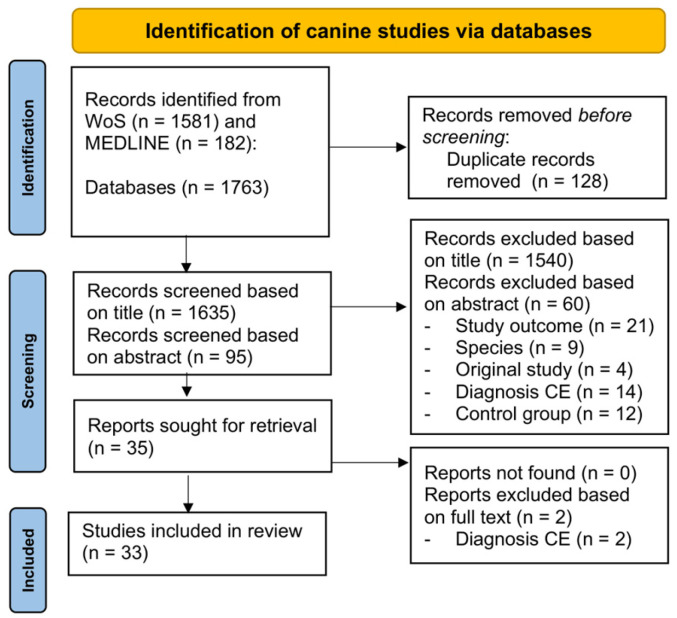
Table 1: Summary of canine studies identified through scoping literature search.
| Reference | Category | Population | Sample Type | Main Findings in CE Dogs | ASM Exposure | Epilepsy Subtypes/Diet | Level of Evidence |
|---|---|---|---|---|---|---|---|
| Kang et al. 2024 [ref. 24] | Immunology | HC = 29; IE = 49 | Serum | ↑ C3 and C4 | Treated: n = 19 untreated: n = 30 | IdiopathicTIER 2Diet not standardised | Prognosis Study: IIRetrospective Study |
| de la Fuente et al. 2012 [ref. 25] | Immunology | HC = 7; CE = 15; SID = 7; SRMA = 11; INF = 37; BT = 38; NON-INF = 40 | CSF; Blood | No pleocytosis or protein elevation; bloodand CSF CRP and D-dimers not significantly different | Unknown | IdiopathicTIER 2Diet not standardised | Prognosis Study: IIICase-control study |
| Kostic et al. 2019[ref. 26] | Inflammation | HC = 6; CE = 30; SE = 21 | CSF; Serum | ↑ serum IL-1β | Unknown | IdiopathicTIER 2Diet not standardised | Prognosis Study: IIICase-control study |
| Despa et al. 2024[ref. 27] | Inflammation | HC = 8; CE = 29; SE = 10; RE = 10 | Blood | ↑ NLR | Variable | IdiopathicTIER > 1Diet not standardized | Prognosis Study: IIRetrospective Study |
| Koo et al. 2020[ref. 28] | Inflammation | HC = 26; CE = 14; epileptic SE (MUO, BT) = 14; non epileptic SE (MUO, BT) = 12 | Serum | ↑ HMGB1; higher in >3 vs. ≤3 months | Treated with only ASM (phenobarbital, potassium bromide, or zonisamide) | IdiopathicTIER > 1Diet not standardized | Prognosis Study: IIICase-control study |
| von Rüden et al. 2020[ref. 29] | Inflammation | HC = 28; CE = 9; SE = 12 | Brain tissue | ↑ TLR4 expression; ↑ HMGB1; ↑ HSP70 (piriform lobe) | Unknown | Convulsive seizures (focal and generalized)Diet not standardized | Prognosis Study: IIICase-control study |
| Segers et al. 2017[ref. 30] | Inflammation | HC = 28; CE = 38 | Serum | No consistent ↑ CRP | Treated | IdiopathicTIER 2Diet not standardized | Prognosis Study: IIICase-control study |
| Paltrinieri et al. 2017[ref. 31] | Inflammation | HC = 10; CE = 14; dogs with other CNS disorders = 20 | Serum | ↑ total CK and CK-BB activity↓ Macro-CK2 activity | Unknown | Diet not standardised | Prognosis Study: IIRetrospective Study |
| Merbl et al. 2014[ref. 32] | Inflammation | HC = 12; CE = 7, SE (MUO, BT) = 10 | CSF; Serum | ↑ TNF-α and ↑ IL-6 | Unknown | IdiopathicTIER 2Diet not standardized | Prognosis Study: IIICase-control study |
| Hemmeter et al. 2023[ref. 33] | Immunology | HC = 57; IE/dyskinesia = 58 | Serum, CSF | No specific human or murine neural autoantibodies detected | Unknown | Idiopathic subtype: unknown cause.Diet not standardised | Prognosis Study: IIICase-control study |
| Knebel et al. 2022[ref. 34] | Immunology | HC = 10; IE = 57 | CSF; Serum (IL-17) and whole blood (Th17) | ↑ Th17 cells and ↑ IL-17 | Variable | IdiopathicDiet not standardised | Prognosis Study: IIICase-control study |
| Baka et al. 2021[ref. 35] | Proteome | HC = 9; CE = 17; SE = 7 | CSF | Immune/ECM pathway alterations Differential expression ofprotein markers | 9 treated 8 untreated | IdiopathicTIER > 1Diet not standardized | Prognosis Study: IIICase-control study |
| Phochantachinda et al. 2023 [ref. 36] | Proteome | HC = 4; drug resistance CE = 4 | Plasma | Altered amyloid/inflammatory proteins | Treated | TIER > 1Diet not standardized | Prognosis Study: IIICase-control study |
| Baka et al. 2024[ref. 37] | Proteome | HC = 9; CE untreated = 8 CE treated= 9 SE = 8 | Serum | Differential expression ofprotein markers | Variable | IdiopathicTIER > 1Diet not standardized | Prognosis Study: IIICase-control study |
| Verdoodt et al. 2025[ref. 38] | AA, inflam, OS, vit | HC = 39; CE = 49 | Plasma | Oxidative stress signature | Treated with ASM | IdiopathicTIER 2Diet standardized | Prognosis Study: IIICase-control study |
| Hasegawa et al. 2014 [ref. 39] | AA | HC = 18; CE = 16; SE = 19 | CSF | Altered amino-acid profile (e.g., ↑ glutamic acid) | Unknown | IdiopathicTIER 2Diet not standardized | Prognosis Study: IIRetrospective Study |
| Weber et al. 2012 [ref. 40] | Energy | HC = 7, CE = 101, SRMA = 95, CNS neoplasia = 39, IVDD = 61, MUO = 19, BM = 6 | CSF, blood | ↑ CSF:serum glucose ratio & ↓ CSF protein, cell count vs. SRMA | Unknown | Diet not standardised | Prognosis Study: IIICase-control study |
| Verdoodt et al. 2025 [ref. 41] | MGBA | HC = 39; CE = 49 | Feces | Dysbiosis; altered SCFA pathways | Treated with ASM | IdiopathicTIER 2Diet standardized | Prognosis Study: IIICase-control study |
| García-Belenguer et al. 2021 [ref. 42] | MGBA | HC = 12; CE before treatment = 10; CE aftter treatment = 9 | Feces | ↓ GABA- and SCFA-producing bacteriaASM no impact | Single treatment with phenobarbital or imepitoin >30 days | IdiopathicTIER = 1Diet standardized | Prognosis Study: IIICase-control study |
| Muñana et al. 2020 [ref. 43] | MGBA | HC = 13; CE = 13 | Feces | No major Lactobacillus differences | Not treated | IdiopathicTIER > 1Diet standardized | Prognosis Study: IIICase-control study |
| Silvestrino et al. 2025[ref. 44] | MGBA | HC = 17; CE = 19 | Feces | ↓ Microbial diversity; ↑ pro-inflammatory taxa | Unknown | IdiopathicTIER > 1Diet not standardized but monitored | Prognosis Study: IIICase-control study |
| Yonezawa et al. 2024[ref. 45] | Lipid & OS | HC = 9; CE = 11; MUO = 12 | CSF; Plasma | Dysregulated lipid metabolites | 7 CE treated with ASM 1 CE under prednisolone | IdiopathicTIER 2Diet not standardized | Prognosis Study: IIRetrospective Study |
| Kluger et al. 2008[ref. 46] | Lipid & OS | HC = 57; CE = 57 | Serum | ↑ Triglycerides (Tx CE) | 28 CE under phenobarbital 29 CE phenobarbital and bromide | IdiopathicTIER 2Diet not standardizedbut monitored | Prognosis Study: IIICase-control study |
| Radaković et al. 2023[ref. 47] | Lipid & OS | HC = 15; CE = 15 | Blood | Systemic oxidative stress | Untreated | TIER > 1Diet not standardized | Prognosis Study: IIICase-control study |
| Rosendahl et al. 2023[ref. 48] | Minerals & vit | HC = 19; CE = 19 | Blood | ↑ Cu, Se, Cr | 18 treated | IdiopathicTIER > 1Diet not standardized but monitored | Prognosis Study: IIICase-control study |
| Rosendahl et al. 2023[ref. 49] | Minerals & vit | HC = 42; CE = 63 | Hair | Mineral imbalances | treated (n = 53); untreated (n = 10) | IdiopathicTIER > 1Diet not standardizedbut monitor | Prognosis Study: IIICase-control study |
| Vitale et al. 2019[ref. 50] | Minerals & vit | HC = 50; CE = 92 | Serum | Altered Se, Cu | Controlled CE (12), uncontrolled CE (42), and untreated CE (13) | IdiopathicTIER > 1Diet not standardizedbut monitor | Prognosis Study: IIICase-control study |
| Schmidt et al. 2022[ref. 51] | Neurotransmitters | HC = 127; CE = 63 | Urine | Distinct neurotransmitter profile (e.g., glycine, serotonin, norepinephrine/epinephrine ratio) | Treated with ASM (e.g.,phenobarbital, potassium bromide) and other variable therapy | Idiopathic TIER I (n = 15)TIER II (n = 48)Diet not standardized | Prognosis Study: IIICase-control study |
| Ellenberger et al. 2004[ref. 52] | Neurotransmitters | HC = 20; CE = 94; GE = 35 | CSF | ↓ GABA and ↓ Aspartate | Variable42 CE treated with phenobarbital | IdiopathicTIER > 1Diet not standardized | Prognosis Study: IIRetrospective Study |
| Morita et al. 2005[ref. 53] | Neurotransmitters | HC = 3; IE = 4 + 4 | Blood | ↑ extracellular Glu and Asp levels (during epileptiform activity) ↓ GLT-1 expression (cortex, thalamus)-perineuronal Glu accumulation (cortex) | Unknown | Idiopathic, subtype: genetic causeDiet not standardised | Prognosis Study: IIICase-control study |
| Gesell et al. 2013[ref. 54] | Endocannabinoid | HC = 16; CE = 40 | CSF | ↑ AEA, 2-AG | Unknown | IdiopathicDiet not standardized | Prognosis Study: IIRetrospective Study |
| Kostic et al. 2023[ref. 55] | Endocannabinoid | HC = 7; CE = 5; SE = 7 | Brain tissue | ↓ CB1R in CE dogs hippocampus vs. HC and SE | Unknown | Idiopathic, subtype: unknown cause. With cluster before euthanasia.Diet not standardised | Prognosis Study: IIRetrospective Study |
| García-Gracia et al. 2024[ref. 56] | miRNA | HC = 8; CE = 15 (drug-sensitive = 9; drug-resisance = 6) | Plasma | Distinct miRNA profile | Treated | TIER > 1Diet not standardized | Prognosis Study: IIICase-control study |
Summary of the 33 original research studies identified through the PRISMA-based literature search, each comparing metabolic features of dogs with idiopathic epilepsy to healthy controls across cerebrospinal fluid, blood, brain tissue, or fecal samples. Abbreviations: 2-AG = 2-arachidonoylglycerol; AA = amino acids; AEA = anandamide; BM = bacterial meningoencephalomyelitis; BT= brain tumor; C3/4 = complement factor 3/4; CB1R = cannabinoid 1 receptor; CK (-BB) = creatine kinase–(brain isoform); CNS = central nervous system; CRP = C-reactive protein; CSF = cerebrospinal fluid; Ctl = controlled; ECM = extracellular matrix; HC = healthy controls; HMGB1 = high mobility group box 1; CE = canine idiopathic epilepsy; GE= genetic idiopathic epilepsy; IVDD = intervertebral disc disease; MGBA = microbiota–gut–brain axis; miRNA = micro-RNA; MUO = meningoencephalomyelitis of unknown origin; NLR = neutrophil-to-lymphocyte ratio; OS = oxidative stress; SE = structural epilepsy; SCFA = short chain fatty acids; SRMA = steroid-responsive meningitis-arteritis; TLR4 = Toll-like receptor 4; Tx = treated; vit = vitamins.
In the second step, metabolic pathways, systems and/or states of interest identified in the canine literature were further examined for their relevance to human epilepsy. For each of these, a narrative synthesis of the human literature complemented the CE data to determine whether similar alterations have been reported in human epilepsy studies. The identified metabolic pathways, systems and/or states will be discussed in this manuscript, organized according to the affected functional aspects.
It is important to note that no systematic search strategy was applied specifically to the human literature. Consequently, a selection bias toward the metabolic pathways previously identified in CE is inherent in this work. However, the primary objective of this study was to serve as a reference for future researchers by summarizing the current knowledge in CE and providing insights into how these findings relate to human epilepsy. A comprehensive systematic description of metabolic alterations in human epilepsy was deemed beyond the scope of the present work.
3. Results
The literature retrieved on CE through our scoping search strategy yielded 33 publications (summarized in Table 1), encompassing analyses of cerebrospinal fluid (CSF), brain tissue, blood, and fecal samples. All studies were conducted on client-owned dogs affected by CE in comparison with a healthy control group. Some studies additionally included extra control groups, like dogs with structural epilepsy or meningoencephalitis of unknown origin. Several studies further stratified the CE population into subcategories, such as ‘controlled’ or ‘mild phenotype’ versus ‘uncontrolled’ or ‘drug-resistant,’ and ‘treated’ versus ‘untreated.’ The number of dogs with CE enrolled in individual studies ranged from 4 to 92, while healthy control groups included 4 to 127 dogs. Across these investigations, specific metabolic and physiological mechanisms were identified, many of which appear interrelated and intricately involved in the pathogenesis of CE, with potential relevance to human epilepsy.
The data and results retrieved in this review are presented as different sections based on the functional categories of metabolic alterations described in CE: inflammatory and immune pathways, microbiota–gut–brain axis, oxidative stress, lipid metabolism, amino acid and protein metabolism, minerals, trace elements and vitamins, neurotransmission, endocannabinoid system, and microRNA. In each section, we first summarize the existing research on CE and subsequently compare these findings with evidence from studies on people with epilepsy (PWE), with the aim of delineating cross-species similarities and divergences.
4. Inflammatory and Immune Pathways
4.1. Neuroinflammation
4.1.1. Canine Epilepsy
Although CE is traditionally considered a non-inflammatory epilepsy subtype [ref. 13], emerging evidence suggests a more nuanced pathophysiological profile, with each epileptic seizure inducing secondarily inflammatory processes [ref. 57]. Regarding epilepsy neuroinflammation, three canine studies were identified. One study revealed increased Heat Shock Protein 70 (HSP70) expression, a key damage-associated molecular pattern (DAMP), in the piriform lobe in CE [ref. 29]. Second, the CSF concentration of TNF-α, a pro-inflammatory cytokine, was increased in CE versus healthy dogs [ref. 32]. Third, serum creatine kinase (CK), a biomarker for tissue damage, was shown to be increased in CE [ref. 31]. Notably, this increase was primarily caused by an increase in the isoenzyme predominantly found in the brain, i.e., CK-BB, thus revealing neuron damage and leakage via the BBB. However, CE could not be differentiated from other CNS diseases, like inflammatory or degenerative disorders based on CK [ref. 31].
4.1.2. Human Epilepsy
Similarly to dogs, human research highlighted an important role for neuroinflammation within the pathophysiology of epilepsy [ref. 58,ref. 59], for which similarities and divergences described in the literature are depicted in Table 2. Similar to CE, increased HSP70 levels have been identified in the CNS of PWE. One study found overexpressed HSP70 in epileptic vs. non-epileptic brain tissue from PWE with a focal drug-resistant epilepsy type [ref. 60], while another study identified increased HSP70 immunoreactivity in the surgically removed hippocampi of PWE with mesial temporal lobe epilepsy [ref. 61]. Additionally, the CSF of PWE following status epilepticus showed significantly higher HSP70 levels compared to either controls or PWE experiencing epileptic seizures not including status epilepticus [ref. 62]. Moreover, increased CSF pro-inflammatory cytokines, including TNF-α, like in CE, have been detected in multiple types of PWE [ref. 58,ref. 63,ref. 64].
Table 2: Summary of metabolic system and pathway alterations in canine idiopathic epilepsy in comparison with human epilepsy.
| Metabolic System/Pathway | Similarities Canine—Human | Divergence Canine—Human | Research Gaps or Limitations |
|---|---|---|---|
| Acute phase response | ↑ CRP, neutrophil-to-lymphocyte ratio, CSF TNF-α, IL-1β; BBB dysfunction; ↑ xanthurenic acid | — | CRP variability across subtypes and age groups; specific plasma lipid alterations; ASM influence unclear |
| DAMPs and tissue damage markers | ↑ Serum HMGB1 and CNS HSP70 | ↑ CK-BB CE, but not PWE | CK could not differentiate CE from other CNS diseases |
| Innate immune system | Complement involvement in both species | Complement cascade: ↑ C3/C4 in CE vs. ↓ C3/C4 in PWE | Functional impact on epileptogenesis unclear |
| Adaptive immune system | Th17 cells involved and ↑ IL-17 | — | Role of coagulation markers (D-dimer); Importance of autoimmune encephalitis unclear; ASM influence unclear |
| MGBA | Tryptophan–kynurenine pathway alterations; ↓ fecal Phascolarctobacterium | Phylogeny of some microbial alterations differs | Peripheral histamine role in humans; alterations of Prevotella and Escherichia-Shigella in both directions for PWE; variability due to 16S rRNA sequencing |
| Oxidative stress | ↑ AOPP | Lipid peroxidation inconsistent; selenium status opposite | ASM influence on oxidative markers unclear; driver for epileptogenesis or consequence? |
| Lipid metabolism | Therapeutic use of MCT diets | Specific lipid metabolites (OEA, 11,12-DHET) detected in dogs, not humans | ASM influence on lipid profile unclear |
| Amino acid metabolism | ↑ Blood leucine; lysine acetylation parallels | Blood threonine: ↑ CE vs. ↓ PWE | Influence of timing post-seizure and ASM unclear |
| Protein metabolism | ↑ Amyloid-β | Proteomic patterns differ (e.g., ↓ MMP-2 in CE) | Functional role of haptoglobin unclear |
| Vitamins | ↓ Vitamin B6 | ASM impact on vitamin B6 more documented in PWE | Optimal dosing and safety margins; ASM influence unclear |
| Minerals | ↑ Cu/Zn ratio | Selenium: ↑ CE vs. ↓ PWE | Clinical relevance of manganese and chromium unclear |
| Neurotransmission | Serotonin involvement; GABA/glutamate ratio altered | Urinary NE/E ratio changes in CE; ↑ Plasma and CSF NE in PWE | Correlation with comorbidities unclear |
| Endocannabinoid system | Therapeutic use of CBD | CSF anandamide: ↑ CE vs. ↓ PWELocalisation CB1R in the brain | CB1 receptor modulation complexity |
| Post-transcriptional regulation | ↑ miR-134 and miR-129 in drug-resistant cases | Limited canine data, mainly pilot studies | Inconsistent alterations for miR-223; different subtypes tested in CE vs. PWE |
Abbreviations: 11,12-DHET = 11,12-dihydroxyeicosatrienoic acid; AOPP = advanced oxidation protein products; ASM = antiseizure medication; BBB = blood-brain barrier; C3/4 = complement factor 3/4; CB = cannabinoid; CBD = cannabidiol; CE = canine idiopathic epilepsy; CK-BB = creating kinase-brain isoform; CNS = central nervous system; CRP = C-reactive protein; CSF = cerebrospinal fluid; DAMPs = damage associated molecular patterns; E = epinephrine; HMGB1 = high mobility group box 1; HSP70 = heat shock protein 70; MCT = medium chain triglycerides; miR = microRNA; MMP-2 = matrix metallopeptidase 2; NE = norepinephrine; OEA = oleoylethanolamide; PWE = people with epilepsy; TNF = tumor necrosis factor.
4.1.3. General Remarks on Neuroinflammation
Neuroinflammation has indeed long been recognized as a contributing factor in epilepsy, first as a consequence of recurrent seizures, perpetuating a self-reinforcing inflammatory cycle [ref. 65]. Additionally, evidence from experimental animal models indicated that neuroinflammation also precedes seizure onset, implicating it as a potential initiator of epileptogenesis [ref. 66]. In the brain, microglia activation results in the release of DAMPs [ref. 67], cytokines [ref. 68] and effector pathways, like COX-2 [ref. 69]. Among the DAMPs implicated in epilepsy, High-Mobility Group Box 1 (HMGB1) and HSP70 have emerged as key mediators in experimental rodent models and PWE [ref. 70]. Moreover, the blood–brain barrier (BBB) function is hampered in PWE [ref. 71] and CE [ref. 72], causing peripheral immune cells and molecules to enter the brain. The literature on CE seems to support this theory of a self-perpetuating inflammatory cycle; however, no causation has been studied yet. The current studies only reveal correlations between inflammatory markers and CE, while ASM management was not described nor included as confounder, which is considered an important limitation.
4.2. Peripheral Inflammation
As highlighted by the primary aim of this review, disturbances in epilepsy extend beyond the CNS, and this is equally true for inflammatory processes [ref. 73]. Accordingly, peripheral inflammatory markers may offer complementary insights. Similar to neuroinflammatory processes, peripheral inflammation typically begins with the recruitment of inflammatory cells and the release of pro-inflammatory cytokines, which in turn drive the synthesis of acute-phase proteins (APP) such as C-reactive protein (CRP), haptoglobin, and ceruloplasmin [ref. 74]. To avoid redundancy, APP other than CRP will be addressed under ‘protein metabolism’. The release of APP is ultimately followed by metabolic alterations, which is hypothesized to further reinforce the self-perpetuating inflammatory cycle.
4.2.1. Canine Epilepsy
The inflammatory hematological cells in CE are characterized by a significant greater neutrophil to lymphocyte ratio [ref. 27], revealing primarily neutrophil-mediated inflammation. Additionally, increased DAMPs, i.e., serum HMGB1 [ref. 28], and cytokines, serum IL-1β [ref. 26], have been identified in CE. Regardless of epilepsy etiology, IL-1β was increased, showing no differences between idiopathic and structural epilepsy [ref. 26]. While serum CRP did not significantly increase in CE versus healthy dogs [ref. 30], elevated serum CRP levels were detected in dogs with structural epilepsy, suggesting seizure-induced inflammation may differ between epilepsy subtypes [ref. 27,ref. 75]. However, seizure severity in CE had an influence, with dogs experiencing cluster seizures showing higher serum CRP levels than dogs without clusters [ref. 27].
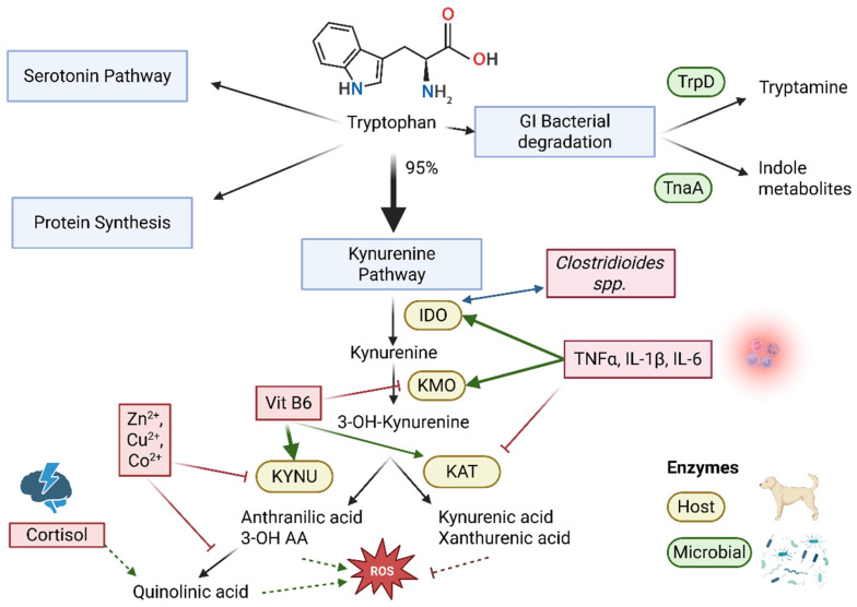
Metabolic studies further support an inflammatory component in CE by identifying elevated plasma lipids, i.e., oleoylethanolamide (OEA), a PPAR agonist with anti-inflammatory properties, and 11,12-DHET [ref. 45]. For the latter, bioactivity is not well documented, while 11,12 epoxyeicosatrienoic acid (11,12 EET), a substrate for 11,12-DHET, has been shown to suppress seizures in mouse hippocampus [ref. 76]. Furthermore, drug-resistant CE was characterized by elevated plasma xanthurenic acid and reduced vitamin B6 levels [ref. 38]. These suggest inflammatory activation of the kynurenine pathway, which is recognized as the major metabolic pathway for tryptophan [ref. 77,ref. 78,ref. 79], and is schematically displayed in Figure 2. Additionally, 4-guanidinobutanoic acid, a proinflammatory substrate for the blood–brain barrier creatine transporter [ref. 80,ref. 81], was increased in drug-resistant CE [ref. 38]. Fecal metabolomics of the same study population revealed increased histamine in drug-resistant CE, together with higher serotonin in mild CE [ref. 41], indicating altered tryptophan metabolism and intestinal inflammation potentially linked with the severity of disease [ref. 82,ref. 83].
4.2.2. Human Epilepsy
Starting with the inflammatory hematological cell types, similar to CE, higher neutrophil to lymphocyte ratios in PWE were indicated by recent meta-analyses [ref. 84,ref. 85]. Moreover, this ratio was significantly elevated in PWE with same-day seizure recurrence [ref. 86]. However, the relationship between epileptic seizures and neutrophil to lymphocyte ratio does not appear linear [ref. 87].
At the cytokine level, like in CE, increased IL-1β and TNFA-α were detected in the serum of PWE [ref. 88]. In addition, significantly higher blood CRP levels were found in adults with epilepsy, but not children [ref. 89]. In contrast, a study in 2022 did find significantly higher serum CRP levels in childhood epilepsy, and decreased CRP following treatment with levetiracetam [ref. 90].
The specific plasma lipids detected in CE, i.e., 11,12 DHET and OEA, have not yet been identified in PWE. Conversely, xanthurenic acid, a kynurenine pathway metabolite, was also increased in children with epileptic spasm syndrome [ref. 91], with a significant increase in children non-responsive to adrenocorticotropic hormone [ref. 92]. These findings suggest a link between epilepsy, the kynurenine and corticoid stress metabolism in both species.
4.2.3. General Remarks on Peripheral Inflammation
The CE literature, similar to the literature on PWE, indicates a correlation with inflammation. For CE, this is based on four studies with a level of evidence of III, and one level II study. However, no causal links have been examined, leaving open the question as to whether inflammation is a cause, consequence, or both. Additionally, details of ASM management were included in only two of the CE studies, while its influence remains inconclusive. The potential influence of ASM on inflammation in PWE is considered mixed and ASM-specific [ref. 93,ref. 94,ref. 95], warranting further investigation. Rodent models showed that the efficacy of ASM was hampered with induced inflammation in one study [ref. 38,ref. 96], while chronic levetiracetam administration stimulated xanthurenic acid production by brain cells in another study [ref. 97]. In the canine study detecting increased plasma xanthurenic acid, it was increased only in drug-resistant CE, of which only 7/27 received levetiracetam [ref. 38]. This suggests that levetiracetam administration is unlikely to account for the observed increase. Conversely, valproic acid has been shown to reduce inflammatory microglia overaction via histone deacetylase inhibition in vitro [ref. 98] and, together with carbamazepine, reduce the levels of proinflammatory cytokines produced by human peripheral immune cells in vitro [ref. 99]. To the best of our knowledge, these anti-inflammatory effects have not been corroborated in a clinical setting. The specific interaction between ASM and inflammations remains a significant research gap in both CE and PWE.
4.3. Innate Immune System
4.3.1. Canine Epilepsy
The complement cascade, a key interface between innate immunity and inflammation, revealed significantly increased serum C3 and C4 levels in CE, regardless of ASM status or seizure timing [ref. 24]. Notably, dogs with a mean seizure frequency ≥ 3/month exhibited higher C3 levels, implicating complement activation in disease severity [ref. 24].
4.3.2. Human Epilepsy
The blood complement cascade was shown to be involved in the discrimination of PWE vs. controls [ref. 100], as well as in children suffering from febrile seizures [ref. 101]. In adults with idiopathic generalized epilepsy specifically, decreased C3 and C4 serum levels were detected [ref. 102]. More recently, lower complement components were detected in patients with DR epilepsy, together with a sex-dependent effect [ref. 103]. These findings are contrasting the increased C3 levels detected in CE [ref. 24], potentially revealing a species-specific complement response.
4.4. Adaptive Immune System
4.4.1. Canine Epilepsy
In CE, one study reported significantly higher serum and CSF IL-17 concentrations, elevated stimulated Th17 cell counts, and a slight positive correlation between Th17 cell count and seizure severity [ref. 34]. Another study on CE reported undetectable concentrations of D-dimers in the CSF [ref. 25], while more recently, hyperfibrinolysis in CE treated with phenobarbital [ref. 104] was detected. One canine case of epilepsy caused by an autoimmune encephalitis has been described [ref. 105]. However, a prospective study evaluating paired CSF and serum samples could not detect neuronal autoantibodies in CE using murine and human antigens [ref. 33]. Conversely, a retrospective study additionally evaluating paired CE CSF and serum samples detected CSF-specific immunoglobulin G-type oligoclonal bands in 21% of CE cases with drug resistance. However, no evidence of association with ASM response could be detected [ref. 106]. These outcomes may reflect the true limited importance of autoimmune encephalitis in CE and/or methodological limitations due to non-homologous antigen use.
4.4.2. Human Epilepsy
In humans, IL-17A promotes hippocampal damage, disrupts BBB integrity, and contributes to epileptogenesis [ref. 107]. Certain ASM (carbamazepine, levetiracetam) can also influence coagulation parameters, including D-dimer, PT, and APTT [ref. 108].
Regarding autoantibodies, PWE shows variable prevalence among patients. Well-characterized antibodies are primarily observed in human autoimmune encephalitis (e.g., anti-NMDAR, LGI1, CASPR2) [ref. 109]. Notably, in cats, an LGI1-autoantibody limbic encephalitis, paralleling its human counterpart, is recognized [ref. 110], while the role for autoimmune encephalitis in CE is limited. Human non-encephalitic epilepsy cohorts on the other hand often exhibit low prevalence of antibodies, influenced by antigen selection and laboratory techniques [ref. 111].
4.4.3. General Remarks on Immune System Involvement
In recent years, research has strengthened the hypothesis that immuno-inflammatory mechanisms, including both the innate and adaptive immune system, contribute to both the onset of epileptic seizures as well as epileptogenesis [ref. 112]. In this context, the complement cascade, pro-inflammatory cytokines and T-cells, and neuronal autoantibodies have been investigated. Observations in CE and PWE indeed highlight a role for the immune system in epilepsy. However, specific pathways, like the complement cascade [ref. 24], appear to diverge between species, whereas IL-17 may be relevant in both, although in CE, only one level III study has been described, with variability in ASM management [ref. 34].
5. Microbiota–Gut–Brain Axis
The microbiota–gut–brain axis (MGBA) provides a bidirectional communication pathway between the gastrointestinal (GI) tract, the enteric nervous system (ENS) and the central nervous system (CNS) via the vagal and spinal afferent nerves, the GI immune system, the hypothalamic–pituitary–adrenal cortical axis and bacterial metabolites in the circulation [ref. 113]. The GI microbiota, i.e., the collection of microorganisms residing in the GI tract, represent an important factor within this axis [ref. 114]. The MGBA is moreover greatly influenced by nutrition in both dogs and people [ref. 19,ref. 115]. Recently, interest in the MGBA in the context of epilepsy has been increasing rapidly, both in human [ref. 116] and veterinary medicine [ref. 117,ref. 118]. The pathways involved in the MGBA are not yet fully elucidated, but important interactions with oxidative stress, inflammatory and immune pathways are recognized.
5.1. Canine Epilepsy
Studies in CE related to the MGBA have primarily studied and identified microbial changes. Overlapping findings have been detected, although all studies relied on 16S rRNA sequencing, which is inherently prone to interstudy variability caused by sample preparation as well as the bioinformatics and taxonomic databases used [ref. 119,ref. 120,ref. 121]. Hereby, CE showed lower fecal Prevotella spp. and Phascolarctobacterium [ref. 41,ref. 42,ref. 44,ref. 122], while an additional increase in fecal Escherichia-Shigella and Cl. sensu stricto 1 was noted compared to healthy dogs [ref. 41,ref. 44]. Importantly, the functional impact of the GI microbiota on host metabolism is expected to be more relevant than compositional changes alone. In this context, metabolomics provides a powerful approach to elucidate these functional interactions [ref. 123]. This has been implemented in CE too, revealing alterations in histamine and tryptophan metabolism likely related to changes in the GI microbiota, in addition to the tryptophan and kynurenine metabolites discussed earlier (Figure 2). In one CE population studied, fecal indole-3-carboxylic acid, which is a bacterial metabolite of tryptophan, was reduced [ref. 41], together with reduced plasma 2,6-dihydroxybenzoic acid [ref. 38], which is a diet-associated metabolite microbially derived from phenolic compounds [ref. 124]. These findings suggest a different intestinal microbial function in CE.
5.2. Human Epilepsy
Previously, the structural and functional similarity of the canine and human GI microbiota, as well as similar reactions to diet, were shown [ref. 19]. Indeed, PWE showed some overlapping GI microbiota alterations with CE. Two studies identified decreased fecal Phascolarctobacterium in PWE compared to healthy controls [ref. 125,ref. 126], while an increase in fecal Escherichia-Shigella was additionally noticed [ref. 125], paralleling the findings in CE. In contrast, another study identified lower fecal Escherichia-Shigella and higher Prevotella spp. in PWE compared to healthy controls [ref. 127], while the same research group later identified a restorative effect of ketogenic diet in children with epilepsy on the GI microbiota composition, including increased fecal Alloprevotella spp. [ref. 128]. The Alloprevotella genus is closely related to Prevotella and exerts similar functions, like the production of short-chain fatty acids [ref. 129]. Additionally, an earlier diet intervention trial in children with epilepsy revealed increased fecal Prevotella spp. following a ketogenic diet [ref. 130]. Like for many bacterial genera, considerable functional species and even subspecies strain-level variation exists within the genus Prevotella [ref. 129], which could explain these apparent contrasting findings in different epilepsy studies.
Additional alterations, not paralleled (yet) in CE, have been detected in PWE. A Mendelian randomization study detected a causal relationship with increased epilepsy risk for bacteria from the Class Betaproteobacteria and Order Burkholderiales [ref. 131], while the elevation of fecal bacterial genera promoting neuroinflammation in PWE was additionally highlighted in a recent meta-analysis [ref. 132]. These bacterial alterations highlight the relevance of the MGBA in the pathophysiology of PWE, which is, at least partially, mirrored in CE.
5.3. Pre-Clinical Rodent Models
Different rodent studies further established a role for the GI microbiota in epileptogenesis and seizure susceptibility. One study in mice receiving a ketogenic diet, i.e., a low-carbohydrate and high-fat diet resulting in the production of ketone bodies [ref. 133], showed that the anticonvulsive effect of this diet was mediated by GI microbiota alterations in Akkermansia and Parabacteroides [ref. 134]. Similarly, another preclinical study showed that fecal microbial transplantation (FMT) from stressed to non-stressed rats accelerated kindling and increased the duration of induced seizures, while, vice versa, fecal transplants from non-stressed to stressed rats could counteract the proepileptic effects of stress [ref. 135]. Lastly, in a rat model for posttraumatic epilepsy, the pre-existent fecal microbial abundancies of specific members of the Lachnospiraceae family could predict the risk for developing epilepsy following traumatic brain injury [ref. 136].
5.4. General Remarks on Microbiota–Gut–Brain Axis
Studies show a potentially important role for GI microbiota in the pathogenesis and management of epilepsy. Differences in GI microbiota composition have been demonstrated in CE [ref. 41,ref. 42,ref. 44] and PWE [ref. 132] compared to healthy controls. Moreover, interventions altering the composition of the GI microbiota, aiming to reduce the epileptic seizure frequency, have been described in CE [ref. 117] and PWE [ref. 132]. These findings support a potential role for dysbiosis, i.e., disease-promoting imbalance in the GI microbiota composition [ref. 137], in epileptogenesis despite the fact that inconsistent GI microbiota alterations were revealed in CE and PWE. These inconsistencies between studies likely reflect technical limitations of 16S rRNA sequencing data, in addition to potential biological variation. Moreover, the GI microbiota of drug-resistant PWE was significantly altered compared to drug-sensitive PWE, indicating a potential role for the normalization of the GI microbiota towards that of healthy individuals in the management of epilepsy [ref. 128,ref. 138].
A recent metabolomics study in pediatric epilepsy additionally highlighted a role for tryptophan metabolism and the MGBA by identifying lower plasma indole levels, another microbial tryptophan metabolite, in children with epilepsy compared to age-matched healthy controls [ref. 5]. Notably, in CE, higher fecal indole was associated with reduced seizure frequency, potentially revealing similar mechanisms [ref. 41]. In children with cerebral palsy, on the other hand, increased fecal indole concentrations were observed in drug-resistant cases [ref. 139]. Together, these findings seem to support a role for tryptophan metabolism as a link between the GI microbiota and the brain [ref. 140] in both CE and PWE, with potential species- and epilepsy subtype-specific metabolite alterations.
5.5. Peripheral Histamine
While peripheral histamine involvement could be hypothesized for CE, based on the increased fecal histamine in these dogs [ref. 41], human studies on this remain limited. The existing literature for PWE primarily focuses on central histaminergic signalling, including one study showing higher 1-methylhistamine in the brain tissue of humans with temporal lobe epilepsy [ref. 141] and case reports or small-scale clinical studies investigating the effects of histamine receptor antagonists [ref. 142]. Two rat models indicate a seizure protective effect for histamine and its precursor [ref. 143,ref. 144]. Generally, a complex interaction between central histamine, neural excitability and epilepsy exists, whereby the effect is mediated by the location and type of receptors (H1,2,3 or H4-R) [ref. 142]. However, histamine levels in feces are unlikely to be related to the central histaminergic system. Interestingly, high GI concentrations of histamine have been shown to disrupt tight junctions, and thus hamper intestinal barrier integrity [ref. 145,ref. 146]. Therefore, the findings in CE are suspected to be related to an indirect effect via inflammatory signalling and the MGBA, while this has not (yet) been detected in PWE.
6. Oxidative Stress
Oxidative stress is defined as an imbalance between oxidants and antioxidants in favour of the former, leading to disruption of redox signalling and control and/or molecular damage [ref. 147]. It is closely associated with mitochondrial function and plays a significant role in the pathogenesis of CNS disorders such as Parkinson’s disease, Alzheimer’s disease, Huntington’s disease, Friedreich’s ataxia, and amyotrophic lateral sclerosis [ref. 147]. Moreover, oxidative stress has been implicated in the mechanisms underlying epilepsy and epileptogenesis in a rat model [ref. 148].
6.1. Canine Epilepsy
In veterinary neurology, recent studies have documented an altered serum oxidative profile in CE compared to healthy controls, with increased levels of advanced oxidation protein products (AOPP) and reduced antioxidant markers such as thiol groups (R-SH), glutathione, paraoxonase-1 (PON-1), and butyrylcholinesterase (BChE) activity [ref. 47]. A more recent CE metabolomic study confirmed significant alterations in oxidative stress–related plasma metabolites, i.e., elevated gluconic acid and xanthurenic acid, and decreased carnosine and 2,6-dihydroxybenzoic acid [ref. 38]. Consistently, Yonezawa et al. (2024) [ref. 45] reported significantly increased concentrations of nitric oxide metabolites (nitrite + nitrate; NOx) in CSF for CE and meningoencephalitis of unknown origin (MUO) compared to healthy controls. In contrast, plasma NOx levels did not differ significantly among healthy controls, CE, and MUO [ref. 45].
6.2. Human Epilepsy
In PWE, increased AOPP levels, as seen in CE, were associated with increased ROS and myeloperoxidase activity [ref. 149]. Notably, AOPP levels have been shown to decrease following the surgical resection of epileptogenic foci in humans [ref. 150] further supporting their role as potential biomarkers. The role for lipid peroxidation in PWE, like CE, shows inconsistent results [ref. 151,ref. 152,ref. 153], suggesting the need for more sensitive or specific lipid peroxidation markers in epilepsy research. In addition, natural antioxidants like resveratrol, N-acetylcysteine, and sulforaphane have shown neuroprotective and anticonvulsant properties in PWE and rodent models [ref. 153,ref. 154,ref. 155].
6.3. General Remarks on Oxidative Stress
Oxidative stress, with AOPP emerging as potential consistent biomarker between species, may represent an area of convergence in the pathophysiology of CE (one level III study) and PWE. A primary limitation of current evidence is the lack of established causation, as most studies, including all CE studies, are observational and cannot definitively determine if oxidative stress is a driver or a consequence of epileptogenesis.
6.4. Oxidative Stress and Antiseizure Medication
Although oxidative stress is not considered a primary mechanism of action for currently available ASM, several studies demonstrate that ASM can modulate redox pathways. Valproate, for example, has been shown to reverse glutathione depletion and lipid peroxidation in PTZ-induced rodent seizure models [ref. 156,ref. 157,ref. 158,ref. 159]. Similar antioxidant activity has been observed for phenytoin, phenobarbital, carbamazepine [ref. 159], lamotrigine [ref. 160] and diazepam [ref. 161]. Therefore, studies on oxidative stress in CE and PWE should be interpreted with caution, given the often-variable ASM management in the studied populations.
7. Lipid Metabolism
Possible disruptions in lipid metabolism in epilepsy primarily involve triglycerides, cholesterol, sphingolipids, and fatty acids [ref. 162]. In CE, one study identified significantly higher fasting serum triglyceride concentrations in ASM-treated dogs [ref. 46]. Likewise, several observational studies in PWE have documented elevated plasma triglyceride levels in subsets of patients, particularly those with drug-resistant epilepsy, suggesting a potential association between lipid dysregulation and drug resistance [ref. 163,ref. 164]. However, such associations may be confounded by the metabolic effects of chronic ASM therapy [ref. 165]. In CE, phenobarbital administration has been shown to significantly decrease the fecal abundance of Clostridiales and increase fecal SCFAs [ref. 166].
Growing evidence supports a role for SCFAs, including acetate, propionate, and butyrate, in mechanisms relevant to epilepsy. Supporting this hypothesis, fecal supernatants from dogs with idiopathic epilepsy have been demonstrated to activate enteric neurons, suggesting that microbiota-derived metabolites, potentially including SCFAs, may exert direct neuroactive effects along the MGBA [ref. 167]. Experimental studies in rodent models indicate that reduced SCFA availability is associated with increased seizure susceptibility, whereas supplementation with specific SCFAs, particularly butyrate, can exert anticonvulsant and neuroprotective effects [ref. 135]. Consistent with these findings, both PWE and CE exhibit alterations in gut microbiota composition that may affect SCFA-producing taxa, as discussed in the preceding sections [ref. 41,ref. 128,ref. 138,ref. 168].
Both in PWE and CE, dietary interventions that modify triglyceride composition, particularly ketogenic diets or those based on medium-chain triglycerides, have shown potential antiepileptic benefits and an acceptable safety profile [ref. 18].
General Remarks on Lipid Metabolism
Lipid metabolism dysregulation is increasingly recognized as a relevant component of epilepsy pathophysiology in both PWE and CE, involving multiple lipid classes and interacting with oxidative stress, inflammatory pathways, and gut–brain axis signalling [ref. 46,ref. 162,ref. 163,ref. 164]. Although alterations in triglycerides, cholesterol, sphingolipids, and fatty acids have been reported across studies, their causal contribution to epileptogenesis remains uncertain and may be influenced by antiseizure medication effects as well as other metabolic confounders [ref. 46,ref. 163,ref. 165,ref. 166,ref. 167]. In addition, emerging evidence linking short-chain fatty acids to neuronal excitability suggests a potential mechanistic link between microbiota-derived metabolites and seizure susceptibility, although current data remain largely indirect [ref. 36,ref. 135,ref. 164,ref. 169]. Dietary lipid-modifying interventions, including ketogenic and medium-chain triglyceride diets, further support a functional role of lipid metabolism in seizure modulation in both species [ref. 18].
Overall, CE mirrors several lipid-related alterations observed in PWE and well-controlled mechanistic studies are required to clarify causal pathways and the therapeutic relevance of lipid metabolism in epilepsy. In this context, the dog represents a valuable translational model for investigating lipid-mediated neuronal mechanisms and for refining therapeutic strategies [ref. 18], owing to the feasibility of dietary standardization combined with environmental exposure patterns comparable to those of PWE.
These findings support the presence of overlapping alterations in triglyceride metabolism and SCFA-associated pathways in CE and PWE, while some discrepancies across studies may indicate species-specific metabolic responses and differential effects of ASM treatment and diet.
8. Amino Acid and Protein Metabolism
8.1. Protein Metabolism
8.1.1. Amyloid-β
In drug-resistant CE, systemic alterations in amyloid metabolism have been reported, as evidenced by significantly increased plasma β-amyloid (Aβ) 42 concentrations compared with healthy dogs [ref. 36]. Similarly, elevated levels of Aβ have been associated with epilepsy in PWE, as well as in murine models [ref. 36,ref. 169,ref. 170]. Furthermore, an early-onset canine neurodegenerative disorder associated with PITRM1 (a mitochondrial protease involved in peptide degradation) dysfunction has been described, characterized by mitochondrial impairment, Aβ accumulation, and fatal epilepsy, although this is currently only reported in a single breed with a specific epilepsy syndrome [ref. 171]. Moreover, it has been hypothesized that the Aβ precursor protein contributes to the pathophysiological mechanisms underlying drug-resistance in PWE, supported by its increased expression in temporal lobe and hippocampal tissue from drug-resistant PWE [ref. 170,ref. 172]. Recent evidence suggests that elevated Aβ levels in adulthood represent a risk factor for late-onset epilepsy, while amyloid pathology also correlates with cognitive impairment in PWE [ref. 169]. In line with these clinical observations, in APP/PS1 transgenic mice, the presence of Aβ plaques has been correlated with an increased frequency and duration of epileptiform discharges, further reinforcing the link between Aβ pathology and seizure susceptibility [ref. 173].
The increase in Aβ levels in both species may explain the observed correlation of PWE and CE with Alzheimer’s disease in humans and its equivalent in dogs: canine cognitive dysfunction (CCD) syndrome [ref. 174,ref. 175]. In humans, epileptic seizure activity itself has been shown to trigger neurodegenerative processes by generating aberrant electrical currents and promoting both the production and release of Aβ [ref. 173,ref. 176]. Moreover, the presence of Aβ42 has been demonstrated to enhance neuronal excitability in a rodent model of Alzheimer’s disease, thereby facilitating the development and progression of epilepsy [ref. 177]. Canine studies have further supported these associations. The dynamics of plasma Aβ concentrations in dogs mirror those observed in ageing humans, both with and without AD [ref. 178,ref. 179]. Additionally, dogs affected by drug-resistant epilepsy have been shown to exhibit an increased risk of developing CCD at a younger age compared with neurologically normal dogs [ref. 180].
8.1.2. Haptoglobin
Baka et al. conducted two distinct studies on CE, performing proteomic analyses on both serum and CSF from the same cohort. Epileptic dogs were divided into three groups: CE treated with ASM, untreated CE, and structural epilepsy. Comparison with healthy dogs revealed significant alterations in both CSF and serum across all groups for several proteins, including haptoglobin (HP) [ref. 35,ref. 37]. The multifunctional protein HP exhibits anti-inflammatory properties that facilitate Aβ clearance [ref. 181,ref. 182] and exerts antioxidant effects by binding free hemoglobin, thereby preventing oxidative tissue damage [ref. 183]. Its role in epilepsy remains to be fully elucidated, as current evidence in PWE remains inconsistent. Previous studies in PWE have reported an association between reduced plasma HP levels and the occurrence of epileptic seizures [ref. 184,ref. 185,ref. 186], whereas others have shown increased HP levels associated with refractory epilepsy in children [ref. 187] and idiopathic epilepsy in adults [ref. 186]. Conversely, some investigations have found no significant association between HP and seizure occurrence [ref. 188]. Experimental evidence suggests that decreased HP may impair the clearance of free hemoglobin in the CNS, potentially contributing to epileptogenesis, while elevated HP may reflect immune activation and BBB dysfunction [ref. 186,ref. 189].
8.1.3. Matrix Metallopeptidase 2
The zinc-dependent endopeptidase MMP-2, which is decreased in treated CE, is known to be involved in extracellular matrix degradation and BBB integrity [ref. 190,ref. 191]. It also exhibits anti-inflammatory effects via interactions with interleukin-4 and interleukin-13 [ref. 192] and is upregulated in various neurological and inflammatory disorders in humans [ref. 190,ref. 193,ref. 194], as well as in human non-infectious conditions with an inflammatory component [ref. 195,ref. 196]. However, MMP-2’s role in epileptogenesis remains unclear [ref. 197]. In the studies by Baka et al. mentioned above, downregulation of MMP-2 was observed in the CSF of treated dogs with CE. This reduction may reflect the effects of ASM, the time elapsed since the last epileptic seizure, or excessive utilization with slow resynthesis [ref. 35].
8.2. Amino Acid Metabolism
Parallel to the proteomic studies, canine metabolomics research revealed alterations in amino acids comparing CE and healthy dogs in multiple biofluids, i.e., CSF, plasma and feces [ref. 38,ref. 39,ref. 41,ref. 198]. Notably, disruptions in the levels of lysine, threonine, leucine, methylated and acetylated amino acids, phenylalanine and glutamate were identified in plasma and CSF using a targeted approach [ref. 38,ref. 39]. Importantly, CSF metabolic differences between idiopathic and structural epilepsy in dogs highlight an impact of the pathophysiological mechanism beyond the occurrence of epileptic seizures [ref. 39]. Additionally, altered urinary levels of glycine and serotonin, as well as a decreased γ-aminobutyric acid (GABA)–glutamate ratio, have been reported in CE [ref. 51]. Similar metabolic disturbances have been identified in PWE and other animal models, including disruptions in alanine, aspartate, and glutamate metabolism, together with disruptions in glycine, serine, and threonine metabolism [ref. 199,ref. 200,ref. 201].
Analyzing individual amino acids in greater detail across studies reveals both convergent and divergent metabolic alterations associated with epilepsy. A human Mendelian randomized study suggested a causal impact on PWE for increased leucine [ref. 202]. This might align with the higher plasma leucine detected in CE [ref. 38]. However, from a therapeutic perspective, in PWE, leucine, together with lysine, could enhance a ketogenic diet’s ability to treat epileptic seizures [ref. 203]. For lysine, an imbalance in acetylation and deacetylation has been described in PWE [ref. 204]. Additionally, lysine metabolism disorders, such as SEDT1B-related syndrome, are associated with epilepsy in humans [ref. 205]. In CE, an increase in plasma N6-acetyl-lysine [ref. 38] might be linked with a similar pathophysiological mechanism. Hypothesized mechanisms causing dysregulation of protein lysine acetylation include altered deacetylase activity or increased mitochondrial acetyl-CoA [ref. 206,ref. 207].
In contrast, threonine alterations appear to differ between species and disease states. While threonine levels were reported to be increased in CE, serum threonine concentrations were decreased in PWE compared with healthy controls [ref. 208]. Moreover, a transient reduction in serum threonine has been observed following seizures in PWE, whereas no significant changes were detected under basal conditions [ref. 209]. These metabolic discrepancies may be species-specific or related to the time elapsed since the last epileptic seizure. The latter could be supported by observations in a rodent model of temporal lobe epilepsy, which demonstrated distinct alterations in glycine, serine, and threonine metabolism at 48 h (i.e., increased glycine) versus 6 weeks (i.e., no significant changes in glycine) after status epilepticus [ref. 210].
Tryptophan
As addressed in the preceding sections and schematically illustrated in Figure 2, tryptophan metabolism (particularly the kynurenine pathway) lies at the interface of inflammation, oxidative stress, and the microbiota–gut–brain axis. Comparative evidence from canine and human studies consistently highlights the involvement of this pathway in epilepsy across species.
The kynurenine pathway generates bioactive metabolites that modulate immune responses and neuroactive signalling, thereby linking peripheral metabolic alterations to CNS function. Alterations in tryptophan–kynurenine metabolism have been reported in adults with status epilepticus [ref. 211], PWE [ref. 208], drug-resistant epilepsy [ref. 212], and children with epilepsy [ref. 5]. A metabolic shift toward increased kynurenic acid production has been described in children with drug-resistant epilepsy responding to a ketogenic diet [ref. 213], while elevated fecal kynurenic acid levels were also reported in children with cerebral palsy and epilepsy [ref. 139].
Beyond kynurenine pathway metabolites, serotonin, a major downstream product of tryptophan metabolism, has also been implicated in epilepsy. Serotonin exists in two functionally and anatomically distinct pools: a central serotonergic system, synthesized in the brain by tryptophan hydroxylase 2 (TPH2), and a peripheral serotonergic system, accounting for ~90–95% of total body serotonin and predominantly produced in gastrointestinal enterochromaffin cells via tryptophan hydroxylase 1 (TPH1) [ref. 214,ref. 215]. Peripheral serotonin does not cross the blood–brain barrier; therefore, changes in fecal, urinary, or circulating serotonin primarily reflect gut-derived and microbiota-modulated tryptophan metabolism rather than direct alterations in central serotonergic neurotransmission [ref. 215].
While central serotonergic mechanisms are discussed in detail in the Neurotransmission section, accumulating evidence supports a relevant role for peripheral serotonin dysregulation in epilepsy. In dogs, fecal serotonin was increased in CE with a mild epileptic phenotype, whereas fecal indole-3-carboxylic acid was decreased in both mild and drug-resistant dogs compared to healthy dogs [ref. 41]. Additionally, altered urinary serotonin levels further support the dysregulation of downstream tryptophan metabolism in CE [ref. 51].
In humans, changes in platelet serotonin content, serum serotonin levels, and urinary serotonin metabolites have been reported in PWE and have been linked to seizure burden, autonomic dysfunction, and systemic inflammatory states [ref. 216,ref. 217]. In addition, peripheral serotonergic dysregulation has been implicated in the pathophysiology of sudden unexpected death in epilepsy, highlighting the relevance of non-central serotonin pools in epilepsy-related morbidity [ref. 216]. Collectively, the current data support a potential role of peripheral serotonin dysregulation as part of the broader alterations in tryptophan metabolism observed in PWE and dogs with CE.
Finally, several vitamins and minerals involved as enzymatic cofactors in tryptophan metabolism, including vitamin B6, pantothenic acid, magnesium, iron, and zinc, have been reported to be altered in both CE and PWE, potentially contributing to the dysregulation of downstream metabolic pathways [ref. 218].
8.3. General Remarks on Amino Acid and Protein Metabolism
Collectively, comparative evidence from CE and PWE indicates convergent alterations in amyloid metabolism, amino acid homeostasis, and tryptophan–kynurenine pathway signalling, supporting shared mechanisms linking neuroinflammation, neuronal excitability, and gut–brain axis dysfunction across species. Nevertheless, differences in specific metabolites, including threonine and other amino acid profiles, may reflect species-specific metabolic responses as well as variability related to seizure timing and ASM exposure.
9. Minerals, Trace Elements and Vitamins
9.1. Canine Epilepsy
Evidence for the involvement of vitamins in CE is limited; however, one study mentioned vitamin B6 as a key metabolite, with markedly reduced plasma concentrations observed in CE compared to healthy dogs [ref. 38]. No other studies evaluating vitamins in CE could be retrieved. Conversely, minerals and trace elements have been evaluated in different CE studies. First, a study by Vitale et al. (2019) reported significantly elevated serum copper, manganese, selenium, and zinc concentrations in CE [ref. 50] compared to healthy controls. Increased micromineral levels were subsequently corroborated by Rosendahl et al. (2023), who demonstrated higher whole-blood concentrations of copper and selenium, an increased Cu/Zn ratio, and lower whole-blood chromium concentrations in CE [ref. 48]. Additional evidence from one study assessing trace element concentrations in the hair of dogs with CE further supports the occurrence of such alterations [ref. 49]. From these studies, blood copper and hair arsenic concentrations seem to be linked to ASM rather than epilepsy. Notably, only selenium concentrations in CE were above the established reference range for healthy dogs in all sample types. Surprisingly, negative long-term health effects have solely been associated with selenium deficiency rather than toxicity [ref. 219,ref. 220]. Therefore, future studies should include markers for selenium bioactivity and urinary excretion to provide insights into the biological meaning of selenium alterations in CE.
9.2. Human Epilepsy
9.2.1. Copper and Zinc
In PWE, elevated blood copper levels have been reported, with some evidence linking this increase to specific ASM [ref. 221,ref. 222,ref. 223]. Phenobarbital therapy, for instance, can enhance ceruloplasmin oxidation, thereby elevating serum ceruloplasmin and copper concentrations [ref. 224]. Importantly, the Cu/Zn ratio has been considered a more sensitive biomarker than the individual mineral concentrations, with significant increases documented in children with epilepsy compared to healthy controls [ref. 221,ref. 225]. However, it should be noted that these measurements are limited to the blood, which may not always accurately reflect tissue mineral status due to the body’s homeostatic regulation [ref. 226].
Copper plays an essential role in brain health, contributing to neurotransmitter synthesis, synaptic activity modulation, and nerve myelination. Both Cu deficiency and excess can have deleterious effects on neuronal integrity and function [ref. 227]. Excess Cu can enhance reactive oxygen species (ROS) production, despite the antioxidant properties of Cu-Zn superoxide dismutase, triggering pro-inflammatory responses and potentially increasing the risk of neurotoxicity and epilepsy [ref. 228]. Conversely, Cu deficiency can impair mitochondrial respiration, reduce antioxidant defence, and disrupt neurotransmitter metabolism, ultimately leading to neuronal dysfunction and enhanced seizure susceptibility [ref. 229,ref. 230].
Zinc, on the other hand, exhibits antioxidant and anti-inflammatory properties, and the Cu/Zn ratio has been proposed as a biomarker of oxidative stress and inflammation [ref. 231,ref. 232], both of which, as discussed previously, are implicated in epileptogenesis. Interestingly, zinc supplementation has been associated with reduced epileptic seizure frequency in PWE, while seizure activity returned upon withdrawal of supplementation [ref. 233].
9.2.2. Selenium
Selenium status in PWE remains a subject of debate. One recent meta-analysis reported reduced selenium concentrations in PWE [ref. 234,ref. 235], whereas another study conversely found significantly higher levels in PWE compared with controls [ref. 235]. Earlier studies in pediatric patients with intractable epilepsy additionally showed low serum selenium concentrations [ref. 233], and moreover, clinical improvements have been reported in selenium-deficient PWE following supplementation [ref. 236,ref. 237,ref. 238]. Such discrepancies may be related to selenium’s narrow safety margin in humans, unlike dogs [ref. 239,ref. 240]. Indeed, impaired selenoprotein expression, reduced selenium availability, disruption of sodium selenate biosynthesis, or impaired brain selenium transport have been implicated in the pathogenesis of epilepsy and other neurodevelopmental disorders [ref. 241]. Conversely, excessive selenium can promote oxidative stress through glutathione depletion, suppress cholinergic signalling, and induce cholinergic neuron degeneration [ref. 242]. Maintaining optimal selenium levels is critical to harness its antioxidant benefits without incurring neurotoxic risks [ref. 243].
9.2.3. Vitamin B6
The current literature suggests that vitamin B6, more specifically its active form pyridoxal-5′-phosphate (PLP), may modulate the neurotoxic triad of excitotoxicity, oxidative stress, and inflammation that underlies epileptogenesis [ref. 244], as schematically illustrated in Figure 3. First, PLP is hypothesized to restore the excitatory–inhibitory balance by facilitating the conversion of glutamate into GABA [ref. 245,ref. 246,ref. 247]. Second, PLP may reduce homocysteine levels, theoretically preventing NMDA receptor overactivation and excessive neuronal excitation [ref. 248,ref. 249]. Third, PLP is proposed to enhance glutathione synthesis, thereby protecting neurons from oxidative damage [ref. 250,ref. 251,ref. 252,ref. 253]. Lastly, PLP has been suggested to enhance epileptic seizure control by modulating cytokine production, exerting anti-inflammatory effects [ref. 254,ref. 255].
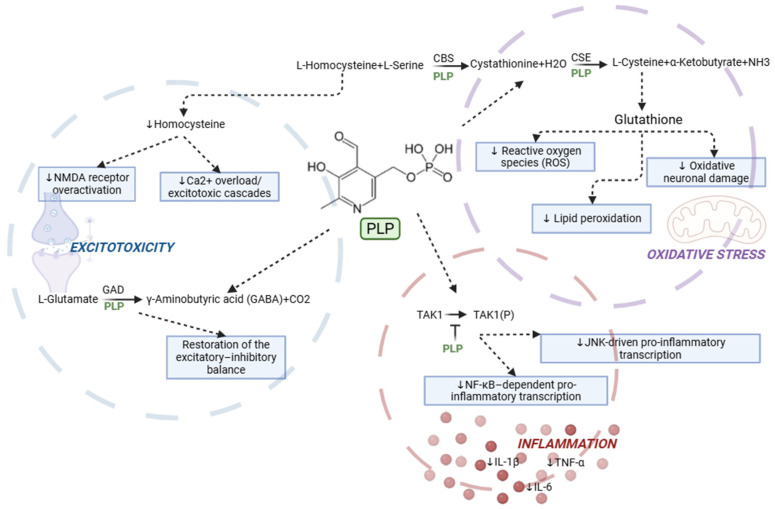
However, a water-soluble, high-dose administration of vitamin B6 can additionally cause reversible neurological side effects in humans and dogs [ref. 256,ref. 257]. Therefore, optimal dosing would be primordial to attain therapeutic benefits. The levels of PLP may be influenced by ASM in PWE, although findings vary across drug types and study designs [ref. 258,ref. 259,ref. 260,ref. 261,ref. 262]. Most studies report significantly lower PLP levels in PWE, particularly in those receiving enzyme-inducing ASM, like phenytoin or carbamazepine [ref. 261,ref. 262].
9.3. General Remarks on Vitamins, Minerals and Trace Elements
Emerging research highlights that trace elements, particularly selenium, iron, copper and zinc, are associated with both the development of epilepsy and changes in seizure susceptibility in PWE, CE and rodent models [ref. 263]. Antiseizure interventions, including ASM and the ketogenic diet, have been shown to modify the serum concentrations of these elements, suggesting that the maintenance of trace element homeostasis may play a role in both the prevention and the therapeutic management of epilepsy [ref. 264]. In addition to trace elements, several vitamins have also been implicated in PWE, with deficiencies or imbalances potentially influencing neuronal excitability and seizure thresholds [ref. 265,ref. 266]. In CE, vitamin B6 remains the only vitamin directly linked to epilepsy in one level II study, while the status and potential involvement of other vitamins have not yet been systematically investigated in this population [ref. 38].
10. Neurotransmission
10.1. Canine Epilepsy
Urinary neurotransmitter profiling in CE revealed a decreased GABA/glutamate and norepinephrine/epinephrine (NE/E) ratio [ref. 51]. The same study further indicated that sex and ASM therapy may modulate these concentrations [ref. 51]. Furthermore, two earlier studies investigated neurotransmitter alterations in CE. Among these, Ellenberger et al. (2004) confirmed altered CSF concentrations of GABA and glutamate, as well as changes in their ratio [ref. 52]. Similarly, Morita et al. (2005), employing cerebral microdialysis, electroencephalographic recordings, and immunohistochemical analyses, documented elevated extracellular concentrations of glutamate and aspartate in CE brains, which were associated with an increased spike frequency [ref. 53].
10.2. Human Epilepsy
Although the function for GABA and glutamate in maintaining the brain’s inhibitory–excitatory balance is well established, studies in PWE on glutamate and GABA are limited to small cohorts with suboptimal statistical power, and an incomplete understanding of how central and peripheral concentrations relate to each other [ref. 201]. The observation of analogous neurochemical alterations in CE suggests that dogs may constitute a translational model for investigating glutamate–GABA dysregulation and guiding the development of targeted interventions [ref. 38,ref. 201].
10.3. Glutamate and Glutamic Acid
Across human and animal studies, altered glutamate and glutamic acid levels have been consistently documented in the blood [ref. 38,ref. 200,ref. 209] and CSF [ref. 39,ref. 201,ref. 267,ref. 268,ref. 269] of individuals with epilepsy. Under physiological conditions, excess glutamate is actively cleared from the CNS into systemic circulation. However, the relationship between central and peripheral glutamate is complex: while blood glutamate concentrations may partially reflect brain metabolism, they are also influenced by peripheral sources (e.g., muscle, liver, and gut) and by the selective transport properties of the blood–brain barrier [ref. 270,ref. 271]. As the primary excitatory neurotransmitter in the CNS [ref. 271], glutamate accumulation is central to epileptogenesis. Perturbations in the excitatory–inhibitory equilibrium, particularly altered GABA–glutamate ratios, constitute a core mechanism underlying seizure initiation [ref. 201]. Beyond its central actions, glutamate also serves as an excitatory neurotransmitter within the enteric nervous system, where enteric neurons express glutamatergic receptors and transporters analogous to those found in the central nervous system [ref. 272,ref. 273]. Glutamatergic signalling along the MGBA has been proposed as a mechanism by which metabolic and neuroactive signals in the periphery may influence brain function [ref. 274]. Furthermore, studies in rodent models indicate that gastric glutamate can activate afferent pathways that are suppressed by vagotomy, consistent with the vagal modulation of central responses to gut stimuli [ref. 275].
10.4. Epinephrine and Norepinephrine
Epinephrine and norepinephrine are closely linked to epilepsy in both humans and dogs. In PWE, noradrenergic system dysregulation has been hypothesized to be correlated with common comorbidities such as sleep disturbances and cognitive deficits [ref. 276,ref. 277,ref. 278,ref. 279]. Similarly, in CE, increased urinary epinephrine concentrations likely cause the altered urinary norepinephrine/epinephrine ratio [ref. 51] and, like in PWE, these are associated with common comorbidities, including sleep disturbances, ADHD-like behaviours, and anxiety [ref. 51,ref. 180,ref. 280,ref. 281].
10.5. Glycine
Glycine is an inhibitory neurotransmitter that, when dysregulated, can induce epileptic seizures through multiple mechanisms at both low and high concentrations [ref. 282,ref. 283]. Elevated urinary glycine levels detected in CE may be associated with seizure onset, cognitive impairment, and hyperactivity, consistent with clinical observations in PWE [ref. 51,ref. 180,ref. 284,ref. 285]. Evidence from human and murine studies indeed indicates that dysfunction in glycine receptors is associated with epileptic phenotypes, and several receptor subtypes have been proposed as potential therapeutic targets [ref. 286,ref. 287,ref. 288,ref. 289]. Further evidence supporting the involvement of glycine in epilepsy pathogenesis comes from the existence of a hereditary disorder of glycine metabolism in humans, known as nonketotic hyperglycinaemia [ref. 290]. This condition leads to the accumulation of glycine in the body, resulting in refractory epileptic seizures, hyperactivity, elevated levels of glycine in the urine, and, in adults, cognitive impairment [ref. 291]. In contrast to PWE, in whom valproate, an ASM, has been shown to markedly increase both urinary and plasma glycine concentrations [ref. 292], no comparable effect has been documented in CE treated with first- or second-line ASM (i.e., phenobarbital or potassium bromide) approved for the management of CE [ref. 51].
10.6. Serotonin
As mentioned in previous sections, serotonin represents a key downstream product of tryptophan metabolism; however, beyond its peripheral metabolic role, it also exerts critical functions as a central neurotransmitter relevant to epilepsy. In humans, reduced serotonin levels are associated with various psychiatric disorders. Furthermore, in PWE, alterations in the serotonergic system have been shown to lower the epileptic seizure threshold and contribute to frequently co-occurring neurobehavioral comorbidities [ref. 293,ref. 294]. The International League Against Epilepsy has suggested that selective serotonin reuptake inhibitors (SSRIs) may be used with caution for the treatment of anxiety in certain PWE. These drugs are already widely used to manage behavioural and psychological disorders in both species [ref. 284,ref. 295,ref. 296].
Experimental evidence in PWE indicates that SSRIs or serotonin–norepinephrine reuptake inhibitors (SNRIs) can reduce the severity of epilepsy by alleviating depressive symptoms [ref. 297,ref. 298]. Additionally, in dogs, fluoxetine (an SSRI) has been reported to be effective in treating fly-snapping syndrome, a condition considered to represent limbic epilepsy by some but a compulsive behavioural disorder by others [ref. 299].
10.7. General Remarks on Neurotransmission
Overall, converging evidence in both PWE and CE supports a consistent dysregulation of excitatory and inhibitory neurotransmission, particularly involving glutamatergic, GABAergic, glycinergic, and monoaminergic systems [ref. 38,ref. 39,ref. 200,ref. 201,ref. 209]. Despite this general agreement, the current findings are limited by small sample sizes, methodological heterogeneity, and an incomplete understanding of the relationship between central and peripheral neurotransmitter levels [ref. 201,ref. 270,ref. 271]. The observed similarities between species may further support the translational relevance of CE as a natural model of epilepsy [ref. 38,ref. 201]. Nevertheless, causal mechanisms remain largely undefined, and future studies with standardized approaches and appropriate control of confounders, including ASM, are required to better define the role of neurotransmitter alterations in epileptogenesis [ref. 38,ref. 51,ref. 201,ref. 282].
11. Endocannabinoid System
11.1. Endogenous Endocannabinoid Metabolism
The endocannabinoid system is a neuromodulatory network composed of endocannabinoids, like anandamide, 1- and 2-arachidonoyl glycerol, their receptors (CB1, CB2), and associated enzymes, which regulates synaptic transmission and maintains homeostasis in the central and peripheral nervous system. It plays a key role in modulating neuronal excitability, inflammation, and seizure susceptibility [ref. 300]. Importantly, species-specific differences in CB1 expression have been documented, particularly regarding regional distribution, cellular localization, and density. In dogs, CB1 is widely expressed in the cerebral cortex, hippocampus, basal ganglia, cerebellum, hindbrain, spinal cord, peripheral nerves, and glial cells [ref. 301,ref. 302,ref. 303], whereas in humans CB1 is predominantly neuronal and concentrated in the hippocampus, cortex, amygdala, basal ganglia, and cerebellum [ref. 304,ref. 305]. Moreover, CB1R expression in humans is dynamic rather than static: it varies during development, with ageing, and in response to stress or environmental experiences [ref. 306]. Therefore, while dogs could serve as a model to study differential receptor modulation between healthy and CE, they are not suitable for the direct comparison of CB1 receptor localization across species.
11.1.1. Canine Epilepsy
In CE, higher CSF anandamide was detected, and this increase was even more pronounced in dogs with a severe phenotype, i.e., status epilepticus and/or cluster seizures, or long disease history [ref. 54]. More recently, a distinct modification in the hippocampal CB1 receptors was highlighted for CE compared to structural epilepsy. Dogs with CE showed decreased CB1 receptor expression in the CA1 region, in contrast to dogs with structural epilepsy, showing increased expression [ref. 55].
11.1.2. Human Epilepsy
In humans with newly diagnosed temporal lobe epilepsy, decreased CSF anandamide was detected [ref. 307]. This seems to contrast with the increased CSF anandamide in CE; however, the authors of the CE study suggest a physiological counter-mechanism to attempt to regulate epileptic seizure thresholds. This would indeed not yet be present in the acute phase of the disease studied in PWE. The alteration in CB1 receptors is considered complex and depending on the etiology in PWE [ref. 308], with, e.g., a downregulation in the hippocampus of people with temporal lobe epilepsy [ref. 309], similar to the distinct receptor modification seen in CE. However, for CB1 expression no direct cross-species comparison is possible, as stressed by the differences in regional distribution, cellular localization, and density [ref. 302].
11.2. Phytocannabinoids
Both in PWE and CE, cannabidiol, i.e., a phytocannabinoid, emerged as a novel management strategy, highlighting potential efficacy in epileptic seizure reduction, but also showed adverse effects like somnolence and gastro-intestinal symptoms [ref. 310,ref. 311]. Most research to date argues against the relevance of CB1 and CB2 receptors in the anticonvulsive effect of phytocannabinoids [ref. 312]. Other receptors, including the G protein receptor 55 (GPR55) and transient receptor potential Vannilloid-1 (TRPV1) [ref. 313,ref. 314], together with an interaction via adenosine signalling [ref. 315], are hypothesized as the major pathways leading to its anticonvulsive effects.
11.3. General Remarks on the Endocannabinoid System
The endocannabinoid system represents a key neuromodulatory pathway implicated in the regulation of neuronal excitability and seizure susceptibility in both PWE and CE [ref. 300]. Evidence from both species indicates alterations in endocannabinoid signalling, including changes in anandamide levels and CB1 receptor expression, although these appear to be context-dependent and influenced by disease type and stage [ref. 54,ref. 55,ref. 307,ref. 308]. However, interpretation is limited by interspecies differences in CB1 distribution and regulation, as well as by variability in study design and clinical populations [ref. 302,ref. 304,ref. 305]. While CE shares several biochemical and receptor-level similarities with PWE, direct anatomical and functional comparisons remain constrained. In addition, although cannabidiol shows anticonvulsant effects in both species, current evidence suggests these are largely mediated through non-CB1/CB2 mechanisms [ref. 310,ref. 311,ref. 312,ref. 313,ref. 314,ref. 315]. Further studies are required to clarify the precise role of endocannabinoid signalling in epileptogenesis and its translational therapeutic potential.
12. MicroRNA
MicroRNAs (miRNAs) play a crucial role in the post-transcriptional regulation of genes involved in neuronal metabolic pathways, including mitochondrial function, glycolysis, and oxidative stress responses. Emerging evidence indicates that altered miRNA expression contributes to the metabolic dysregulation observed in epilepsy, affecting neuronal energy homeostasis and excitability. These findings suggest that miRNA–metabolism interactions represent a key molecular link between bioenergetic imbalance and epileptogenesis [ref. 316].
12.1. Canine Epilepsy
A recent study investigated microRNAs (miRNAs) in CE [ref. 56]. The study focused on seven miRNAs previously identified as dysregulated in both human and murine epilepsy [ref. 317,ref. 318,ref. 319,ref. 320,ref. 321]. Six of these (miR-16, miR-27a-3p, miR-93-5p, miR-132, miR-142, and miR-574-3p) were found to be altered in CE, with five showing significant downregulation. Notably, a panel combining miR-93-5p, miR-142, and miR-574 demonstrated promising diagnostic performance [ref. 56].
In earlier work on CE, Gutierrez-Quintana et al. (2022) [ref. 322] investigated miR-134, a miRNA upregulated in rodent models of drug-resistant epilepsy and in human temporal lobe epilepsy. Plasma levels of miR-134 were significantly higher in drug-resistant CE compared to controls and drug-responsive dogs [ref. 322]. Additionally, Pasierbinska et al. (2025) [ref. 323] profiled blood miRNAs in drug-naive and treated CE in a pilot study, identifying miRNAs that distinguish CE from healthy dogs (e.g., miR-381, miR-214, miR-224), and those differentiating drug-resistant versus drug-responsive cases (e.g., miR-223, miR-129, miR-210). These results support a potential role of miRNAs in the molecular mechanisms underlying CE and drug-resistance, establishing a basis for further biomarker discovery and therapeutic studies.
12.2. Human Epilepsy
In PWE, several miRNAs have been implicated in neuronal plasticity, dendritic spine morphology, synaptic regulation, and excitability. Similar to CE, miR-134 (a neuron-enriched miRNA involved in dendritic spine development through targets such as LIMK1) is consistently upregulated in the brain tissue of patients with drug-resistant temporal lobe epilepsy, reflecting synaptic remodelling associated with hyperexcitability [ref. 324,ref. 325]. Circulating levels of miR-134 are also elevated in plasma or serum of patients with drug-resistant epilepsy, suggesting potential as a biomarker for pharmacoresistance [ref. 318]. Additionally, miR-129-2-3p is upregulated in cortical tissue and plasma of patients with refractory temporal lobe epilepsy. ROC analyses indicate miR-129-2-3p can discriminate PWE from controls with good sensitivity and specificity [ref. 325].
For miR-223, findings in PWE are inconsistent across studies, but it has been reported to be overexpressed in the serum of patients with temporal lobe epilepsy and may differentiate drug-resistant from drug-responsive cases. Similarly, miR-210, a hypoxia-regulated miRNA, shows limited and inconsistent evidence in PWE [ref. 317,ref. 326], with most functional data derived from animal models, where it modulates neuronal survival and GABAergic signalling post-seizure [ref. 317].
In summary, studies in PWE strongly support a role for miR-134 and miR-129-2-3p in pathophysiology and drug resistance, while canine evidence remains preliminary. Systematic cross-species studies are needed to establish the translational relevance for biomarker discovery and targeted interventions.
13. Limitations
This review is subject to several limitations that warrant critical consideration. Firstly, an inherent selection bias arises from the hybrid review design, wherein the canine literature search was systematic, but the synthesis of the human literature was non-systematic and narrative. This approach introduced a bias towards metabolic pathways and markers previously identified in dogs, rather than providing a comprehensive systematic overview of human metabolic epilepsy research—in line with the proposed research aim. Secondly, the current body of evidence is predominantly associative, with most retrieved studies being observational in nature. Consequently, it remains unclear whether metabolic alterations, such as neuroinflammation or oxidative stress, are primary drivers of epileptogenesis or secondary consequences of recurrent seizures. Thirdly, confounding variables, particularly the influence of ASM management, pose significant challenges. Many studies failed to adequately account for or standardize ASM management, yet ASM may modulate redox pathways, influence lipid profiles, and affect inflammatory markers, leaving their metabolic impact inconclusive. Additionally, variability in dietary intake and fasting conditions at the time of sampling further complicates metabolic research, particularly in human studies, where dietary standardization is difficult. Finally, cohort and methodological limitations, including small and heterogeneous sample sizes might reduce statistical power and generalisability. Technological biases, such as those inherent in 16S rRNA sequencing, introduce interstudy variability, while biological inconsistencies, including the use of non-homologous antigens for autoantibody testing in CE and inconsistent markers for lipid peroxidation, highlight the need for more sensitive and specific analytical tools in epilepsy research for both species.
14. Conclusions and Future Insights
The present review underscores a substantial convergence in the metabolic alterations associated with CE and PWE, while acknowledging species-specific divergences that warrant further investigation. Shared inflammatory pathways, including, e.g., increased IL-17 and TNF-α, oxidative stress markers like AOPP, amyloid pathology, and metabolic responses to dietary interventions—especially MCT diets—highlight the translational potential of the CE model. However, differences in complement cascade activation, specific amino acid alterations like threonine, and cannabinoid receptor localisation emphasize the need for the cautious extrapolation of findings between species.
In both CE and PWE, inflammatory and immune pathways, together with mitochondrial function and related oxidative stress, are hypothesized to emerge as central mechanisms in epilepsy and appear to form a self-perpetuating cycle (Figure 4). Metabolic disturbances in amino acids, lipids, minerals, trace elements, and vitamins may act as triggers within this cycle. Additional alterations in the endocannabinoid system, microbiota–gut–brain axis, neurotransmission, and post-transcriptional regulation further appear to contribute to epileptogenesis and may modulate these interactions.
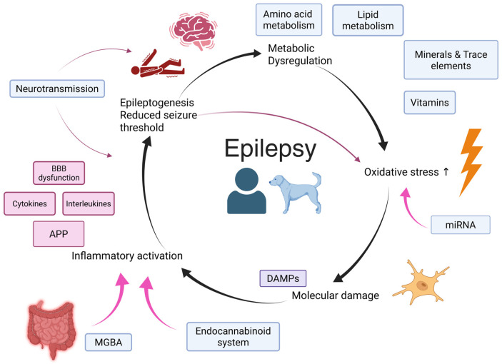
Priorities for future research include: (1) harmonized cross-species, longitudinal cohorts with standardized phenotyping, including epileptic seizure semiology, comorbidities, diet information, and ASM exposure, to address current study heterogeneity and common confounding factors; (2) multi-omics pipelines integrating metabolomics, proteomics, trace-element and vitamin status, microbiome and microRNAs to derive robust biomarker panels and responder stratifiers; (3) randomized, clinical trials of nutritional and metabolic interventions, e.g., the MCT-diet, and GI microbiome modulation (pre- and probiotics, FMT); and (4) the translational validation of emerging targets, like specific miRNAs, immunomodulation, and endocannabinoid regulation, with attention to the species-specific characteristics highlighted in this review. Taken together, CE has the potential to bridge experimental models and clinical practice, enhancing epilepsy research in both species.
References
- F. Boele, C. Jensen, G. Madigan Johnson, A. Lammers-Spijker, A. Altinbas, L. van den Berg, K. Broekman-Labinac, R. Fronczek, M. Schuur, G. Vonk. Health-Related Quality of Life and Unmet Needs of People with Epilepsy and Their Family Caregivers: A Systematic Scoping Review. Epilepsy Behav., 2025. [DOI | PubMed]
- 2. World Health Organization EPILEPSY: A Public Health Imperative International League Against EpilepsyWHOGeneva, Switzerland2019
- M. Syvertsen, K.O. Nakken, A. Edland, G. Hansen, M.K. Hellum, J. Koht. Prevalence and Etiology of Epilepsy in a Norwegian County—A Population Based Study. Epilepsia, 2015. [DOI | PubMed]
- W. Löscher, H. Potschka, S.M. Sisodiya, A. Vezzani. Drug Resistance in Epilepsy: Clinical Impact, Potential Mechanisms, and New Innovative Treatment Options. Pharmacol. Rev., 2020. [DOI | PubMed]
- K. Chojnowski, M. Opiełka, K. Urbanowicz, M. Zawadzka, K. Wangin, R.T. Smoleński, M. Mazurkiewicz-Bełdzińska. Untargeted Metabolomics Reveals Key Metabolic Alterations in Pediatric Epilepsy with Insights into Tryptophan Metabolism and the Gut–Brain Axis. Sci. Rep., 2025. [DOI | PubMed]
- A. Fujita, M. Ota, K. Kato. Urinary Volatile Metabolites of Amygdala-Kindled Mice Reveal Novel Biomarkers Associated with Temporal Lobe Epilepsy. Sci. Rep., 2019. [DOI | PubMed]
- Ł. Boguszewicz, E. Jamroz, M. Ciszek, E. Emich-Widera, M. Kijonka, T. Banasik, A. Skorupa, M. Sokół. NMR-Based Metabolomics in Pediatric Drug Resistant Epilepsy—Preliminary Results. Sci. Rep., 2019. [DOI | PubMed]
- E. Maa, J. Arnold, K. Ninedorf, H. Olsen. Canine Detection of Volatile Organic Compounds Unique to Human Epileptic Seizure. Epilepsy Behav., 2021. [DOI | PubMed]
- T. Eid. Harnessing Metabolomics to Advance Epilepsy Research. Epilepsy Curr., 2022. [DOI | PubMed]
- E. Hirsch, J. French, I.E. Scheffer, A. Bogacz, T. Alsaadi, M.R. Sperling, F. Abdulla, S.M. Zuberi, E. Trinka, N. Specchio. ILAE Definition of the Idiopathic Generalized Epilepsy Syndromes: Position Statement by the ILAE Task Force on Nosology and Definitions. Epilepsia, 2022. [DOI | PubMed]
- R.J. McGinn, E.L. Von Stein, J.E. Summers Stromberg, Y. Li. Precision Medicine in Epilepsy. Prog. Mol. Biol. Transl. Sci., 2022. [PubMed]
- M. Berendt, R.G. Farquhar, P.J.J. Mandigers, A. Pakozdy, S.F.M. Bhatti, L. De Risio, A. Fischer, S. Long, K. Matiasek, K. Muñana. International Veterinary Epilepsy Task Force Consensus Report on Epilepsy Definition, Classification and Terminology in Companion Animals. BMC Vet. Res., 2015. [DOI | PubMed]
- L. De Risio, S. Bhatti, K. Muñana, J. Penderis, V. Stein, A. Tipold, M. Berendt, R. Farqhuar, A. Fischer, S. Long. International Veterinary Epilepsy Task Force Consensus Proposal: Diagnostic Approach to Epilepsy in Dogs. BMC Vet. Res., 2015. [DOI | PubMed]
- M. Charalambous, A. Fischer, H. Potschka, M.C. Walker, R. Raedt, K. Vonck, P. Boon, H. Lohi, W. Löscher, G. Worrell. Translational Veterinary Epilepsy: A Win-Win Situation for Human and Veterinary Neurology. Vet. J., 2023. [DOI | PubMed]
- W. Löscher. Dogs as a Natural Animal Model of Epilepsy. Front. Vet. Sci., 2022. [DOI | PubMed]
- H. Potschka, A. Fischer, E.L. Von Rüden, V. Hülsmeyer, W. Baumgärtner. Canine Epilepsy as a Translational Model?. Epilepsia, 2013. [DOI | PubMed]
- O. Devinsky, J.M. Boesch, S. Cerda-Gonzalez, B. Coffey, K. Davis, D. Friedman, B. Hainline, K. Houpt, D. Lieberman, P. Perry. A Cross-Species Approach to Disorders Affecting Brain and Behaviour. Nat. Rev. Neurol., 2018. [DOI | PubMed]
- F.Y. Han, L. Conboy-Schmidt, G. Rybachuk, H.A. Volk, B. Zanghi, Y. Pan, K. Borges. Dietary Medium Chain Triglycerides for Management of Epilepsy: New Data from Human, Dog, and Rodent Studies. Epilepsia, 2021. [DOI | PubMed]
- L.P. Coelho, J.R. Kultima, P.I. Costea, C. Fournier, Y. Pan, G. Czarnecki-Maulden, M.R. Hayward, S.K. Forslund, T.S.B. Schmidt, P. Descombes. Similarity of the Dog and Human Gut Microbiomes in Gene Content and Response to Diet. Microbiome, 2018. [DOI | PubMed]
- K. Wagstyl, K. Kobow, P.M. Casillas-Espinosa, A.J. Cole, D. Jiménez-Jiménez, H. Nariai, S. Baulac, T. O’Brien, D.C. Henshall, O. Akman. WONOEP 2022: Neurotechnology for the Diagnosis of Epilepsy. Epilepsia, 2024. [DOI | PubMed]
- D. Obeso, E. Zubeldia-Varela, A. Villaseñor. Uncovering the Influence of Diet and Gut Microbiota in Human Serum Metabolome. Allergy, 2021. [DOI | PubMed]
- A.C. Tricco, E. Lillie, W. Zarin, K.K. O’Brien, H. Colquhoun, D. Levac, D. Moher, M.D.J. Peters, T. Horsley, L. Weeks. PRISMA Extension for Scoping Reviews (PRISMA-ScR): Checklist and Explanation. Ann. Intern. Med., 2018. [DOI | PubMed]
- M.J. Page, J.E. McKenzie, P.M. Bossuyt, I. Boutron, T.C. Hoffmann, C.D. Mulrow, L. Shamseer, J.M. Tetzlaff, E.A. Akl, S.E. Brennan. The PRISMA 2020 statement: An updated guideline for reporting systematic reviews. BMJ, 2021. [DOI | PubMed]
- S. Kang, Y. Koo, T. Yun, Y. Chae, D. Lee, H. Kim, M.P. Yang, B.T. Kang. Serum Concentrations of Complement C3 and C4 in Dogs with Idiopathic Epilepsy. J. Vet. Intern. Med., 2024. [DOI | PubMed]
- C. de la Fuente, L. Monreal, J. Cerón, J. Pastor, J. Viu, S. Añor. Fibrinolytic Activity in Cerebrospinal Fluid of Dogs with Different Neurological Disorders. J. Vet. Intern. Med., 2012. [DOI | PubMed]
- D. Kostic, R. Carlson, D. Henke, K. Rohn, A. Tipold. Evaluation of IL-1β Levels in Epilepsy and Traumatic Brain Injury in Dogs. BMC Neurosci., 2019. [DOI | PubMed]
- A. Despa, M. Musteata, G. Solcan. Evaluation of Blood C Reactive Protein (CRP) and Neutrophil-to-Lymphocyte Ratio (NLR) Utility in Canine Epilepsy. Vet. Sci., 2024. [DOI | PubMed]
- Y. Koo, H. Kim, T. Yun, D. Jung, J. Kang, D. Chang, K. Na, M. Yang, B. Kang. Evaluation of Serum High-mobility Group Box 1 Concentration in Dogs with Epilepsy: A Case-control Study. J. Vet. Intern. Med., 2020. [DOI | PubMed]
- E.-L. von Rüden, F. Gualtieri, K. Schönhoff, M. Reiber, F. Wolf, W. Baumgärtner, F. Hansmann, A. Tipold, H. Potschka. Molecular Alterations of the TLR4-Signaling Cascade in Canine Epilepsy. BMC Vet. Res., 2020. [DOI | PubMed]
- E. Segers, V. Martlé, S. Piepers, L. Van Ham, S.F.M. Bhatti. C-Reactief Proteïneconcentraties in Het Serum van Honden Met Idiopathische Epilepsie. Vlaams Diergeneeskd. Tijdschr., 2017. [DOI]
- S. Paltrinieri, L. Pintore, F. Balducci, A. Giordano, A. Costabile, M. Bernardini. Serum Creatine Kinase Isoenzymes and Macroenzymes in Dogs with Different Neurologic Diseases. Vet. Clin. Pathol., 2017. [DOI | PubMed]
- Y. Merbl, A. Sommer, O. Chai, I. Aroch, G. Zimmerman, A. Friedman, H. Soreq, M.H. Shamir. Tumor Necrosis Factor-α and Interleukin-6 Concentrations in Cerebrospinal Fluid of Dogs After Seizures. J. Vet. Intern. Med., 2014. [DOI | PubMed]
- L. Hemmeter, C.G. Bien, C.I. Bien, A. Tipold, J. Neßler, A. Bathen-Nöthen, K. Matiasek, M. Dahlhoff, C. Rusbridge, C. Rotter Black. Investigation of the Presence of Specific Neural Antibodies in Dogs with Epilepsy or Dyskinesia Using Murine and Human Assays. J. Vet. Intern. Med., 2023. [DOI | PubMed]
- A. Knebel, A. Kämpe, R. Carlson, K. Rohn, A. Tipold. Th17 Cell-Mediated Immune Response in a Subpopulation of Dogs with Idiopathic Epilepsy. PLoS ONE, 2022. [DOI | PubMed]
- R. Baka, D. Eckersall, A. Horvatic, A. Gelemanovic, V. Mrljak, M. McLaughlin, L.V. Athanasiou, N. Papaioannou, I. Stylianaki, H.Q. Hanh. Quantitative Proteomics of Cerebrospinal Fluid Using Tandem Mass Tags in Dogs with Recurrent Epileptic Seizures. J. Proteom., 2021. [DOI]
- S. Phochantachinda, B. Chantong, O. Reamtong, D. Chatchaisak. Protein Profiling and Assessment of Amyloid Beta Levels in Plasma in Canine Refractory Epilepsy. Front. Vet. Sci., 2023. [DOI | PubMed]
- R.D. Baka, J. Kuleš, A. Beletić, V. Farkaš, D. Rešetar Maslov, B.B. Ljubić, I. Rubić, V. Mrljak, M. McLaughlin, D. Eckersall. Quantitative Serum Proteome Analysis Using Tandem Mass Tags in Dogs with Epilepsy. J. Proteom., 2024. [DOI]
- F. Verdoodt, S.F.M. Bhatti, J. Molina, L. Van Ham, L. Vanhaecke, G. Junius, L.Y. Hemeryck, M. Hesta. Plasma Metabolome Reveals Altered Oxidative Stress, Inflammation, and Amino Acid Metabolism in Dogs with Idiopathic Epilepsy. Epilepsia, 2025. [DOI | PubMed]
- T. Hasegawa, M. Sumita, Y. Horitani, R. Tamai, K. Tanaka, M. Komori, S. Takenaka. Gas Chromatography-Mass Spectrometry-Based Metabolic Profiling of Cerebrospinal Fluid from Epileptic Dogs. J. Vet. Med. Sci., 2014. [DOI | PubMed]
- J. Weber, A. Maiolini, A. Tipold. Untersuchungen zu erniedrigten Glukosewertenim Liquor cerebrospinalis des Hundes [Evaluation of decreased glucose levels in the cerebrospinal fluid of dogs]. Tierarztl. Prax. Ausg. K. Kleintiere Heimtiere, 2012. [PubMed]
- F. Verdoodt, M. Hesta, E. Goossens, F. Van Immerseel, J. Molina, L. Van Ham, L. Vanhaecke, L.Y. Hemeryck, S.F.M. Bhatti. The Fecal Metabolome and Microbiome Are Altered in Dogs with Idiopathic Epilepsy Compared to Healthy Dogs. Sci. Rep., 2025. [DOI | PubMed]
- S. García-Belenguer, L. Grasa, O. Valero, J. Palacio, I. Luño, B. Rosado. Gut Microbiota in Canine Idiopathic Epilepsy: Effects of Disease and Treatment. Animals, 2021. [DOI | PubMed]
- K.R. Muñana, M.E. Jacob, B.J. Callahan. Evaluation of fecal Lactobacillus populations in dogs with idiopathic epilepsy: A pilot study. Anim. Microbiome, 2020. [DOI | PubMed]
- M. Silvestrino, M. Pirolo, A. Bianco, S. Castellana, L. Del Sambro, V.D. Tarallo, L. Guardabassi, A. Zatelli, F. Gernone. Idiopathic Epilepsy in Dogs Is Associated with Dysbiotic Faecal Microbiota. Anim. Microbiome, 2025. [DOI | PubMed]
- T. Yonezawa, S. Takenouchi, T. Motegi, M. Miyazaki, N. Nagata, K. Kobayashi, M. Yamada, T. Murata. Lipid Metabolites and Nitric Oxide Production in the Cerebrospinal Fluid and Plasma of Dogs with Meningoencephalitis of Unknown Origin and Idiopathic Epilepsy: A Pilot Study. Front. Vet. Sci., 2024. [DOI | PubMed]
- E.K. Kluger, R. Malik, W.J. Ilkin, D. Snow, D.R. Sullivan, M. Govendir. Serum Triglyceride Concentration in dogs with Epilepsy Treated with phenobarbital or with Phenobarbital and Bromide. J. Am. Vet. Med. Assoc., 2008. [DOI | PubMed]
- M. Radaković, J.F. Andrić, K. Spariosu, B. Vejnović, M.K. Filipović, N. Andrić. Serum Oxidant–Antioxidant Status and Butyrylcholinesterase Activity in Dogs with Idiopathic Epilepsy—A Pilot Study. Res. Vet. Sci., 2023. [DOI | PubMed]
- S. Rosendahl, J. Anturaniemi, T.-K. Kukko-Lukjanov, K.A. Vuori, R. Moore, M. Hemida, A. Muhle, A. Hielm-Björkman. Whole Blood Trace Element and Toxic Metal Concentration in Dogs with Idiopathic Epilepsy and Healthy Dogs: A Case-Control Study. Front. Vet. Sci., 2023. [DOI | PubMed]
- S. Rosendahl, J. Anturaniemi, T.K. Kukko-Lukjanov, K.A. Vuori, R. Moore, M. Hemida, A. Muhle, A. Hielm-Björkman. Mineral, Trace Element, and Toxic Metal Concentration in Hair from Dogs with Idiopathic Epilepsy Compared to Healthy Controls. J. Vet. Intern. Med., 2023. [DOI | PubMed]
- S. Vitale, D.W. Hague, K. Foss, M.C. de Godoy, L.E. Selmic. Comparison of Serum Trace Nutrient Concentrations in Epileptics Compared to Healthy Dogs. Front. Vet. Sci., 2019. [DOI | PubMed]
- T. Schmidt, S. Meller, S.R. Talbot, B.A. Berk, T.H. Law, S.L. Hobbs, N. Meyerhoff, R.M.A. Packer, H.A. Volk. Urinary Neurotransmitter Patterns Are Altered in Canine Epilepsy. Front. Vet. Sci., 2022. [DOI | PubMed]
- C. Ellenberger, M. Mevissen, M. Doherr, G. Scholtysik, A. Jaggy. Inhibitory and Excitatory Neurotransmitters in the Cerebrospinal Fluid of Epileptic Dogs. Am. J. Vet. Res., 2004. [DOI | PubMed]
- T. Morita, M. Takahashi, T. Takeuchi, Y. Hikasa, S. Ikeda, M. Sawada, K. Sato, T. Shibahara, A. Shimada. Changes in Extracellular Neurotransmitters in the Cerebrum of Familial Idiopathic Epileptic Shetland Sheepdogs Using an Intracerebral Microdialysis Technique and Immunohistochemical Study for Glutamate Metabolism. J. Veter. Med. Sci., 2005. [DOI]
- F.K. Gesell, A.A. Zoerner, C. Brauer, S. Engeli, D. Tsikas, A. Tipold. Alterations of Endocannabinoids in Cerebrospinal Fluid of Dogs with Epileptic Seizure Disorder. BMC Vet. Res., 2013. [DOI | PubMed]
- D. Kostic, M. Nowakowska, J. Freundt Revilla, F. Attig, K. Rohn, F. Gualtieri, W. Baumgärtner, H. Potschka, A. Tipold. Hippocampal Expression of the Cannabinoid Receptor Type 1 in Canine Epilepsy. Sci. Rep., 2023. [DOI | PubMed]
- M. García-Gracia, L. Moreno-Martinez, A. Hernaiz, S. Usón, J. Moral, D. Sanz-Rubio, P. Zaragoza, J. Palacio, B. Rosado, R. Osta. Analysis of Plasma-Derived Exosomal MicroRNAs as Potential Biomarkers for Canine Idiopathic Epilepsy. Animals, 2024. [DOI | PubMed]
- E.-L. von Rüden, H. Potschka, A. Tipold, V.M. Stein. The Role of Neuroinflammation in Canine Epilepsy. Vet. J., 2023. [DOI]
- M.J. Aguilar-Castillo, P. Cabezudo-García, G. García-Martín, Y. Lopez-Moreno, G. Estivill-Torrús, N.L. Ciano-Petersen, B. Oliver-Martos, M. Narváez-Pelaez, P.J. Serrano-Castro. A Systematic Review of the Predictive and Diagnostic Uses of Neuroinflammation Biomarkers for Epileptogenesis. Int. J. Mol. Sci., 2024. [DOI | PubMed]
- G. Costagliola, G. Depietri, A. Michev, A. Riva, T. Foiadelli, S. Savasta, A. Bonuccelli, D. Peroni, R. Consolini, G.L. Marseglia. Targeting Inflammatory Mediators in Epilepsy: A Systematic Review of Its Molecular Basis and Clinical Applications. Front. Neurol., 2022. [DOI | PubMed]
- M. Hossain, S. Williams, L. Ferguson, W. Bingaman, A. Ghosh, I.M. Najm, C. Ghosh. Heat Shock Proteins Accelerate the Maturation of Brain Endothelial Cell Glucocorticoid Receptor in Focal Human Drug-Resistant Epilepsy. Mol. Neurobiol., 2020. [DOI | PubMed]
- N. Dericioglu, F. Soylemezoglu, Y. Gursoy-Ozdemir, N. Akalan, S. Saygi, T. Dalkara. Cell Death and Survival Mechanisms Are Concomitantly Active in the Hippocampus of Patients with Mesial Temporal Sclerosis. Neuroscience, 2013. [DOI | PubMed]
- K. Rejdak, J. Kuhle, S. Rüegg, R.L.P. Lindberg, A. Petzold, D. Sulejczak, E. Papuc, R. Rejdak, Z. Stelmasiak, P. Grieb. Neurofilament Heavy Chain and Heat Shock Protein 70 as Markers of Seizure-Related Brain Injury. Epilepsia, 2012. [DOI | PubMed]
- X.G. Ye, S.W. Dong, B.Z. Wu, Z.G. Liu. Increased Expression of Proinflammatory Cytokines in the Cerebrospinal Fluid of Patients with a History of Electrical Status Epilepticus in Sleep. Neurol. Sci., 2025. [DOI | PubMed]
- R.A. Kalsariya, D. Kavila, S. Shorter, D. Negi, I.C.A. Goodall, S. Boussios, S. Ovsepian. V Molecular Biomarkers of Glial Activation and Injury in Epilepsy. Drug Discov. Today, 2025. [DOI | PubMed]
- A. Vezzani, T. Granata. Brain Inflammation in Epilepsy: Experimental and Clinical Evidence. Epilepsia, 2005. [DOI | PubMed]
- A. Vezzani, J. French, T. Bartfai, T.Z. Baram. The Role of Inflammation in Epilepsy. Nat. Rev. Neurol., 2011. [DOI | PubMed]
- A. Klegeris. Regulation of Neuroimmune Processes by Damage- and Resolution-Associated Molecular Patterns. Neural Regen. Res., 2021. [DOI | PubMed]
- R.G. Giffard, R.-Q. Han, J.F. Emery, M. Duan, J. Francois. Regulation of Apoptotic and Inflammatory Cell Signaling in Cerebral Ischemia The Complex Roles of Heat Shock Protein 70. Anesthesiology, 2008. [DOI | PubMed]
- C. Rawat, S. Kukal, U.R. Dahiya, R. Kukreti. Cyclooxygenase-2 (COX-2) Inhibitors: Future Therapeutic Strategies for Epilepsy Management. J. Neuroinflamm., 2019. [DOI]
- M. Maroso, S. Balosso, T. Ravizza, J. Liu, E. Aronica, A.M. Iyer, C. Rossetti, M. Molteni, M. Casalgrandi, A.A. Manfredi. Toll-like Receptor 4 and High-Mobility Group Box-1 Are Involved in Ictogenesis and Can Be Targeted to Reduce Seizures. Nat. Med., 2010. [DOI | PubMed]
- G. Bar-Klein, S. Lublinsky, L. Kamintsky, I. Noyman, R. Veksler, H. Dalipaj, V.V. Senatorov, E. Swissa, D. Rosenbach, N. Elazary. Imaging Blood-Brain Barrier Dysfunction as a Biomarker for Epileptogenesis. Brain, 2017. [DOI | PubMed]
- E. Hanael, R. Veksler, A. Friedman, G. Bar-Klein, V.V. Senatorov, D. Kaufer, L. Konstantin, M. Elkin, O. Chai, D. Peery. Blood-Brain Barrier Dysfunction in Canine Epileptic Seizures Detected by Dynamic Contrast-Enhanced Magnetic Resonance Imaging. Epilepsia, 2019. [DOI | PubMed]
- A. Vezzani. Brain Inflammation and Seizures: Evolving Concepts and New Findings in the Last 2 Decades. Epilepsy Curr., 2020. [DOI | PubMed]
- I. Kushner. Regulation of the Acute Phase Response by Cytokines. Perspect. Biol. Med., 1993. [DOI | PubMed]
- E.K. Mahon, T.L. Williams, L. Alves. Serum C-Reactive Protein Concentrations in Dogs with Structural and Idiopathic Epilepsy. Vet. Rec., 2023. [DOI | PubMed]
- N.K. Mule, A.C. Orjuela Leon, J.R. Falck, M. Arand, A. Marowsky. 11,12 -Epoxyeicosatrienoic Acid (11,12 EET) Reduces Excitability and Excitatory Transmission in the Hippocampus. Neuropharmacology, 2017. [DOI | PubMed]
- L. Rios-Avila, H.F. Nijhout, M.C. Reed, H.S. Sitren, J.F. Gregory. A Mathematical Model of Tryptophan Metabolism via the Kynurenine Pathway Provides Insights into the Effects of Vitamin B-6 Deficiency, Tryptophan Loading, and Induction of Tryptophan 2,3-Dioxygenase on Tryptophan Metabolites. J. Nutr., 2013. [DOI | PubMed]
- A. Agus, J. Planchais, H. Sokol. Gut Microbiota Regulation of Tryptophan Metabolism in Health and Disease. Cell Host Microbe, 2018. [DOI | PubMed]
- C. Xue, G. Li, Q. Zheng, X. Gu, Q. Shi, Y. Su, Q. Chu, X. Yuan, Z. Bao, J. Lu. Tryptophan Metabolism in Health and Disease. Cell Metab., 2023. [DOI | PubMed]
- E. Schepers, G. Glorieux, L. Dou, C. Cerini, N. Gayrard, L. Louvet, C. Maugard, P. Preus, M. Rodriguez-Ortiz, A. Argiles. Guanidino Compounds as Cause of Cardiovascular Damage in Chronic Kidney Disease: An in Vitro Evaluation. Blood Purif., 2010. [DOI | PubMed]
- M. Tachikawa, K. Hosoya. Transport Characteristics of Guanidino Compounds at the Blood-Brain Barrier and Blood-Cerebrospinal Fluid Barrier: Relevance to Neural Disorders. Fluids Barriers CNS, 2011. [DOI | PubMed]
- M.P. Blasco, A. Chauhan, P. Honarpisheh, H. Ahnstedt, J. d’Aigle, A. Ganesan, S. Ayyaswamy, F. Blixt, S. Venable, A. Major. Age-Dependent Involvement of Gut Mast Cells and Histamine in Post-Stroke Inflammation. J. Neuroinflamm., 2020. [DOI]
- M.P.B. Conesa, F.W. Blixt, P. Peesh, R. Khan, J. Korf, J. Lee, G. Jagadeesan, A. Andersohn, T.K. Das, C. Tan. Stabilizing Histamine Release in Gut Mast Cells Mitigates Peripheral and Central Inflammation after Stroke. J. Neuroinflamm., 2023. [DOI]
- T. Li, D. Wei, H. Zhang, A. Aihemaitiniyazi, C. Liu. Clinical Utility of Neutrophil to Lymphocyte Ratio (NLR) for Epileptic Seizures: A Systematic Review and Meta-Analysis. Clin. Neurol. Neurosurg., 2025. [DOI | PubMed]
- S. Hosseini, A.M.E. Mofrad, P. Mokarian, S. Nourigheimasi, A. Azarhomayoun, S. Khanzadeh, S. Habibzadeh, A. Ghaedi. Neutrophil to Lymphocyte Ratio in Epilepsy: A Systematic Review. Mediat. Inflamm., 2022. [DOI]
- M. Alemdar, E. Sarıca Darol, A.K. Polat. Predictive Value of Traditional Laboratory Parameters and Inflammatory Indices for Same-Day Seizure Recurrence in Patients with Epileptic Seizures. Epilepsia Open, 2025. [DOI | PubMed]
- X. Wang, Y. Zhang, Y. Ye, L. Wang, Y. Xu, S. Ren, L. Wang, G. Wu. Neutrophil-to-Lymphocyte Ratio as a Potential Predictive Marker for Epileptic Seizures: Unveiling the “V”-Shaped Link. Mediat. Inflamm., 2025. [DOI]
- C. Cao, J. Mu, G. Hu, Y. Wang, Y. Gong. A Correlation between Inflammatory Factors and Epileptic Seizures: A Meta-Analysis. Actas Esp. Psiquiatr., 2025. [DOI | PubMed]
- R. Zhong, Q. Chen, M. Li, X. Zhang, W. Lin. Elevated Blood C-Reactive Protein Levels in Patients With Epilepsy: A Systematic Review and Meta-Analysis. Front. Neurol., 2019. [DOI | PubMed]
- Y.F. Zhou, Y. Huang, G.H. Liu. Effects of Levetiracetam on the Serum C-Reactive Protein in Children With Epilepsy: A Meta-Analysis. Front. Pharmacol., 2022. [DOI | PubMed]
- J. Yan, K. Kothur, E.A. Innes, V.X. Han, H.F. Jones, S. Patel, E. Tsang, R. Webster, S. Gupta, C. Troedson. Decreased Cerebrospinal Fluid Kynurenic Acid in Epileptic Spasms: A Biomarker of Response to Corticosteroids. EBioMedicine, 2022. [DOI | PubMed]
- L. Wan, X. Shi, H. Yan, Y. Liang, X. Liu, G. Zhu, J. Zhang, J. Wang, M. Wang, G. Yang. Abnormalities in Clostridioides and Related Metabolites before ACTH Treatment May Be Associated with Its Efficacy in Patients with Infantile Epileptic Spasm Syndrome. CNS Neurosci. Ther., 2024. [DOI | PubMed]
- G. Zaccara, S. Lattanzi, F. Brigo. Cardiac Adverse Effects of Antiseizure Medications. Expert Opin. Drug Saf., 2022. [DOI | PubMed]
- A. Butera, M. Pirrone, A.S. Accetta, C. Consoli, A.G. Nicotera, L. Turriziani. Unravelling the Impact: Pulmonary Side Effects of Anti-Seizure Medications. Curr. Respir. Med. Rev., 2025. [DOI]
- Y.Q. Shi, H.C. Yang, C. He, Y.H. Wang, J. Zheng, X.Y. Wang, F.Y. Hao, C.W. Feng, L. Ma, Y.H. Zhang. Inflammatory Links between Epilepsy and Depression: A Review of Mechanisms and Therapeutic Strategies. Front. Neurosci., 2025. [DOI | PubMed]
- M. Yan, X. Qiu, S. Zhang, X. Yu, M. Sun, Y. Yang, Y. Gong, S. Zou, M. Li, F. Fei. Neuroinflammation Leads to Pharmacoresistance in Temporal Lobe Epilepsy via Promoting Spermine Degradation. Acta Pharmacol. Sin., 2025. [DOI | PubMed]
- K. Fukuyama, M. Okada. Effects of Levetiracetam on Astroglial Release of Kynurenine-Pathway Metabolites. Br. J. Pharmacol., 2018. [DOI | PubMed]
- P.S. Chen, C.C. Wang, C.D. Bortner, G.S. Peng, X. Wu, H. Pang, R.B. Lu, P.W. Gean, D.M. Chuang, J.S. Hong. Valproic Acid and Other Histone Deacetylase Inhibitors Induce Microglial Apoptosis and Attenuate Lipopolysaccharide-Induced Dopaminergic Neurotoxicity. Neuroscience, 2007. [DOI | PubMed]
- G. Popović, S. Rakočević, M. Čolić, L. Kozić, M. Drakul, V. Mališ, D. Bokonjić, D. Mihajlović. Anti-Inflammatory and Immunomodulatory Effects of Valproate and Carbamazepine Involve Distinct Signaling in Human Peripheral Blood Mononuclear Cells. Immunopharmacol. Immunotoxicol., 2025. [DOI | PubMed]
- M. Kopczynska, W.M. Zelek, S. Vespa, S. Touchard, M. Wardle, S. Loveless, R.H. Thomas, K. Hamandi, B.P. Morgan. Complement System Biomarkers in Epilepsy. Seizure, 2018. [DOI | PubMed]
- S. Şahin, E. Şimşek, S. Özer Yaman, S.C. Karahan, M. Kalyoncu. Vascular Cell Adhesion Molecule-1 and Complement C3 Involvement in Febrile Seizures in Children. J. Mol. Neurosci., 2025. [DOI | PubMed]
- C. Liguori, A. Romigi, F. Izzi, F. Placidi, M. Nuccetelli, A. Cordella, S. Bernardini, M.N. Biagio. Complement System Dysregulation in Patients Affected by Idiopathic Generalized Epilepsy and the Effect of Antiepileptic Treatment. Epilepsy Res., 2017. [DOI | PubMed]
- N. Pinzon-Hoyos, Y. Li, M. McGee, N.P. Poolos, N. Marchi, A.L. Brewster. Drug-Resistant Epilepsy Associated with Peripheral Complement Decreases and Sex-Specific Cytokine Imbalances: A Pilot Study. Sci. Rep., 2025. [DOI | PubMed]
- R. García, J. Pastor, C. de la Fuente, S. Añor. Thromboelastography in Dogs with Idiopathic Epilepsy Treated with Phenobarbital Monotherapy. Vet. Clin. Pathol., 2024. [DOI | PubMed]
- E.I. Huenerfauth, C.G. Bien, C. Bien, H.A. Volk, N. Meyerhoff. Case Report: Anti-GABAA Receptor Encephalitis in a Dog. Front. Vet. Sci., 2022. [DOI | PubMed]
- J. Föhr, J.K. Prümmer, A. Maiolini, E. Marti, I. Jelcic, B. Vidondo, M. Ziegler, A. Bathen-Nöthen, A. Tipold, H.A. Volk. Cerebrospinal Fluid-specific Oligoclonal Bands in Dogs with Idiopathic Epilepsy. J. Vet. Intern. Med., 2025. [DOI | PubMed]
- Y. Lu, P. Zhang, F. Xu, Y. Zheng, H. Zhao. Advances in the Study of IL-17 in Neurological Diseases and Mental Disorders. Front. Neurol., 2023. [DOI | PubMed]
- J. Li, C. Zhong, G. Bian. Effect of Carbamazepine and Levetiracetam on Coagulation Parameters: Prothrombin Time, Activated Partial Thromboplastin Time, D-Dimer, and Fibrinogen Levels. J. Med. Biochem., 2025. [DOI | PubMed]
- N. Çarçak, S. Al Maawal, P. Thergarajan, T.H.L. Tan, V.K. Mehta, T. Baker, D.C. Henshall, A.S. Galanopoulou, Ö. Akman, P.M. Casillas-Espinosa. WONOEP XVII Appraisal: The Immunopathogenesis of Epilepsy. Epilepsia, 2025. [DOI | PubMed]
- U. Glantschnigg-Eisl, A. Klang, S. Kneissl, B. Lang, P. Waters, S.R. Irani, S.N.M. Binks, A. Pakozdy. A Feline Model of Spontaneously Occurring Autoimmune Limbic Encephalitis. Vet. J., 2023. [DOI]
- P. Cabezudo-García, N.L. Ciano-Petersen, N. Mena-Vázquez, J. Ortega-Pinazo, M.J. Postigo-Pozo, G. García-Martín, H. Antolí-Martínez, V. Sánchez-Sánchez, P. Quiroga-Subirana, P.J. Serrano-Castro. Prevalence of Neural Autoantibodies in Paired Serum and Cerebrospinal Fluid in Adult Patients with Drug-Resistant Temporal Lobe Epilepsy of Unknown Etiology. J. Clin. Med., 2021. [DOI | PubMed]
- M. Levite, H. Goldberg. Autoimmune Epilepsy—Novel Multidisciplinary Analysis, Discoveries and Insights. Front. Immunol., 2022. [DOI | PubMed]
- G. Agirman, K.B. Yu, E.Y. Hsiao. Signaling Inflammation across the Gut-Brain Axis. Science, 2021. [DOI | PubMed]
- S.M. O’Mahony, G. Clarke, Y.E. Borre, T.G. Dinan, J.F. Cryan. Serotonin, Tryptophan Metabolism and the Brain-Gut-Microbiome Axis. Behav. Brain Res., 2015. [DOI | PubMed]
- E. Schneider, K.J. O’Riordan, G. Clarke, J.F. Cryan. Feeding Gut Microbes to Nourish the Brain: Unravelling the Diet–Microbiota–Gut–Brain Axis. Nat. Metab., 2024. [DOI | PubMed]
- H. Zhu, W. Wang, Y. Li. The Interplay between Microbiota and Brain-Gut Axis in Epilepsy Treatment. Front. Pharmacol., 2024. [DOI | PubMed]
- L. Blanquet, D. Serra, C. Marrinhas, A. Almeida. Exploring Gut Microbiota-Targeted Therapies for Canine Idiopathic Epilepsy. Int. J. Mol. Sci., 2025. [DOI | PubMed]
- F. Gernone, A. Uva, M. Silvestrino, M.A. Cavalera, A. Zatelli. Role of Gut Microbiota through Gut–Brain Axis in Epileptogenesis: A Systematic Review of Human and Veterinary Medicine. Biology, 2022. [DOI | PubMed]
- H. Roume, S. Mondot, A. Saliou, S. Le Fresne-Languille, J. Doré. Multicenter Evaluation of Gut Microbiome Profiling by Next-Generation Sequencing Reveals Major Biases in Partial-Length Metabarcoding Approach. Sci. Rep., 2023. [DOI | PubMed]
- A.J. Hoisington, C.E. Stamper, J.C. Ellis, C.A. Lowry, L.A. Brenner. Quantifying Variation across 16S RRNA Gene Sequencing Runs in Human Microbiome Studies. Appl. Microbiol. Biotechnol., 2024. [DOI | PubMed]
- S.P. Forry, S.L. Servetas, J.G. Kralj, K. Soh, M. Hadjithomas, R. Cano, M. Carlin, M.G. de Amorim, B. Auch, M.G. Bakker. Variability and Bias in Microbiome Metagenomic Sequencing: An Interlaboratory Study Comparing Experimental Protocols. Sci. Rep., 2024. [DOI | PubMed]
- S. García-Belenguer, B. Rosado, A. Hernaiz, J. Moral, I. Martín-Burriel, J. Palacio. Effect of Lactobacillus Supplementation on Seizure Control, Gut Microbiota, and Blood Neurotransmitters in Dogs with Idiopathic Epilepsy. Res. Vet. Sci., 2025. [DOI | PubMed]
- J. Zierer, M.A. Jackson, G. Kastenmüller, M. Mangino, T. Long, A. Telenti, R.P. Mohney, K.S. Small, J.T. Bell, C.J. Steves. The Fecal Metabolome as a Functional Readout of the Gut Microbiome. Nat. Genet., 2018. [DOI | PubMed]
- M.L.Y. Wan, V.A. Co, H. El-Nezami. Dietary Polyphenol Impact on Gut Health and Microbiota. Crit. Rev. Food Sci. Nutr., 2021. [DOI | PubMed]
- G. Cui, S. Liu, Z. Liu, Y. Chen, T. Wu, J. Lou, H. Wang, Y. Zou, Y. Sun, B. Rao. Gut Microbiome Distinguishes Patients With Epilepsy From Healthy Individuals. Front. Microbiol., 2022. [DOI | PubMed]
- K. Zhou, L. Jia, Z. Mao, P. Si, C. Sun, Z. Qu, W. Wang. Integrated Macrogenomics and Metabolomics Explore Alterations and Correlation between Gut Microbiota and Serum Metabolites in Adult Epileptic Patients: A Pilot Study. Microorganisms, 2023. [DOI | PubMed]
- X. Gong, X. Liu, C. Chen, J. Lin, A. Li, K. Guo, D. An, D. Zhou, Z. Hong. Alteration of Gut Microbiota in Patients With Epilepsy and the Potential Index as a Biomarker. Front. Microbiol., 2020. [DOI | PubMed]
- X. Gong, Q. Cai, X. Liu, D. An, D. Zhou, R. Luo, R. Peng, Z. Hong. Gut Flora and Metabolism Are Altered in Epilepsy and Partially Restored after Ketogenic Diets. Microb. Pathog., 2021. [DOI | PubMed]
- A. Tett, E. Pasolli, G. Masetti, D. Ercolini, N. Segata. Prevotella Diversity, Niches and Interactions with the Human Host. Nat. Rev. Microbiol., 2021. [DOI | PubMed]
- G. Xie, Q. Zhou, C.Z. Qiu, W.K. Dai, H.P. Wang, Y.H. Li, J.X. Liao, X.G. Lu, S.F. Lin, J.H. Ye. Ketogenic Diet Poses a Significant Effect on Imbalanced Gut Microbiota in Infants with Refractory Epilepsy. World J. Gastroenterol., 2017. [DOI | PubMed]
- Y. Zeng, S. Cao, H. Yang. Roles of Gut Microbiome in Epilepsy Risk: A Mendelian Randomization Study. Front. Microbiol., 2023. [DOI | PubMed]
- S.M. Mousavi, S. Younesian, H.S. Ejtahed. The Alteration of Gut Microbiota Composition in Patients with Epilepsy: A Systematic Review and Meta-Analysis. Microb. Pathog., 2025. [DOI | PubMed]
- J.M. Rho. How Does the Ketogenic Diet Induce Anti-Seizure Effects?. Neurosci. Lett., 2017. [DOI | PubMed]
- C.A. Olson, H.E. Vuong, J.M. Yano, Q.Y. Liang, D.J. Nusbaum, E.Y. Hsiao. The Gut Microbiota Mediates the Anti-Seizure Effects of the Ketogenic Diet. Cell, 2018. [DOI | PubMed]
- J.S. Medel-Matus, D. Shin, E. Dorfman, R. Sankar, A. Mazarati. Facilitation of Kindling Epileptogenesis by Chronic Stress May Be Mediated by Intestinal Microbiome. Epilepsia Open, 2018. [DOI | PubMed]
- J.S. Medel-Matus, V. Lagishetty, C. Santana-Gomez, D. Shin, W. Mowrey, R.J. Staba, A.S. Galanopoulou, R. Sankar, J.P. Jacobs, A.M. Mazarati. Susceptibility to Epilepsy after Traumatic Brain Injury Is Associated with Preexistent Gut Microbiome Profile. Epilepsia, 2022. [DOI | PubMed]
- G.A. Weiss, T. Hennet. Mechanisms and Consequences of Intestinal Dysbiosis. Cell. Mol. Life Sci., 2017. [DOI | PubMed]
- A. Peng, X. Qiu, W. Lai, W. Li, L. Zhang, X. Zhu, S. He, J. Duan, L. Chen. Altered Composition of the Gut Microbiome in Patients with Drug-Resistant Epilepsy. Epilepsy Res., 2018. [DOI | PubMed]
- Y. Peng, A.T.G. Chiu, V.W.Y. Li, X. Zhang, W.L. Yeung, S.H.S. Chan, H.M. Tun. The Role of the Gut-Microbiome-Brain Axis in Metabolic Remodeling amongst Children with Cerebral Palsy and Epilepsy. Front. Neurol., 2023. [DOI | PubMed]
- K. Gao, C.L. Mu, A. Farzi, W.Y. Zhu. Tryptophan Metabolism: A Link between the Gut Microbiota and Brain. Adv. Nutr., 2020. [DOI | PubMed]
- I. Bañuelos-Cabrera, M. Cuéllar-Herrera, A.L. Velasco, F. Velasco, M. Alonso-Vanegas, F. Carmona, R. Guevara, J.A. Arias-Montaño, L. Rocha. Pharmacoresistant Temporal Lobe Epilepsy Modifies Histamine Turnover and H3 Receptor Function in the Human Hippocampus and Temporal Neocortex. Epilepsia, 2016. [DOI | PubMed]
- L. Yang, Y. Wang, Z. Chen. Central Histaminergic Signalling, Neural Excitability and Epilepsy. Br. J. Pharmacol., 2022. [DOI | PubMed]
- Z. Chen, W.-D. Li, L.-J. Zhu, Y.-J. Shen, E.-Q. Wei. Effects of Histidine, a Precursor of Histamine, on Pentylenetetrazole-Induced Seizures in Rats. Acta Pharmacol. Sin., 2002. [PubMed]
- J. Ago, T. Ishikawa, N. Matsumoto, A. Rahman, C. Kamei. Mechanism of Imipramine-Induced Seizures in Amygdala-Kindled Rats. Epilepsy Res., 2006. [DOI | PubMed]
- B. Schirmer, L. Lindemann, K.S. Bittkau, R. Isaev, D. Bösche, M. Juchem, R. Seifert, D. Neumann. Mouse Colonic Epithelial Cells Functionally Express the Histamine H4 Receptor. J. Pharmacol. Exp. Ther., 2020. [DOI | PubMed]
- I. Grosheva, D. Zheng, M. Levy, O. Polansky, A. Lichtenstein, O. Golani, M. Dori-Bachash, C. Moresi, H. Shapiro, S. Del Mare-Roumani. High-Throughput Screen Identifies Host and Microbiota Regulators of Intestinal Barrier Function. Gastroenterology, 2020. [DOI | PubMed]
- M.F. Beal. Mitochondrial Dysfunction in Neurodegenerative Diseases. Biochim. Biophys. Acta (BBA)-Bioenerg., 1998. [DOI]
- L.P. Liang, Y.S. Ho, M. Patel. Mitochondrial Superoxide Production in Kainate-Induced Hippocampal Damage. Neuroscience, 2000. [DOI | PubMed]
- M. Ercegovac, N. Jovic, T. Simic, L. Beslac-Bumbasirevic, D. Sokic, T. Djukic, A. Savic-Radojevic, M. Matic, J. Mimic-Oka, M. Pljesa-Ercegovac. Byproducts of Protein, Lipid and DNA Oxidative Damage and Antioxidant Enzyme Activities in Seizure. Seizure, 2010. [DOI | PubMed]
- J. López, M.E. González, L. Lorigados, L. Morales, G. Riverón, J.Y. Bauzá. Oxidative Stress Markers in Surgically Treated Patients with Refractory Epilepsy. Clin. Biochem., 2007. [DOI | PubMed]
- C.-S. Liu, H.-M. Wu, S.-H. Kao, Y.-H. Wei. Phenytoin-Mediated Oxidative Stress in Serum of Female Epileptics: A Possible Pathogenesis in the Fetal Hydantoin Syndrome. Hum. Exp. Toxicol., 1997. [DOI | PubMed]
- H. Sun, J. Li, B. Maimaiti, J. Liu, Z. Li, Y. Cheng, W. Zhao, S. Mijiti, T. Jiang, Q. Meng. Circulating Malondialdehyde Level in Patients with Epilepsy: A Meta-Analysis. Seizure Eur. J. Epilepsy, 2022. [DOI]
- K.K. Borowicz-Reutt, S.J. Czuczwar. Role of Oxidative Stress in Epileptogenesis and Potential Implications for Therapy. Pharmacol. Rep., 2020. [DOI | PubMed]
- A. Pauletti, G. Terrone, T. Shekh-Ahmad, A. Salamone, T. Ravizza, M. Rizzi, A. Pastore, R. Pascente, L.-P. Liang, B.R. Villa. Targeting Oxidative Stress Improves Disease Outcomes in a Rat Model of Acquired Epilepsy. Brain, 2019. [DOI | PubMed]
- J. Folbergrová, P. Ješina, J. Otáhal. Protective Effect of Sulforaphane on Oxidative Stress and Mitochondrial Dysfunction Associated with Status Epilepticus in Immature Rats. Mol. Neurobiol., 2023. [DOI | PubMed]
- P.U. Devi, K.K. Pillai, D. Vohora. Facilitation Action of N-Acetylcysteine on the Anticonvulsant Effect of Sodium Valproate in Mice. Basic Clin. Pharmacol. Toxicol., 2006. [DOI | PubMed]
- J.G. Luft, L. Steffens, A.M. Morás, M.S. da Rosa, G. Leipnitz, G.G. Regner, P.F. Pflüger, D. Gonçalves, D.J. Moura, P. Pereira. Rosmarinic Acid Improves Oxidative Stress Parameters and Mitochondrial Respiratory Chain Activity Following 4-Aminopyridine and Picrotoxin-Induced Seizure in Mice. Naunyn-Schmiedeb. Arch. Pharmacol., 2019. [DOI]
- M. Pahuja, J. Mehla, Y. Kumar Gupta. Anticonvulsant and Antioxidative Activity of Hydroalcoholic Extract of Tuber of Orchis Mascula in Pentylenetetrazole and Maximal Electroshock Induced Seizures in Rats. J. Ethnopharmacol., 2012. [DOI | PubMed]
- F. Mazhar, S.M. Malhi, S.U. Simjee. Comparative Studies on the Effects of Clinically Used Anticonvulsants on the Oxidative Stress Biomarkers in Pentylenetetrazole-Induced Kindling Model of Epileptogenesis in Mice. J. Basic Clin. Physiol. Pharmacol., 2017. [DOI | PubMed]
- R. Goel, A. Goel, A. Manocha, K. Pillai, R. Srivastava. Influence of Nebivolol on Anticonvulsant Effect of Lamotrigine. Indian J. Pharmacol., 2009. [DOI | PubMed]
- K. Rajasekaran. Seizure-Induced Oxidative Stress in Rat Brain Regions: Blockade by NNOS Inhibition. Pharmacol. Biochem. Behav., 2005. [DOI | PubMed]
- L. Meier, E. Bruginski, J.R. Marafiga, L.B. Caus, M.V. Pasquetti, M.E. Calcagnotto, F.R. Campos. Hippocampal Metabolic Profile during Epileptogenesis in the Pilocarpine Model of Epilepsy. Biomed. Chromatogr., 2024. [DOI | PubMed]
- H. Guo, W.J. Wang, N. Dong, Y.T. Zhao, H.R. Dai, Y.H. Hu, Y.Y. Zhang, J. Wang, J.C. Qiu, X.P. Lu. Integrating Metabolomics and Lipidomics Revealed a Decrease in Plasma Fatty Acids but an Increase in Triglycerides in Children with Drug-Refractory Epilepsy. Epilepsia Open, 2023. [DOI | PubMed]
- B. Sarecka-Hujar, I. Szołtysek-Bołdys, I. Kopyta. Do Antiepileptic Drugs Change the Levels of Arginine Derivatives in Epileptic Children Treated with Polytherapy? The Results of a Case–Control Study. Children, 2022. [DOI | PubMed]
- P. Chochoł, N. Arturo, P.M. Łajczak, A. Rizwan Ahmed, A. Koppanatham, T.C. Varkey. Effects of Antiseizure Medications on Lipid Profile and Weight in Patients with Epilepsy: A Systematic Review with Meta-Analysis. CNS Drugs, 2025. [DOI | PubMed]
- A. Watanangura, S. Meller, J.S. Suchodolski, R. Pilla, M.R. Khattab, S. Loderstedt, L.F. Becker, A. Bathen-Nöthen, G. Mazzuoli-Weber, H.A. Volk. The Effect of Phenobarbital Treatment on Behavioral Comorbidities and on the Composition and Function of the Fecal Microbiome in Dogs with Idiopathic Epilepsy. Front. Vet. Sci., 2022. [DOI | PubMed]
- K. Elfers, A. Watanangura, P. Hoffmann, J.S. Suchodolski, M.R. Khattab, R. Pilla, S. Meller, H.A. Volk, G. Mazzuoli-Weber. Fecal Supernatants from Dogs with Idiopathic Epilepsy Activate Enteric Neurons. Front. Neurosci., 2024. [DOI | PubMed]
- C. Zhou, S. Gong, S. Xiang, L. Liang, X. Hu, R. Huang, Z. Liao, Y. Ma, Z. Xiao, J. Qiu. Changes and Significance of Gut Microbiota in Children with Focal Epilepsy before and after Treatment. Front. Cell. Infect. Microbiol., 2022. [DOI | PubMed]
- R. Farhan, S.A. Hashmi, J. Kapur, A. D’Abreu, V. Punia, C. Manning, V.L. Smith, I. Zawar. Exploring Biomarkers of Neurodegeneration in Epilepsy: Critical Insights. Epileptic Disord., 2025. [DOI | PubMed]
- A. Aroor, P. Nguyen, Y. Li, R. Das, J.N. Lugo, A.L. Brewster. Assessment of Tau Phosphorylation and β-Amyloid Pathology in Human Drug-Resistant Epilepsy. Epilepsia Open, 2023. [DOI | PubMed]
- M.K. Hytönen, R. Sarviaho, C.B. Jackson, P. Syrjä, T. Jokinen, K. Matiasek, M. Rosati, C. Dallabona, E. Baruffini, I. Quintero. In-Frame Deletion in Canine PITRM1 Is Associated with a Severe Early-Onset Epilepsy, Mitochondrial Dysfunction and Neurodegeneration. Hum. Genet., 2021. [DOI | PubMed]
- X. Sima, J. Xu, J. Li, W. Zhong, C. You. Expression of β-Amyloid Precursor Protein in Refractory Epilepsy. Mol. Med. Rep., 2014. [DOI | PubMed]
- I. Gureviciene, I. Ishchenko, S. Ziyatdinova, N. Jin, A. Lipponen, K. Gurevicius, H. Tanila. Characterization of Epileptic Spiking Associated With Brain Amyloidosis in APP/PS1 Mice. Front. Neurol., 2019. [DOI | PubMed]
- L.B. Hickman, J.M. Stern, D.H.S. Silverman, N. Salamon, K. Vossel. Clinical, Imaging, and Biomarker Evidence of Amyloid- and Tau-Related Neurodegeneration in Late-Onset Epilepsy of Unknown Etiology. Front. Neurol., 2023. [DOI | PubMed]
- I. Dolev, H. Fogel, H. Milshtein, Y. Berdichevsky, N. Lipstein, N. Brose, N. Gazit, I. Slutsky. Spike Bursts Increase Amyloid-β 40/42 Ratio by Inducing a Presenilin-1 Conformational Change. Nat. Neurosci., 2013. [DOI | PubMed]
- S. Gourmaud, H. Shou, D.J. Irwin, K. Sansalone, L.M. Jacobs, T.H. Lucas, E.D. Marsh, K.A. Davis, F.E. Jensen, D.M. Talos. Alzheimer-like Amyloid and Tau Alterations Associated with Cognitive Deficit in Temporal Lobe Epilepsy. Brain, 2020. [DOI | PubMed]
- R. Minkeviciene, S. Rheims, M.B. Dobszay, M. Zilberter, J. Hartikainen, L. Fülöp, B. Penke, Y. Zilberter, T. Harkany, A. Pitkänen. Amyloid β-Induced Neuronal Hyperexcitability Triggers Progressive Epilepsy. J. Neurosci., 2009. [DOI | PubMed]
- W.K. Panek, D.M. Murdoch, M.E. Gruen, F.M. Mowat, R.D. Marek, N.J. Olby. Plasma Amyloid Beta Concentrations in Aged and Cognitively Impaired Pet Dogs. Mol. Neurobiol., 2021. [DOI | PubMed]
- A. Alsulami, S.W. Boland, S. McGrath, E.L. MacLean, C.S. Latimer, M. Darvas, J.A. Moreno. Plasma and Cerebrospinal Fluid Biomarkers in Aged Dogs with Cognitive Decline. BMC Vet. Res., 2025. [DOI | PubMed]
- R.M.A. Packer, P.D. McGreevy, H.E. Salvin, M.J. Valenzuela, C.M. Chaplin, H.A. Volk. Cognitive Dysfunction in Naturally Occurring Canine Idiopathic Epilepsy. PLoS ONE, 2018. [DOI | PubMed]
- I.U. Song, Y.-D. Kim, S.W. Chung, H.J. Cho. Association between Serum Haptoglobin and the Pathogenesis of Alzheimer’s Disease. Intern. Med., 2015. [DOI | PubMed]
- H. Bai, A.C. Naj, P. Benchek, L. Dumitrescu, T. Hohman, K. Hamilton-Nelson, A.R. Kallianpur, A.J. Griswold, B. Vardarajan, E.R. Martin. A Haptoglobin (HP) Structural Variant Alters the Effect of APOE Alleles on Alzheimer’s Disease. Alzheimers Dement., 2023. [DOI | PubMed]
- M. Melamed-Frank, O. Lache, B.I. Enav, T. Szafranek, N.S. Levy, R.M. Ricklis, A.P. Levy. Structure-Function Analysis of the Antioxidant Properties of Haptoglobin. Blood, 2001. [DOI | PubMed]
- S.S. Panter, S.M. Sadrzadeh, P.E. Hallaway, J.L. Haines, V.E. Anderson, J.W. Eaton. Hypohaptoglobinemia Associated with Familial Epilepsy. J. Exp. Med., 1985. [DOI | PubMed]
- P. Saccucci, M. Verdecchia, A. Piciullo, N. Bottini, R. Rizzo, F. Gloria-Bottini, P. Lucarelli, P. Curatolo. Convulsive Disorder and Genetic Polymorphism. Association of Idiopathic Generalized Epilepsy with Haptoglobin Polymorphism. Neurogenetics, 2004. [DOI | PubMed]
- S.M.H. Sadrzadeh, Y. Saffari, J. Bozorgmehr. Haptoglobin Phenotypes in Epilepsy. Clin. Chem., 2004. [DOI | PubMed]
- V.E. Saengow, W. Chiangjong, C. Khongkhatithum, C. Changtong, D. Chokchaichamnankit, C. Weeraphan, P. Kaewboonruang, L. Thampratankul, W. Manuyakorn, S. Hongeng. Proteomic Analysis Reveals Plasma Haptoglobin, Interferon-γ, and Interleukin-1β as Potential Biomarkers of Pediatric Refractory Epilepsy. Brain Dev., 2021. [DOI | PubMed]
- G.D. Anderson, N.R. Temkin, S.S. Dikmen, R. Diaz-Arrastia, J.E. Machamer, C. Farhrenbruch, J.W. Miller, S.M.H. Sadrzadeh. Haptoglobin Phenotype and Apolipoprotein E Polymorphism: Relationship to Posttraumatic Seizures and Neuropsychological Functioning after Traumatic Brain Injury. Epilepsy Behav., 2009. [DOI | PubMed]
- T.-S. Lee, A.Y. Li, A. Rapuano, J. Mantis, T. Eid, T.N. Seyfried, N.C. de Lanerolle. Gene Expression in the Epileptic (EL) Mouse Hippocampus. Neurobiol. Dis., 2021. [DOI | PubMed]
- P. Dixit, R.K. Garg, H.S. Malhotra, A. Jain, R. Verma, P.K. Sharma, N. Kumar. Cytokines and Matrix Metalloproteinases in the Cerebrospinal Fluid of Patients with Acute Transverse Myelitis: An Outcome Analysis. Inflamm. Res., 2016. [DOI | PubMed]
- F.H. Duits, M. Hernandez-Guillamon, J. Montaner, J.D.C. Goos, A. Montañola, M.P. Wattjes, F. Barkhof, P. Scheltens, C.E. Teunissen, W.M. van der Flier. Matrix Metalloproteinases in Alzheimer’s Disease and Concurrent Cerebral Microbleeds. J. Alzheimers Dis., 2015. [DOI | PubMed]
- E. Kovacs. Serum Levels of IL-12 and the Production of IFN-Gamma, IL-2 and IL-4 by Peripheral Blood Mononuclear Cells (PBMC) in Cancer Patients Treated with Viscum Album Extract. Biomed. Pharmacother., 2000. [DOI | PubMed]
- Y. Kanoh, T. Ohara, T. Tadano, M. Kanoh, T. Akahoshi. Changes to N-Linked Oligosaccharide Chains of Human Serum Immunoglobulin G and Matrix Metalloproteinase-2 with Cancer Progression. Anticancer Res., 2008. [PubMed]
- T. Patil, R.K. Garg, A. Jain, M.M. Goel, H.S. Malhotra, R. Verma, G.P. Singh, P.K. Sharma. Serum and CSF Cytokines and Matrix Metalloproteinases in Spinal Tuberculosis. Inflamm. Res., 2015. [DOI | PubMed]
- I. Niebroj-Dobosz, P. Janik, B. Sokołowska, H. Kwiecinski. Matrix Metalloproteinases and Their Tissue Inhibitors in Serum and Cerebrospinal Fluid of Patients with Amyotrophic Lateral Sclerosis. Eur. J. Neurol., 2010. [DOI | PubMed]
- A. Trentini, M. Castellazzi, C. Cervellati, M.C. Manfrinato, C. Tamborino, S. Hanau, C.A. Volta, E. Baldi, V. Kostic, J. Drulovic. Interplay between Matrix Metalloproteinase-9, Matrix Metalloproteinase-2, and Interleukins in Multiple Sclerosis Patients. Dis. Markers, 2016. [DOI | PubMed]
- A. Beroun, S. Mitra, P. Michaluk, B. Pijet, M. Stefaniuk, L. Kaczmarek. MMPs in Learning and Memory and Neuropsychiatric Disorders. Cell. Mol. Life Sci., 2019. [DOI | PubMed]
- F. Verdoodt, S.F.M. Bhatti, K. Kragic, L. Van Ham, L. Vanhaecke, M. Hesta, L.Y. Hemeryck. Towards a Better Understanding of Idiopathic Epilepsy through Metabolic Fingerprinting of Cerebrospinal Fluid in Dogs. Sci. Rep., 2024. [DOI | PubMed]
- W. Lai, D. Du, L. Chen. Metabolomics Provides Novel Insights into Epilepsy Diagnosis and Treatment: A Review. Neurochem. Res., 2022. [DOI | PubMed]
- H.-X. Wang, Y.-P. Wang. Gut Microbiota-Brain Axis. Chin. Med. J., 2016. [DOI | PubMed]
- G.L. Sarlo, K.F. Holton. Brain Concentrations of Glutamate and GABA in Human Epilepsy: A Review. Seizure, 2021. [DOI | PubMed]
- C. Zhang, L. Li, W. Li, J. Fu, L. Wu, L. Sun, L. Yao. Association between Branched-Chain Amino Acids and Epilepsy: A Mendelian Randomized Study. Epilepsy Behav., 2024. [DOI | PubMed]
- R.M. Clanton, G. Wu, G. Akabani, R. Aramayo. Control of Seizures by Ketogenic Diet-Induced Modulation of Metabolic Pathways. Amino Acids, 2017. [DOI | PubMed]
- J. Wang, F. Yun, J. Sui, W. Liang, D. Shen, Q. Zhang. HAT- and HDAC-Targeted Protein Acetylation in the Occurrence and Treatment of Epilepsy. Biomedicines, 2023. [DOI]
- M.J.A. Weerts, K. Lanko, F.J. Guzmán-Vega, A. Jackson, R. Ramakrishnan, K.J. Cardona-Londoño, K.A. Peña-Guerra, Y. van Bever, B.W. van Paassen, A. Kievit. Delineating the Molecular and Phenotypic Spectrum of the SETD1B-Related Syndrome. Genet. Med., 2021. [DOI | PubMed]
- C. Choudhary, B.T. Weinert, Y. Nishida, E. Verdin, M. Mann. The Growing Landscape of Lysine Acetylation Links Metabolism and Cell Signalling. Nat. Rev. Mol. Cell Biol., 2014. [DOI | PubMed]
- D. Guo, N. Li, X. Zhang, R. Zhou, J. He, X.P. Ding, W. Yu, F. Tong, S. Yin, Y. Wang. Co-Translational Deposition of N6-Acetyl-L-Lysine in Nascent Proteins Contributes to the Acetylome in Mammalian Cells. Adv. Sci., 2025. [DOI]
- C. Wei, Y. Li, H. Yao, H. Liu, X. Zhang, R. Guo. A Metabonomics Study of Epilepsy in Patients Using Gas Chromatography Coupled with Mass Spectrometry. Mol. Biosyst., 2012. [DOI | PubMed]
- J.C. Avalos, L. Pellizza, M. Aran. Metabolic Alterations Associated with Epileptic Seizures Detected by NMR Spectroscopy. Sci. Rep., 2025. [DOI | PubMed]
- F.M. Antmen, E. Matpan, E.D. Dayanc, E.O. Savas, Y. Eken, D. Acar, A. Ak, B. Ozefe, D. Sakar, U. Canozer. The Metabolic Profile of Plasma During Epileptogenesis in a Rat Model of Lithium–Pilocarpine-Induced Temporal Lobe Epilepsy. Mol. Neurobiol., 2025. [DOI | PubMed]
- A. Hanin, C. Chollet, S. Demeret, L. Di Meglio, F. Castelli, V. Navarro. Metabolomic Changes in Adults with Status Epilepticus: A Human Case–Control Study. Epilepsia, 2024. [DOI | PubMed]
- H. Zhu, S. Zheng, L. Xie, Y. Yun, P. Kwan, B. Rollo, H. Huang. Identification and Enrichment of Potential Pathways in the Buffy Coat of Patients with DRE Using Non-Targeted Metabolomics Integrated with GEO Datasets. Eur. J. Med. Res., 2025. [DOI | PubMed]
- I. Żarnowska, D. Wróbel-Dudzińska, M. Tulidowicz-Bielak, T. Kocki, K. Mitosek-Szewczyk, M. Gasior, W.A. Turski. Changes in Tryptophan and Kynurenine Pathway Metabolites in the Blood of Children Treated with Ketogenic Diet for Refractory Epilepsy. Seizure, 2019. [DOI | PubMed]
- M.D. Gershon, J. Tack. The Serotonin Signaling System: From Basic Understanding To Drug Development for Functional GI Disorders. Gastroenterology, 2007. [DOI | PubMed]
- M. Berger, J.A. Gray, B.L. Roth. The Expanded Biology of Serotonin. Annu. Rev. Med., 2009. [DOI | PubMed]
- A.N. Petrucci, K.G. Joyal, B.S. Purnell, G.F. Buchanan. Serotonin and Sudden Unexpected Death in Epilepsy. Exp. Neurol., 2020. [DOI | PubMed]
- D.C. Hesdorffer, G. Logroscino, E.K.T. Benn, N. Katri, G. Cascino, W.A. Hauser. Estimating Risk for Developing Epilepsy. Neurology, 2011. [DOI | PubMed]
- M. Majewski, A. Kozlowska, M. Thoene, E. Lepiarczyk, W.J. Grzegorzewski. Overview of the Role of Vitamins and Minerals on the Kynurenine Pathway in Health and Disease. J. Physiol. Pharmacol., 2016. [PubMed]
- S.E. Todd, D.G. Thomas, G. Bosch, W.H. Hendriks. Selenium Status in Adult Cats and Dogs Fed High Levels of Dietary Inorganic and Organic Selenium1. J. Anim. Sci., 2012. [DOI | PubMed]
- M. van Zelst, M. Hesta, K. Gray, R. Staunton, G. Du Laing, G.P.J. Janssens. Biomarkers of Selenium Status in Dogs. BMC Vet. Res., 2016. [DOI | PubMed]
- D.K.V. Prasad, U. Shaheen, U. Satyanarayana, T. Surya Prabha, A. Jyothy, A. Munshi. Association of Serum Trace Elements and Minerals with Genetic Generalized Epilepsy and Idiopathic Intractable Epilepsy. Neurochem. Res., 2014. [DOI | PubMed]
- M. Seven, S.Y. Basaran, M. Cengiz, S. Unal, A. Yuksel. Deficiency of Selenium and Zinc as a Causative Factor for Idiopathic Intractable Epilepsy. Epilepsy Res., 2013. [DOI | PubMed]
- A. Ilhan, E. Özerol, M. Güleç, B. Işik, N. Ilhan, N. Ilhan, Ö. Akyol. The Comparison of Nail and Serum Trace Elements in Patients with Epilepsy and Healthy Subjects. Prog. Neuropsychopharmacol. Biol. Psychiatry, 2004. [DOI | PubMed]
- M.J. Tutor-Crespo, J. Hermida, J.C. Tutor. Assessment of Copper Status in Epileptic Patients Treated with Anticonvulsant Drugs by Measuring the Specific Oxidase Activity of Ceruloplasmin. Epilepsy Res., 2003. [DOI | PubMed]
- H.M.A. El-Masry, A.A. Sadek, M.H. Hassan, H.H. Ameen, H.A. Ahmed. Metabolic Profile of Oxidative Stress and Trace Elements in Febrile Seizures among Children. Metab. Brain Dis., 2018. [DOI | PubMed]
- M. López-Alonso, I. Rivas, M. Miranda. Trace Mineral Imbalances in Global Health: Challenges, Biomarkers, and the Role of Serum Analysis. Nutrients, 2025. [DOI | PubMed]
- G. Gromadzka, B. Tarnacka, A. Flaga, A. Adamczyk. Copper Dyshomeostasis in Neurodegenerative Diseases—Therapeutic Implications. Int. J. Mol. Sci., 2020. [DOI | PubMed]
- E. Płonka-Półtorak, P. Zagrodzki, F. Nicol, J. Kryczyk, H. Bartoń, T. Westermarck, P. Kaipainen, S. Ounjaijean, M. Kaski, F. Atroshi. Antioxidant Agents and Physiological Responses in Adult Epileptic Patients Treated with Lamotrigine. Pharmacol. Rep., 2013. [DOI | PubMed]
- J.R. Prohaska, T.L. Smith. Effect of Dietary or Genetic Copper Deficiency on Brain Catecholamines, Trace Metals and Enzymes in Mice and Rats. J. Nutr., 1982. [DOI | PubMed]
- N. Kumar, J.B. Gross. Mutation in the ATP7A Gene May Not Be Responsible for Hypocupraemia in Copper Deficiency Myelopathy. Postgrad. Med. J., 2006. [DOI | PubMed]
- S.Y. Jeong, H.Y. Shim, Y.J. Lee, B. Park. Association between Copper–Zinc Ratio in Hair and Neutrophil–Lymphocyte Ratio within the Context of a Normal White Blood Cell Count among Overweight or Obese Korean Individuals: A Pilot Study. Korean J. Fam. Med., 2021. [DOI | PubMed]
- M. Malavolta, R. Giacconi, F. Piacenza, L. Santarelli, C. Cipriano, L. Costarelli, S. Tesei, S. Pierpaoli, A. Basso, R. Galeazzi. Plasma Copper/Zinc Ratio: An Inflammatory/Nutritional Biomarker as Predictor of All-Cause Mortality in Elderly Population. Biogerontology, 2010. [DOI | PubMed]
- H.H. Elshorbagy, H. Hatem, M.B. Mohammed, M.K. Naglaa, A.A. Ahmed, A.G. Ibrahim. Study of Trace Elements and Role of Zinc Supplementation in Children with Idiopathic Intractable Epilepsy. J. Pediatr. Epilepsy, 2015. [DOI]
- W. Jia, Y. Song, L. Yang, J. Kong, T. Boczek, Z. He, Y. Wang, X. Zhang, H. Hu, D. Shao. The Changes of Serum Zinc, Copper, and Selenium Levels in Epileptic Patients: A Systematic Review and Meta-Analysis. Expert Rev. Clin. Pharmacol., 2020. [DOI | PubMed]
- A. Gündoğdu, Ö.F. Bolattürk, R. Aygül, F. Akyürek. The Relationship of Fatigue and Depression with Trace Element Levels in Epileptic Patients. Biol. Trace Elem. Res., 2023. [DOI | PubMed]
- V.A. Yürekli, M. Nazıroğlu. Selenium and Topiramate Attenuates Blood Oxidative Toxicity in Patients with Epilepsy: A Clinical Pilot Study. Biol. Trace Elem. Res., 2013. [DOI | PubMed]
- G.F. Weber, P. Maertens, X. Meng, C.E. Pippenger. Glutathione Peroxidase Deficiency and Childhood Seizures. Lancet, 1991. [DOI | PubMed]
- V. Ramaekers, M. Calomme, D. Vanden Berghe, W. Makropoulos. Selenium Deficiency Triggering Intractable Seizures. Neuropediatrics, 1994. [DOI | PubMed]
- G. Barchielli, A. Capperucci, D. Tanini. The Role of Selenium in Pathologies: An Updated Review. Antioxidants, 2022. [DOI | PubMed]
- V. Zentrichová, A. Pechová, S. Kovaříková. Selenium and Dogs: A Systematic Review. Animals, 2021. [DOI | PubMed]
- U. Schweizer, S. Bohleber, W. Zhao, N. Fradejas-Villar. The Neurobiology of Selenium: Looking Back and to the Future. Front. Neurosci., 2021. [DOI | PubMed]
- B.V.S. Lakshmi, M. Sudhakar, K.S. Prakash. Protective Effect of Selenium Against Aluminum Chloride-Induced Alzheimer’s Disease: Behavioral and Biochemical Alterations in Rats. Biol. Trace Elem. Res., 2015. [DOI | PubMed]
- A.O. Estevez, C.L. Mueller, K.L. Morgan, N.J. Szewczyk, L. Teece, A. Miranda-Vizuete, M. Estevez. Selenium Induces Cholinergic Motor Neuron Degeneration in Caenorhabditis Elegans. Neurotoxicology, 2012. [DOI | PubMed]
- K.F. Holton. Micronutrients May Be a Unique Weapon Against the Neurotoxic Triad of Excitotoxicity, Oxidative Stress and Neuroinflammation: A Perspective. Front. Neurosci., 2021. [DOI | PubMed]
- D.L. Martin. Regulatory Properties of Brain Glutamate Decarboxylase. Cell. Mol. Neurobiol., 1987. [DOI | PubMed]
- W. Löscher, M. Schirmer, C. Freichel, M. Gernert. Distribution of GABAergic Neurons in the Striatum of Amygdala-Kindled Rats: An Immunohistochemical and in Situ Hybridization Study. Brain Res., 2006. [DOI | PubMed]
- V. André, C. Marescaux, A. Nehlig, J.M. Fritschy. Alterations of Hippocampal GABAergic System Contribute to Development of Spontaneous Recurrent Seizures in the Rat Lithium-pilocarpine Model of Temporal Lobe Epilepsy. Hippocampus, 2001. [DOI | PubMed]
- B.L. Zaric, M. Obradovic, V. Bajic, M.A. Haidara, M. Jovanovic, E.R. Isenovic. Homocysteine and Hyperhomocysteinaemia. Curr. Med. Chem., 2019. [DOI | PubMed]
- S.N. Deep, S. Mitra, S. Rajagopal, S. Paul, R. Poddar. GluN2A-NMDA Receptor–Mediated Sustained Ca2+ Influx Leads to Homocysteine-Induced Neuronal Cell Death. J. Biol. Chem., 2019. [DOI | PubMed]
- I.B. Turkyilmaz, N. Altas, I. Arisan, R. Yanardag. Effect of Vitamin B 6 on Brain Damage in Valproic Acid Induced Toxicity. J. Biochem. Mol. Toxicol., 2021. [DOI | PubMed]
- S. Tunali. The Effects of Vitamin B6 on Lens Antioxidant System in Valproic Acid-Administered Rats. Hum. Exp. Toxicol., 2014. [DOI | PubMed]
- M.M. Mahfouz, F.A. Kummerow. Vitamin C or Vitamin B6 Supplementation Prevent the Oxidative Stress and Decrease of Prostacyclin Generation in Homocysteinemic Rats. Int. J. Biochem. Cell Biol., 2004. [DOI | PubMed]
- K. Kannan, S.K. Jain. Effect of Vitamin B666 on Oxygen Radicals, Mitochondrial Membrane Potential, and Lipid Peroxidation in H2O2-Treated U937 Monocytes. Free Radic. Biol. Med., 2004. [DOI | PubMed]
- P.M. Ueland, A. McCann, Ø. Midttun, A. Ulvik. Inflammation, Vitamin B6 and Related Pathways. Mol. Asp. Med., 2017. [DOI]
- N. Al-Daghri, S. Rahman, S. Sabico, S. Yakout, K. Wani, O. Al-Attas, P. Saravanan, G. Tripathi, P. McTernan, M. Alokail. Association of Vitamin B12 with Pro-Inflammatory Cytokines and Biochemical Markers Related to Cardiometabolic Risk in Saudi Subjects. Nutrients, 2016. [DOI | PubMed]
- D. Turck, T. Bohn, M. Cámara, J. Castenmiller, S. De Henauw, Á. Jos, A. Maciuk, I. Mangelsdorf, B. McNulty. Safety of Synthetic Cannabidiol as a Novel Food Pursuant to Regulation (EU) 2015/2283. EFSA J., 2025. [DOI | PubMed]
- S. Yun, W. Kim, M.S. Kang, T.H. Kim, Y. Kim, J.O. Ahn, J.H. Choi, I.K. Hwang, J.Y. Chung. Neuropathological Changes in Dorsal Root Ganglia Induced by Pyridoxine in Dogs. BMC Neurosci., 2020. [DOI | PubMed]
- M. Albersen, M. Bosma, J.J.M. Jans, F.C. Hofstede, P.M. Van Hasselt, M.G.M. De Sain-van Der Velden, G. Visser, N.M. Verhoeven-Duif. Vitamin B6 in Plasma and Cerebrospinal Fluid of Children. PLoS ONE, 2015. [DOI | PubMed]
- E.S.N. Husebye, B. Riedel, A.L. Bjørke-Monsen, O. Spigset, A.K. Daltveit, N.E. Gilhus, M.H. Bjørk. Vitamin B Status and Association with Antiseizure Medication in Pregnant Women with Epilepsy. Epilepsia, 2021. [DOI | PubMed]
- M. Linnebank, S. Moskau, A. Semmler, G. Widman, M. Weller, U. Kallweit, C.E. Elger. Antiepileptic Drugs and Vitamin B6 Plasma Levels in Adult Patients. Epilepsy Res., 2012. [DOI | PubMed]
- S. Mintzer, C.T. Skidmore, M.R. Sperling. B-Vitamin Deficiency in Patients Treated with Antiepileptic Drugs. Epilepsy Behav., 2012. [DOI | PubMed]
- T. Tamura, K. Aiso, K.E. Johnston, L. Black, E. Faught. Homocysteine, Folate, Vitamin B-12 and Vitamin B-6 in Patients Receiving Antiepileptic Drug Monotherapy. Epilepsy Res., 2000. [DOI | PubMed]
- W. Liu, J. Xu, L. Zhang, F. Li, L. Zhang, Z. Tai, J. Yang, H. Zhang, J. Tuo, C. Yu. Research Progress on Correlations between Trace Element Levels and Epilepsy. Front. Cell Dev. Biol., 2023. [DOI | PubMed]
- M. Kawahara, M. Kato-Negishi, K.I. Tanaka. Dietary Trace Elements and the Pathogenesis of Neurodegenerative Diseases. Nutrients, 2023. [DOI | PubMed]
- J.W. Miller, A. Smith, A.M. Troen, J.B. Mason, P.F. Jacques, J. Selhub. Excess Folic Acid and Vitamin B12 Deficiency: Clinical Implications?. Food Nutr. Bull., 2024. [DOI | PubMed]
- L. Ran, M. Xu, Z. Zhang, X. Zeng. The Association of Nutrient Intake with Epilepsy: A Cross-Sectional Study from NHANES, 2013–2014. Epilepsy Res., 2024. [DOI | PubMed]
- Y. Shirayama, S. Takahashi, Y. Minabe, T. Ogino. In Vitro1H NMR Spectroscopy Shows an Increase in N-acetylaspartylglutamate and Glutamine Content in the Hippocampus of Amygdaloid-kindled Rats. J. Neurochem., 2005. [DOI | PubMed]
- H. Yang, W. Ji, M. Guan, S. Li, Y. Zhang, Z. Zhao, L. Mao. Organic Washes of Tissue Sections for Comprehensive Analysis of Small Molecule Metabolites by MALDI MS Imaging of Rat Brain Following Status Epilepticus. Metabolomics, 2018. [DOI | PubMed]
- A.M. Lalwani, A. Yilmaz, H. Bisgin, Z. Ugur, S. Akyol, S.F. Graham. The Biochemical Profile of Post-Mortem Brain from People Who Suffered from Epilepsy Reveals Novel Insights into the Etiopathogenesis of the Disease. Metabolites, 2020. [DOI | PubMed]
- T.S. Rimmele, P.A. Rosenberg. GLT-1: The Elusive Presynaptic Glutamate Transporter. Neurochem. Int., 2016. [DOI | PubMed]
- M.C. McKenna. Glutamate Pays Its Own Way in Astrocytes. Front. Endocrinol., 2013. [DOI]
- M.-T. Liu, J.D. Rothstein, M.D. Gershon, A.L. Kirchgessner. Glutamatergic Enteric Neurons. J. Neurosci., 1997. [DOI | PubMed]
- M. Ding, Y. Lang, H. Shu, J. Shao, L. Cui. Microbiota–Gut–Brain Axis and Epilepsy: A Review on Mechanisms and Potential Therapeutics. Front. Immunol., 2021. [DOI | PubMed]
- A. Baj, E. Moro, M. Bistoletti, V. Orlandi, F. Crema, C. Giaroni. Glutamatergic Signaling Along The Microbiota-Gut-Brain Axis. Int. J. Mol. Sci., 2019. [DOI | PubMed]
- T. Kondoh, T. Tsurugizawa, K. Torii. Brain Functional Changes in Rats Administered with Monosodium l -Glutamate in the Stomach. Ann. N. Y. Acad. Sci., 2009. [DOI | PubMed]
- H.E. Salvin, P.D. McGreevy, P.S. Sachdev, M.J. Valenzuela. The Canine Cognitive Dysfunction Rating Scale (CCDR): A Data-Driven and Ecologically Relevant Assessment Tool. Vet. J., 2011. [DOI | PubMed]
- A.H. Roliz, S. Kothare. The Interaction Between Sleep and Epilepsy. Curr. Neurol. Neurosci. Rep., 2022. [DOI | PubMed]
- M. Gannon, P. Che, Y. Chen, K. Jiao, E.D. Roberson, Q. Wang. Noradrenergic Dysfunction in Alzheimer’s Disease. Front. Neurosci., 2015. [DOI | PubMed]
- O. Borodovitsyna, M. Flamini, D. Chandler. Noradrenergic Modulation of Cognition in Health and Disease. Neural Plast., 2017. [DOI | PubMed]
- S.L. Hobbs, E.J. Blackwell, K.E. Wetz, R.M.A. Packer. Owner Reported Management of Interictal Anxiety Behaviours in Canine Epilepsy. Vet. Rec., 2022. [DOI | PubMed]
- R.M.A. Packer, S.L. Hobbs, E.J. Blackwell. Behavioral Interventions as an Adjunctive Treatment for Canine Epilepsy: A Missing Part of the Epilepsy Management Toolkit?. Front. Vet. Sci., 2019. [DOI | PubMed]
- B.L. Roth, Z. Xia. Molecular and Cellular Mechanisms for the Polarized Sorting of Serotonin Receptors: Relevance for Genesis and Treatment of Psychosis. Crit. Rev. Neurobiol., 2004. [DOI | PubMed]
- M. Pourhamzeh, F.G. Moravej, M. Arabi, E. Shahriari, S. Mehrabi, R. Ward, R. Ahadi, M.T. Joghataei. The Roles of Serotonin in Neuropsychiatric Disorders. Cell. Mol. Neurobiol., 2022. [DOI | PubMed]
- F. Watson, C. Rusbridge, R.M.A. Packer, R.A. Casey, S. Heath, H.A. Volk. A Review of Treatment Options for Behavioural Manifestations of Clinical Anxiety as a Comorbidity in Dogs with Idiopathic Epilepsy. Vet. J., 2018. [DOI | PubMed]
- T. Schmidt, S. Meller, S.R. Talbot, R.M.A. Packer, H.A. Volk. Urinary Neurotransmitter Analysis and Canine Behavior Assessment. Front. Vet. Sci., 2023. [DOI]
- N. Mizzi, R. Blundell. Glycine Receptors: Structure, Function, and Therapeutic Implications. Mol. Asp. Med., 2025. [DOI]
- H.-Y. Shen, E.A. van Vliet, K.-A. Bright, M. Hanthorn, N.K. Lytle, J. Gorter, E. Aronica, D. Boison. Glycine Transporter 1 Is a Target for the Treatment of Epilepsy. Neuropharmacology, 2015. [DOI | PubMed]
- S.D. Fraser, R.J. Harvey. The Emerging Role of Glycine Receptor A2 Subunit Defects in Neurodevelopmental Disorders. Front. Mol. Neurosci., 2025. [DOI | PubMed]
- S.D. Fraser, R.V. Klaassen, C. Villmann, A.B. Smit, R.J. Harvey. Milestone Review: Unlocking the Proteomics of Glycine Receptor Complexes. J. Neurochem., 2025. [DOI | PubMed]
- J. Farris, B. Calhoun, M.S. Alam, S. Lee, K. Haldar. Large Scale Analyses of Genotype-Phenotype Relationships of Glycine Decarboxylase Mutations and Neurological Disease Severity. PLoS Comput. Biol., 2020. [DOI | PubMed]
- C. Krawiec, C. Anastasopoulou. Nonketotic Hyperglycinemia. StatPearls, 2025
- A.G. Chapman. Valproate and Myoclonus. Adv. Neurol., 1986. [PubMed]
- G.F. Buchanan, N.M. Murray, M.A. Hajek, G.B. Richerson. Serotonin Neurones Have Anti-convulsant Effects and Reduce Seizure-induced Mortality. J. Physiol., 2014. [DOI | PubMed]
- G.B. Richerson, G.F. Buchanan. The Serotonin Axis: Shared Mechanisms in Seizures, Depression, and SUDEP. Epilepsia, 2011. [DOI]
- M. DeFilippis, K.D. Wagner. Management of Treatment-Resistant Depression in Children and Adolescents. Pediatr. Drugs, 2014. [DOI]
- B.S. Simpson, G.M. Landsberg, I.R. Reisner, J.J. Ciribassi, D. Horwitz, K.A. Houpt, T.L. Kroll, A. Luescher, K.S. Moffat, G. Douglass. Effects of Reconcile (Fluoxetine) Chewable Tablets plus Behavior Management for Canine Separation Anxiety. Vet. Ther., 2007. [PubMed]
- M. Tallarico, M. Pisano, A. Leo, E. Russo, R. Citraro, G. De Sarro. Antidepressant Drugs for Seizures and Epilepsy: Where Do We Stand?. Curr. Neuropharmacol., 2023. [DOI | PubMed]
- M.J. Maguire, A.G. Marson, S.J. Nevitt. Antidepressants for People with Epilepsy and Depression. Cochrane Database Syst. Rev., 2021. [DOI]
- M. Wrzosek, M. Płonek, J. Nicpoń, S. Cizinauskas, A. Pakozdy. Retrospective Multicenter Evaluation of the “Fly-Catching Syndrome” in 24 Dogs: EEG, BAER, MRI, CSF Findings and Response to Antiepileptic and Antidepressant Treatment. Epilepsy Behav., 2015. [DOI | PubMed]
- C. Zeng, C. Chen. Endocannabinoid Signaling in Epilepsy. Neurobiol. Dis., 2025. [DOI | PubMed]
- J. Freundt-Revilla, K. Kegler, W. Baumgärtner, A. Tipold. Spatial Distribution of Cannabinoid Receptor Type 1 (CB1) in Normal Canine Central and Peripheral Nervous System. PLoS ONE, 2017. [DOI | PubMed]
- A. Di Salvo, E. Chiaradia, M. Sforna, G. della Rocca. Endocannabinoid System and Phytocannabinoids in the Main Species of Veterinary Interest: A Comparative Review. Vet. Res. Commun., 2024. [DOI | PubMed]
- A. Pirone, C. Cantile, V. Miragliotta, C. Lenzi, E. Giannessi, B. Cozzi. Immunohistochemical Distribution of the Cannabinoid Receptor 1 and Fatty Acid Amide Hydrolase in the Dog Claustrum. J. Chem. Neuroanat., 2016. [DOI | PubMed]
- S. Chou, T. Ranganath, K.N. Fish, D.A. Lewis, R.A. Sweet. Cell Type Specific Cannabinoid CB1 Receptor Distribution across the Human and Non-Human Primate Cortex. Sci. Rep., 2022. [DOI | PubMed]
- A. Straiker, J. Wager-Miller, J. Hutchens, K. Mackie. Differential Signalling in Human Cannabinoid CB 1 Receptors and Their Splice Variants in Autaptic Hippocampal Neurones. Br. J. Pharmacol., 2012. [DOI | PubMed]
- M. Zhang, T. Wang, F. Meng, M. Jiang, S. Wu, H. Xu. The Endocannabinoid System in the Brain Undergoes Long-Lasting Changes Following Neuropathic Pain. iScience, 2024. [DOI | PubMed]
- A. Romigi, M. Bari, F. Placidi, M. Grazia Marciani, M. Malaponti, F. Torelli, F. Izzi, C. Prosperetti, S. Zannino, F. Corte. Cerebrospinal Fluid Levels of the Endocannabinoid Anandamide Are Reduced in Patients with Untreated Newly Diagnosed Temporal Lobe Epilepsy. Epilepsia, 2010. [DOI | PubMed]
- Z. Maglóczky, K. Tóth, R. Karlócai, S. Nagy, L. Erocombining Double Acute Accentss, S. Czirják, J. Vajda, G. Rásonyi, A. Kelemen, V. Juhos. Dynamic Changes of CB1-Receptor Expression in Hippocampi of Epileptic Mice and Humans. Epilepsia, 2010. [DOI | PubMed]
- A. Ludányi, L. Eross, S. Czirják, J. Vajda, P. Halász, M. Watanabe, M. Palkovits, Z. Maglóczky, T.F. Freund, I. Katona. Downregulation of the CB1 Cannabinoid Receptor and Related Molecular Elements of the Endocannabinoid System in Epileptic Human Hippocampus. J. Neurosci., 2008. [DOI | PubMed]
- A. Saranti, P. Dragoumi, K. Pavlogiannis, E. Pavlou, D. Zafeiriou. Efficacy and Safety of Cannabidiol in Children with Developmental and Epileptic Encephalopathies: A Systematic Review. Seizure Eur. J. Epilepsy, 2025. [DOI]
- H.A. Jensen, A. Olsen, M. Arendt, P. Sandøe, S.S. Nielsen. Cannabidiol Treatment for Refractory Idiopathic Epilepsy in Dogs: A Systematic Review with Risk of Bias Assessment. Prev. Vet. Med., 2025. [DOI | PubMed]
- H. Potschka, S.F.M. Bhatti, A. Tipold, S. McGrath. Cannabidiol in Canine Epilepsy. Vet. J., 2022. [DOI | PubMed]
- M. Bazelot, B. Whalley. Investigating the Involvement of GPR55 Signaling in the Antiepileptic Effects of Cannabidiol (P5.244). Neurology, 2016. [DOI]
- R.A. Gray, B.J. Whalley. The Proposed Mechanisms of Action of CBD in Epilepsy. Epileptic Disord., 2020. [DOI]
- T.E. Gaston, J.P. Szaflarski. Cannabis for the Treatment of Epilepsy: An Update. Curr. Neurol. Neurosci. Rep., 2018. [DOI | PubMed]
- J. Wang, J. Zhao. MicroRNA Dysregulation in Epilepsy: From Pathogenetic Involvement to Diagnostic Biomarker and Therapeutic Agent Development. Front. Mol. Neurosci., 2021. [DOI | PubMed]
- G.P. Brennan, S. Bauer, T. Engel, E.M. Jimenez-Mateos, F. Del Gallo, T.D.M. Hill, N.M.C. Connolly, L.S. Costard, V. Neubert, B. Salvetti. Genome-Wide MicroRNA Profiling of Plasma from Three Different Animal Models Identifies Biomarkers of Temporal Lobe Epilepsy. Neurobiol. Dis., 2020. [DOI | PubMed]
- S. De Benedittis, F. Fortunato, C. Cava, F. Gallivanone, E. Iaccino, M.E. Caligiuri, I. Castiglioni, G. Bertoli, I. Manna, A. Labate. Circulating MicroRNAs as Potential Novel Diagnostic Biomarkers to Predict Drug Resistance in Temporal Lobe Epilepsy: A Pilot Study. Int. J. Mol. Sci., 2021. [DOI | PubMed]
- E.S. Ioriatti, M.L.A. Cirino, F.S. Lizarte Neto, T.R. Velasco, A.C. Sakamoto, P. Freitas-Lima, D.P.C. Tirapelli, C.G. Carlotti. Expression of Circulating MicroRNAs as Predictors of Diagnosis and Surgical Outcome in Patients with Mesial Temporal Lobe Epilepsy with Hippocampal Sclerosis. Epilepsy Res., 2020. [DOI | PubMed]
- R. Martins-Ferreira, J. Chaves, C. Carvalho, A. Bettencourt, R. Chorão, J. Freitas, R. Samões, D. Boleixa, J. Lopes, J. Ramalheira. Circulating MicroRNAs as Potential Biomarkers for Genetic Generalized Epilepsies: A Three MicroRNA Panel. Eur. J. Neurol., 2020. [DOI | PubMed]
- P. Zheng, H. Bin, W. Chen. Inhibition of MicroRNA-103a Inhibits the Activation of Astrocytes in Hippocampus Tissues and Improves the Pathological Injury of Neurons of Epilepsy Rats by Regulating BDNF. Cancer Cell Int., 2019. [DOI | PubMed]
- R. Gutierrez-Quintana. Proceedings 33rd On-Line Symposium ESVN-ECVN. J. Vet. Intern. Med., 2022. [DOI | PubMed]
- Proceedings 35th Symposium ESVN-ECVN. J. Vet. Intern. Med., 2025. [DOI | PubMed]
- E.M. Jimenez-Mateos, T. Engel, P. Merino-Serrais, R.C. McKiernan, K. Tanaka, G. Mouri, T. Sano, C. O’Tuathaigh, J.L. Waddington, S. Prenter. Silencing MicroRNA-134 Produces Neuroprotective and Prolonged Seizure-Suppressive Effects. Nat. Med., 2012. [DOI | PubMed]
- Y. Sun, X. Wang, Z. Wang, Y. Zhang, N. Che, X. Luo, Z. Tan, X. Sun, X. Li, K. Yang. Expression of MicroRNA-129-2-3p and MicroRNA-935 in Plasma and Brain Tissue of Human Refractory Epilepsy. Epilepsy Res., 2016. [DOI | PubMed]
- K.D. Yakovleva, D.V. Dmitrenko, I.S. Panina, A.A. Usoltseva, K.A. Gazenkampf, O.V. Konovalenko, E.A. Kantimirova, M.A. Novitsky, R.F. Nasyrova, N.A. Shnayder. Expression Profile of MiRs in Mesial Temporal Lobe Epilepsy: Systematic Review. Int. J. Mol. Sci., 2022. [DOI | PubMed]